Top Rated Hyderabad Call Girls Erragadda ⟟ 9332606886 ⟟ Call Me For Genuine ...
Red blood cell formation and fate of RBC
1. Prepared by: Manoj karki (BVSC&AH)
2020/04/25
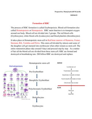
Formation of RBC
The process of RBC formation is called Erythropoiesis. Blood cell formation also
called Hematopoiesis or Hemopoiesis. RBC main function is to transport oxygen
around our body. Blood cell are divided into 3 groups. The red blood cells
(Erythrocytes), white blood cells (Leukocytes) and blood platelets (thrombocytes).
It takes place at Hematopoietic stem cell in Red bone marrow of Humerus, Femur,
Sternum, Rib, Vertebra and Pelvis. This stem cell divided by mitosis and some of
the daughter cell get matured into erythrocyte when other remain as stem cell. The
entire maturation phase take around 4 days and proceed step by step. As a matter
of fact all the blood cell are divided from these stem cell. RBC are formed and
destroyed at breathtaking rate. 200 billion RBC are destroyed and replace.
Hematopoietic stem cell
Pro-Erythroblast
Basophilic Erythroblast
Polychromatic Erythroblast
Reticulocytes
Erythrocytes
2. Area of the body that produce RBC
In early weeks of embryonic life, primitive , nucleated red blood cell are produced
in yolk sac. During the middle trimester of gestation, the liver is the main organ for
production of RBC, but reasonable number are also produced in the spleen and
lymph nodes. Then, during the last month or so of gestation and after birth, RBC
are produced exclusively in bone marrow.
All bone marrow Is red initially but after certain age, some bone marrow like
humerus, femur become fatty (yellow).
In adult, 2.4 million RBC are produced each second. In normal adult, the red cell
of about half a liter (almost one point) of blood is produced by the bone marrow
every week. RBC have a life span of approximately 100-120 days. After they have
completed their life span, they are removed from the blood stream by the spleen.
Regulation of erythropoiesis:
Kidney main function to monitor blood volume and oxygen content of blood. As
necessary kidney release Erythropoietin (EPO). When oxygen concentration in the
blood are low erythropoietin is released from the kidney. Erythropoietin operate in
a negative feedback mechanism to maintain RBC homeostasis.
There are few hormone that secret kidney ie, Thyroxin . The main job of thyroxin
is to increase the ATP production of cells. That means that cell need more oxygen
to make more ATP then they need more RBC. So Thyroxin stimulate the release of
EPO from the kidney.
Man have more Thyroxin then female because Testosterone stimulate Thyroxin. So
man have more RBC in their blood than women because Testosterone stimulated
Thyroxin which stimulate EPO.
3. Fate of RBC/ fate of Haemoglobin
Prepared by: Manoj karki (BVSC&AH)
2020/04/25
RBC
In spleen destroy
Hemoglobin
Phagocyte by macrophage
Fe + porphyrin portion
Converted to
Biliverdin
Bilirubin
Transfer by albumin
Bilirubin reach to liver
Conjugated bilirubin
Passed in the duodenum through
hepato pancreatic duct
Sterobilinogen Urobilinogen
Excreted from faeces
Stercobilin circulate in blood
4. Urobilin (excrete in urine)
As RBC is mature and aged, the metabolic system of the RBC become
progressively less active and the cell become more fragile. Many of the RBC are
self destroyed in the spleen where they squeeze through the red pulp of the spleen.
As the erythrocyte are destroyed the iron containing moiety of hemoglobin is
conserved and the pigmentary part is converted into bile pigment. Hemoglobin is
phagocytosis by macrophage in many part of the body (kupffer cell in liver,
macrophage of spleen and bone marrow). It release iron and prophyrin portion.
The propyrin portion of Hb is converted into biliverdin. The biliverdin is
converted into bilirubin. It is transported by albumin to the liver where it
undergoes conjugation with glucuronyl transferase thus forming conjugated
bilirubin. The conjugated bilirubin is passed into the duodenum through hepato-
pancreatic ducts.
The microbial fermentation of bilirubin convert it into stercobilinogen and
urobilinogen. The urobilinogen is again absorbs in blood and is excreted from
urine as urobilin. The stercobilinogen is excreted from feces as stercobilin.