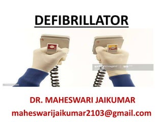
DEFIBRILLATOR
•Als PPTX, PDF herunterladen•
33 gefällt mir•14,855 views

DEFIBRILLATOR

Empfohlen
Weitere ähnliche Inhalte
Was ist angesagt?
Was ist angesagt? (20)
Ähnlich wie DEFIBRILLATOR
Ähnlich wie DEFIBRILLATOR (20)
Mehr von MAHESWARI JAIKUMAR
Mehr von MAHESWARI JAIKUMAR (20)
Kürzlich hochgeladen
Kürzlich hochgeladen (20)
DEFIBRILLATOR
- 3. • Defibrillators are devices that restore a normal heartbeat by sending an electric pulse or shock to the heart • Defibrillators can also restore the heart's beating if the heart suddenly stops
- 4. • Some times defibrillators are used to prevent or correct an arrhythmia (a heartbeat that is uneven or that is too slow or too fast. The parameters affected are rate & rhythm of the heart beat )
- 8. INDICATION FOR DEFIBRILLATION • Defibrillation is a treatment for life- threatening cardiac dysrhythmias, specifically ventricular fibrillation and non-perfusing ventricular tachycardia. • A defibrillator delivers a dose of electric current to the heart and restores the heart’s pulsation
- 10. MECHANISM OF FUNCTIONING • A defibrillator delivers a dose of electric current (often called a counter-shock) to the heart. • This process depolarizes a large amount of the heart muscle, ending the dysrhythmia.
- 11. • Subsequently, the body's natural pacemaker in the sinoatrial node of the heart is able to re-establish normal sinus rhythm. • A heart which is in asystole (flat line) cannot be restarted by a defibrillator, but would be treated by cardiopulmonary resuscitation (CPR).
- 12. TYPES OF DEFIBRILLATOR • Defibrillators can be external, transvenous,or implanted (impla ntable cardioverter-defibrillator), depending on the type of device used or needed
- 13. AUTOMATED EXTERNAL DEFIBRILLATORS • Some external units, known as automated external defibrillators (AEDs), automate the diagnosis of treatable rhythms, meaning that lay responders or by standers to use them successfully with little or no training.
- 15. CONTRA INDICATION FOR DEFIBRILLATION • Defibrillation is also not indicated if the patient is conscious or has a pulse. • Improperly given electrical shocks can cause dangerous dysrhythmias, such as ventricular fibrillation.
- 16. MANUAL EXTERNAL DEFIBRILLATOR • Manual external defibrillators require the expertise of a healthcare professional • They are used in conjunction with an electrocardiogram, which can be separate or built-in
- 17. • A healthcare provider first diagnose the cardiac rhythm and then manually determine the voltage and timing for the electrical shock. These units are primarily found in hospitals and on some ambulances
- 18. MANUAL INTERNAL DEFIBRILLATOR Manual internal defibrillators deliver the shock through paddles placed directly on the heart. • They are mostly used in the operating room and, in rare circumstances, in the emergency room during an open heart procedure.
- 19. AUTOMATED EXTERNAL DEFIBRILLATOR • An AED installed outside is designed for public use. Automated external defibrillators are designed for use by untrained personnel
- 20. • AEDs contain technology for analysis of heart rhythms • As a result, it does not require a trained health provider to determine whether or not a rhythm is shockable. AEDs have improved outcomes for sudden out-of- hospital cardiac arrests.
- 21. • AEDs have set voltages and does not allow the operator to vary voltage according to need. • AEDs may also delay delivery of effective CPR. • AED require the stopping of chest compressions and rescue breathing for diagnosis of heart rhythm
- 22. • AEDs have been incorporated into the algorithm for basic life support (BLS). Many first responders, such as fire fighters, policemen, and security guards, are equipped with them. • AEDs can be fully automatic or semi- automatic.
- 23. SEMI AUTOMATIC AED • A semi-automatic AED automatically diagnoses heart rhythms and determines if a shock is necessary. • If a shock is advised, the user must then push a button to administer the shock.
- 24. FULLY AUTOMATED AED • A fully automated AED automatically diagnoses the heart rhythm and advises the user to stand back while the shock is automatically given. • Some types of AEDs come with advanced features, such as a manual override or an ECG display.
- 25. IMPLANTABLE CARDIOVERTER- DEFIBRILLATOR • Automatic internal cardiac defibrillator (AICD) are implants, similar to pacemakers (and many can also perform the pacemaking function).
- 26. • AICD constantly monitor the patient's heart rhythm, and automatically administer shocks for various life-threatening arrhythmias, according to the device's programming.
- 27. • Many modern AICD devices can distinguish between ventricular fibrillation, ventricular tachycardia, and more benign arrhythmias like supraventricular tachycardia and atrial fibrillation.
- 28. • Some AICD devices may attempt overdrive pacing prior to synchronised cardioversion. When the life-threatening arrhythmia is ventricular fibrillation, the device is programmed to proceed immediately to an unsynchronized shock.
- 29. WEARABLE CARDIOVERTER DEFIBRILLATOR A wearable cardioverter defibrillator is a portable external defibrillator that can be worn by at-risk patients
- 30. • The unit monitors the patient 24 hours a day and can automatically deliver a biphasic shock if VF or VT is detected. • This device is mainly indicated in patients who are not immediate candidates for ICDs
- 31. INTERNAL DEFIBRILLATOR • Internal defibrillator is used to defibrillate the heart during or after cardiac surgery such as a heart bypass. • The electrodes consist of round metal plates that come in direct contact with the myocardium.
- 32. THE PROCEDURE • The connection between the defibrillator and the patient consists of a pair of electrodes, each provided with electrically conductive gel in order to ensure a good connection and to minimize electrical resistance, also called chest impedance (despite the DC discharge) which would burn the patient.
- 33. • Gel may be either wet (similar in consistency to surgical lubricant) or solid (similar to gummi candy). • Solid-gel is more convenient, because there is no need to clean the used gel off the person's skin after defibrillation.
- 34. • The use of solid-gel presents a higher risk of burns during defibrillation, since wet-gel electrodes more evenly conduct electricity into the body.
- 35. PADDLE ELECTRODES An AED with electrodes attached. • The most well-known type of electrode (widely depicted in films and television) is the traditional metal paddle with an insulated (usually plastic) handle.
- 36. • This type must be held in place on the patient's skin with approximately 25 lbs of force while a shock or a series of shocks is delivered. Paddles offer a few advantages over self-adhesive pads. • Many hospitals in the use of paddles, with disposable gel pads attached due to the inherent speed with which these electrodes can be placed and used.
- 37. • This is critical during cardiac arrest, as each second of nonperfusion means tissue loss. Modern paddles allow for monitoring (electrocardiography)
- 38. • Paddles are reusable, being cleaned after use and stored for the next patient. • Gel is therefore not pre applied, and must be added before these paddles are used on the patient. Paddles are generally only found on manual external units.
- 39. SELF-ADHESIVE ELECTRODES Newer types of resuscitation electrodes are designed as an adhesive pad, which includes either solid or wet gel. These are peeled off their backing and applied to the patient's chest when deemed necessary, much the same as any other sticker.
- 40. • The electrodes are then connected to a defibrillator. • If defibrillation is required, the machine is charged, and the shock is delivered, without any need to apply any additional gel or to retrieve and place any paddles.
- 41. • Most adhesive electrodes are designed to be used not only for defibrillation, but also for transcutaneous pacing and synchronized electrical cardioversion
- 42. • These adhesive pads are found on most automated and semi- automated units and are replacing paddles entirely in non-hospital settings. • In hospital, for cases where cardiac arrest is likely to occur , self- adhesive pads may be placed prophylactically.
- 43. • Pads also offer an advantage to the untrained user, and to medics working in the sub-optimal conditions of the field. • Pads do not require extra leads to be attached for monitoring, and they do not require any force to be applied as the shock is delivered.
- 44. • The adhesive electrodes minimize the risk of the operator coming into physical (and thus electrical) contact with the patient as the shock is delivered by allowing the operator to be up to several feet away.
- 45. • The risk of electrical shock to others remains unchanged, as does that of shock due to operator misuse. Self- adhesive electrodes are single-use only. • They may be used for multiple shocks in a single course of treatment, but are replaced if (or in case) the patient recovers then re enters cardiac arrest.
- 46. PLACEMENT OF PADDLES PLACEMENT OF ELECTRODES FOR DEFIBRILLATION • Resuscitation electrodes are placed according to one of two scheme as follows.
- 47. ANTERIOR POSTERIOR SCHEME • The anterior-posterior scheme is the preferred scheme for long-term electrode placement. Where one electrode is placed over the left precordium (the lower part of the chest, in front of the heart).
- 48. The other electrode is placed on the back, behind the heart in the region between the scapula. This placement is preferred because it is best for non-invasive pacing.
- 49. ANTERIOR-APEX SCHEME • The anterior-apex scheme can be used when the anterior-posterior scheme is inconvenient or unnecessary.
- 50. • In this scheme, the anterior electrode is placed on the right, below the clavicle. The apex electrode is applied to the left side of the patient, just below and to the left of the pectoral muscle. • This scheme works well for defibrillation and cardioversion, as well as for monitoring an ECG.
- 52. • Researchers have created a software modeling system capable of mapping an individual's chest and determining the best position for an external or internal cardiac defibrillator.
- 53. MECHANISM OF ACTION • The exact mechanism of defibrillation is not well understood. • One theory is that successful defibrillation affects most of the heart, resulting in insufficient remaining heart muscle to continue the arrhythmia.
- 54. REFERENCE • Ong, ME; Lim, S; Venkataraman, A (2016). "Defibrillation and cardioversion". In Tintinalli JE; et al. (eds.). Tintinalli's Emergency Medicine: A Comprehensive Study Guide, 8e. McGraw-Hill (New York, NY). • ^ Jump up to:a b c d e f Kerber, RE (2011). "Chapter 46. Indications and Techniques of Electrical Defibrillation and Cardioversion". In Fuster V; Walsh RA; Harrington RA (eds.). Hurst's The Heart (13th ed.). New York, NY: McGraw-Hill – via AccessMedicine. • ^ Werman, Howard A.; Karren, K; Mistovich, Joseph (2014). "Automated External Defibrillation and Cardiopulmonary Resuscitation". In Werman A. Howard; Mistovich J; Karren K (eds.). Prehospital Emergency Care, 10e. Pearson Education, Inc. p. 425. • ^ Author:Bradley P Knight, MD, FACCSection Editor:Richard L Page, MDDeputy Editor:Brian C Downey, MD, FACC. "Basic principles and technique of external electrical cardioversion and defibrillation". UpToDate. Retrieved 2019-07-24. • ^ Hoskins, MH; De Lurgio, DB (2012). "Chapter 129. Pacemakers, Defibrillators, and Cardiac Resynchronization Devices in Hospital Medicine". In McKean SC; Ross JJ; Dressler DD; Brotman DJ; Ginsberg JS (eds.). Principles and Practice of Hospital Medicine. New York, NY: McGraw-Hill – via Access Medicine. • ^ Jump up to:a b c Venegas-Borsellino, C; Bangar, MD (2016). "CPR and ACLS Updates". In Orpello JM; et al. (eds.). Critical Care. McGraw-Hill
- 55. THANK YOU