Weitere ähnliche Inhalte
Ähnlich wie Ameloblastoma of gingiva a case report
Ähnlich wie Ameloblastoma of gingiva a case report (20)
Mehr von Quách Bảo Toàn (17)
Kürzlich hochgeladen (20)
Ameloblastoma of gingiva a case report
- 1. ISSN 0975-8437 INTERNATIONAL JOURNAL OF DENTAL CLINICS 2011:3(2):111 -112
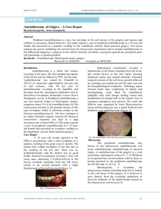
CASE REPORT
Ameloblastoma of Gingiva - A Case Report
Rosaiah Kanaparthy, Aruna Kanaparthy
Abstract
Peripheral ameloblastoma is a rare, but develops in the soft tissues of the gingiva and mucosa and
exhibits an innocuous clinical behavior. Th is paper reports a case of peripheral ameloblastoma in a 35-year-o ld
female that presented as a painless swelling on the mandibular anterior labial attached gingiva . This reports
emphasis the need for submitting all excised tissue for microscopic examination and to include ameloblastoma in
the differential diagnosis a gingival lesion wh ich clin ically resemb les a pyogenic granulo ma, peripheral giant
cell granuloma, or parulis/gumboil.
Keywords: Ameloblastoma; Odontogenic tumor; gingiva
Received on: 28/04/2011 Accepted on: 09/05/2011
Introduction Radio logical examination revealed a
A meloblastoma is a rather rare tumour mu ltilocular cystic lesion extending fro m the lo wer
occurring in the jaws. The first detailed description left central incisor to the first mo lar showing
of this lesion was by Falkson in 1879, but the term displaced canine and second premo lar. Extended
‘ameloblastoma’ was coined by Churchill in surgical excision of the soft tissue growth fo llo wed
1933.(1) It represents approximately one per cent by curettage was carried out and the mass was sent
of oral tu mours, with 80 per cent of for h istopathological evaluation. Pathology report
ameloblastomas occurring in the mandib le, and showed tumor mass comprising of islands and
develops fro m the odontogenic epitheliu m and its interdigitating cords lined by ameloblastic
derivatives or remnants. So met imes it arises fro m a epithelial cells with benign looking nuclei and
dentigerous cyst.(2, 3) Peripheral ameloblastoma, a proliferation of stromal cells in the islands. Foci of
rare and unusual variant of odontogenic tumour, squamous metaplasia were present. The cords and
comprises about 1% of all ameloblastomas.(4) The follicles were separated by loose fibroconnective
extraosseous location is the peculiar feature of this tissue and the diagnosis was a mixed fo llicu lar and
type of tumour, which is otherwise similar to the plexiform type ameloblastoma (Figure 2).
classical ameloblastoma.(5) The best treatment is
an initial extended surgical excision.(6) However
conservative treatment can lead to a high
recurrence rate of about 90%.(1) This paper reports
a case of peripheral ameloblastoma in a 35-year-
old female that presented as a painless swelling on
the mandibular anterior labial attached gingiva.
Case Report
A 35 year o ld wo man reported in the Fig 2 Histopathological slide showing anastomosis and follicles
OPD o f periodontics with a chief co mp laint of a Discussion
painless swelling of the gums since 6 months. The The peripheral ameloblastoma, also
patient had a slight asymmetry of the face due to known as the extraosseous ameloblastoma, soft
the swelling on the left side. There was no tissue ameloblastoma, ameloblastoma of mucosal
ly mphadenopathy and the patient was in good origin, or ameloblastoma of the gingiva is a very
health. Intraoral examination revealed a firm soft uncommon odontogenic tumour. (5, 7, 8) Philipsen
tissue mass measuring 2.5cmX2cmX1cm in the et al reported that several authors refer to Kuru as
buccal vestibule extending fro m the left lo wer having reported on the peripheral ameloblastoma
canine to the second premolar with a slight for the first time in 1911.(5, 7)
displacement of the teeth involved (Figure 1). Histologically, it resemb les the
intraosseous common ameloblastoma but is limited
to the soft tissue of the gingiva. It is believed to
arise directly fro m the overlying epitheliu m or
fro m the remnants of the dental lamina located in
the extraosseous soft tissue.(5, 9)
Fig 1 Preoperative view
©INT ERNA TIONA L JOURNA L OF D ENT AL CL IN ICS VOLU ME 3 ISS UE 2 APRIL-JUN E 2011 111
- 2. ISSN 0975-8437 INTERNATIONAL JOURNAL OF DENTAL CLINICS 2011:3(2):111 -112
Patients have been documented fro m 23 2. Iordanidis S, M akos C, Dimitrakopoulos J, Kariki
years to 82 years of age, and lesions occur on the H. Ameloblastoma of the maxilla. Case report.
mandib le twice as often as on the maxilla. There Australian Dental Journal. 1999;44(1):51-5.
3. Shteyer A, Lustmann J, Lewin-Epstein J. The mural
was no difference in location between the left and
ameloblastoma: a review of the literature. Journal of
right side of the jaws.(4, 5) Ou r patient was 35 oral surgery.1978;36(11):866-72.
years old. The lesion was a 2.5 cm painless, non- 4. Ficarra G, Hansen LS. Peripheral ameloblastoma::
ulcerated growth on the buccal attached gingiva of A case report. Journal of Cranio-M axillofacial
the premolars of the left mandibu lar reg ion. The Surgery. 1987;15:110-2.
lesion was covered by normal mucosa with a 5. Pekiner F, Ozbayrak S, Sener B, Olgac V,
smooth surface. Thus, this lesion is similar to those Sinanoglu A. Peripheral ameloblastoma: a case
described in the literature.(4) There was no report. Dentomaxillofacial Radiology. 2007;36 (3):
radiological evidence of bone involvement. The 183-6.
6. M athur LK, Bhalodi AP, M anohar B, Bhatia A, Rai
lesions had an inferior marg in that was superficial
N, M athur A. Focal fibrous hyperplasia: a case
to the cortical bone. report. International Journal of Dental Clinics.
The types of treatment that can been used 2010; 2(4):56-7.
include both radical and conservative surgical 7. Philipsen H, Reichart P, Nikai H, Takata T, Kudo
excision, curettage, chemical and electrocautery, Y. Peripheral ameloblastoma: biological profile
radiation therapy or a combination of surgery and based on 160 cases from the literature. Oral
radiation.(4, 10) In our case the ameloblastoma was Oncology. 2001;37(1):17-27.
treated conservatively in february2009 and so far 8. Reddy R, Jain U, US S, Agarwal N. Idiopathic
there has been no recurrence. The prognosis of Gingival Enlargement-An Inter-Disciplinary
Approach. International Journal of Dental Clinics.
patients afflicted with this form of neoplastic
2011;3(1):92-3.
disease is favorable, since it is essentially a local 9. Wettan HL, Patella PA, Freedman PD. Peripheral
problem. ameloblastoma: review of the literature and report
Conclusion of recurrence as severe dysplasia. Journal of oral
In conclusion this reports emphasis the need and M axillofacial Surgery. 2001;59(7):811-5.
for submitting all excised tissue for microscopic 10. M arkose E, M ani V. Ameloloblastic carcinoma-a
examination and to include ameloblastoma in the case report. International Journal of Dental Clinics.
differential diagnosis a gingival lesion wh ich 2010;2(4):54-5.
clin ically resembles a pyogenic granulo ma, Address for correspondence
peripheral giant cell granuloma, or parulis/gumboil. Dr. Rosaiah Kanaparthy, M DS,
Authors Affiliations: 1. Dr. Rosaiah Kanaparthy, M DS, Professor and HOD,
Professor and HOD, Dept. of Periodontics, 2. Dr. Aruna Dept. of Periodontics,
Kanaparthy, M DS, Senior Lecturer, Conservative Peoples Dental Academy,
Dentistry, Peoples Dental Academy , Bhopal, M adhya Bhopal, M adhyapradesh, India.
pradesh, India. Ph:+0091.9893050554
References Email: medha98@gmail.com
1. Larsson Å, Almerén H. Ameloblastoma of the jaws.
Acta Pathologica M icrobiologica Scandinavica
Section A Pathology. 1978;86(1‐6):337-49.
Source of Support: Nil, Conflict of Interest: None Declared
©INT ERNA TIONA L JOURNA L OF D ENT AL CL IN ICS VOLU ME 3 ISS UE 2 APRIL-JUN E 2011 112