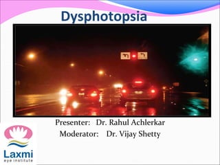
Dysphotopsia
•Als PPTX, PDF herunterladen•
37 gefällt mir•19,040 views

This document summarizes a presentation on dysphotopsia, or unwanted visual images, following cataract surgery. The key points are: - Dysphotopsia is a common complaint following uncomplicated cataract surgery, with 1 in 10 patients experiencing symptoms. Negative dysphotopsia, involving a dark shadow, is most prevalent. - Factors influencing dysphotopsia include intraocular lens (IOL) edge design, material, and coverage of the anterior capsule. Newer IOL designs have reduced symptoms by minimizing light scattering and reflections. - Managing patient expectations before surgery and using techniques like overlapping the capsulorhexis rim over the IOL edge can help reduce dys

Empfohlen
Empfohlen
Weitere ähnliche Inhalte
Was ist angesagt?
Was ist angesagt? (20)
Andere mochten auch
Andere mochten auch (13)
Ähnlich wie Dysphotopsia
Ähnlich wie Dysphotopsia (20)
Mehr von Laxmi Eye Institute
Mehr von Laxmi Eye Institute (20)
Kürzlich hochgeladen
Kürzlich hochgeladen (20)
Dysphotopsia
- 1. Dysphotopsia Presenter: Dr. Rahul Achlerkar Moderator: Dr. Vijay Shetty
- 2. Imagine this You've just performed successful, uncomplicated cataract surgery. Your patient is 20/20 and the surgery looks beautiful you're ready to be congratulated.
- 3. Instead, your patient says, "I hate it! These unwanted images are driving me crazy! You've got to do something about this”
- 4. Of course, this isn't what you want to hear. But the reality is that dysphotopsia has become the number one problem following uncomplicated, successful cataract surgery. And it doesn't go away easily once a patient becomes focused on it.
- 5. Unfortunately, many of these patients are incredibly unhappy. most of whom have told that they're crazy. "Your surgery is perfect,". "There's nothing wrong here." This has entirely the wrong effect, making the patient angrier and even more focused on the unwanted images.
- 6. The Nature of the Problem The no.of patients who actually require an intraocular lens exchange is only about 1 in a 1000 However, the number of patients complaining about dysphotopsia is closer to 1 in 10
- 7. So what's behind the current wave of dysphotopsia complaints? The first element is what the patient is actually seeing. The second element is how the patient reacts to the symptom the patient's reaction can be the most significant factor in resolving (or not resolving) the problem
- 8. What the Patient Sees - temporal darkness - arc - Flare - a central flash
- 9. In the literature, terms such as photopsias, entoptic phenomena photic phenomena have been used to describe these images. In June 2000, the term “dysphotopsia” was first used
- 11. Positive dysphotopsia is usually related to bright artifacts of light on the retina Negative dysphotopsia is manifested by a dark crescent or curved shadow
- 12. The exact etiology of negative dysphotopsia remains an enigma. The question of why this dark shadow of light occurs temporally because the nasal retina may extend further anteriorly than the temporal retina as well as because light coming in nasally may be somewhat tempered by the nose, eyebrow and cheek
- 13. Continued….. However, light coming from the temporal side of the eye that projects to the nasal-most retina may be deflected by the edge of the IOL or even reflected internally by the relatively square edge of the IOL away from the nasal retina. This results in a crescent-shaped shadow noted in the temporal field of vision.
- 14. • Temporal darkness temporal darkness, or negative dysphotopsia, is the most prevalent symptom today.(30 to 40 %)* In this case, the patient detects a black shadow temporally, in the periphery of vision. *Vámosi P, Csákány B, Németh J. Intraocular lens exchange in patients with negative dysphotopsia symptoms. J Cataract Refract Surg. 2010;36(3):418-24.
- 15. Arc patient perceiving the edge of the IOL, which usually only happens at night. It's a common complaint and rarely a serious problem It usually resolves over time—especially if the capsule overlaps the IOL edge.
- 16. Flare This is also a scotopic symptom produced by coma. Correcting minimal cylinder with night driving glasses will often get rid of it. Making the pupil a little smaller at night will also help.
- 17. Central flash this appears to be caused by a peripheral light source reflecting off the internal edge of the IOL Recent advances in edge design have minimized this symptom
- 18. Haloes These may be caused by a multifocal IOL produces haloes around lights from each ring transition zone. Most patients will adapt to this, and a smaller scotopic pupil can help in the meantime.
- 19. night haloes are the number one reason these IOLs are explanted. If patients see haloes with a monofocal IOL, it usually indicates the presence of spherical aberration. new aspheric-optic IOLs will help
- 20. How the Patient Reacts Difficult to eliminate all unwanted images from patient's vision. The brain is adapt at eliminating unwanted visual input by phenomenon of central adaptation. the most obvious example is the hole in our visual field where the optic nerve enters the eye.
- 21. Physiology….. In addition, we get -front- and backscatter off our natural lens, - pupils are irregular, -blood vessels in our retina that we can't see through. there are a lot of unwanted images in our field of vision, but our brain adapts and eliminates them all.
- 22. Managing Dysphotopsia it's inevitable that some patients will experience unwanted images. In these cases, doing the right things before and after surgery can avoid the greater problem
- 23. • Create accurate expectations before surgery Explain to patient about dysphotopsia pre operatively Then, the patient won't be surprised if some new, unwanted visual effect accompanies the new lens
- 24. Minimize the problem surgically 1 Use the right lens Certain IOL characteristics appear to correlate with reduced dysphotopsia. Newer lenses have helped by increasing the front curvature of the lens, which minimizes front and back light scattering
- 25. Optic size is important, because a smaller lens may create more edge problems. d0n't implant a lens any smaller than 6 mm
- 26. All the IOLs studied variably increased internal and external surface reflections when compared to the human crystalline lens.
- 27. SQUARE EDGE DESIGN While the square-edge optic is clearly favored for reducing the risk of PCO, the trade-off for that benefit is an increased rate of pseudophakic dysphotopsia.
- 28. Pseudophakic dysphotopsia. square-edged optic is one responsible factor causing dysphotopsia. The other factors responsible are - the index of refraction of the IOL material, - corneal curvature - pupil size.
- 29. Truncated posterior edge offers barrier effect to lens epithelial cells Sloped edge-minimises internally reflected rays that form arc like images Rounded anterior edge- eliminates mirror effect
- 30. Continued….. Round edge of the optic causes greater dispersion of the internally reflected rays of light, reducing edge glare by 90 percent Increasing the front curvature of the Newer lenses has helped minimize front and back light scattering, reducing glare
- 31. Double square edge When light hits the double-square edge Lens at 23 degrees, little edge glare. at 35 degrees, one begins to see arcs at 55 degrees, transmitted as well as reflected glare becomes significantly more evident.
- 32. Continued…. silicone lens with a rounded edge and lower refractive index seems to be most forgiving, producing the fewest complaints about unwanted images.
- 33. 2. Place the lens carefully A well-centered, in-the-bag lens prevents unnecessary optical problems.
- 34. 3. Overlap the capsulorhexis rim over the edge of the lens The edge of the capsulorhexis will tend to opacify over time the opaque overlap will eliminate many symptoms associated with the edge of the IOL. The brain seems to ignore the edge of the capsule, reacting as it does to the edge of the pupil.
- 35. another major benefit This strategy has another major benefit If we overlap the capsule, we will significantly decrease posterior capsule opacification. Two recent studies, show that overlap of the capsule is more effective at preventing "aftercataract" than switching to an IOL with a truncated edge
- 36. Continued…. Making a smaller capsulorhexis has some potential downsides. It can be more difficult to access the lens, particularly if we use the Phaco technique. Also, we don't want to risk capsular contracture by making the opening too small
- 37. Continued…… To minimize dysphotopsia and PCO, the opening should be roughly 1 mm smaller than the size of the optic, to ensure 360-degree overlap And use at least a 6-mm optic
- 38. After surgery, don't take the wrong attitude if a patient complains The worst thing you can do if a patient complains is to say, "Your result is perfect. Nobody else is complaining. What's your problem?" This virtually guarantees that the patient will "turn up the gain," and fail to adapt to the unwanted images.
- 39. Resolving a Dysphotopsia Crisis Talk to the patient (and say the right thing). First of all, let the patient know that he/she's not crazy. That alone will improve matters. Try night time pupil constriction
- 40. don't open the capsule Whatever you do, don't open the capsule Some ophthalmologists, thinks May be patient got aftercataract. So let's go ahead and do a YAG capsulotomy and see if that will make it better if the problem truly is dysphotopsia, a capsulotomy won't have any positive effect at all
- 41. When we try to take the lens out after a YAG capsulotomy, vitreous comes forward. we often can't put the lens back in the capsule because the capsulotomy tears further. The risk of endophthalmitis and retinal detachment increase dramatically.
- 42. lens exchange ??? • Only resort to lens exchange if it really makes sense. First of all, make sure the patient has had enough time to adapt. If even after six months problems continued then a lens exchange can be consider —only if it improve on the existing lens situation. Otherwise, switching lenses will be a waste of time.
- 43. Factors deciding lens exchange 1. The size of the existing capsulorhexis If the optic is small then larger optic will create more overlap of the edge, this can solve problem 2. Edge design If the current IOL doesn't have an up-to-date edge design, then switching to an updated lens would be helpful
- 44. 3 Refractive index If the current lens has a high refractive index, switching to a rounded-edge silicone lens may be curative, particularly if there is negative dysphotopsia. 4 Condition of the capsule If another surgeon has performed a YAG capsulotomy, a lens exchange will involve more risk.
- 45. If all else fails For some patients,nothing will relieve the symptoms, and IOL exchange may not make sense if the patient already has the most beneficial type and size of IOL. In that case, talk to the patient again and do best to help him or her to relax and adviced to stop thinking about it so much, so the brain has a chance to adapt.
- 46. Pseudophakic Dysphotopsia with Various Intraocular Lens One study was conducted in our institute on Pseudophakic Dysphotopsia with Various Intraocular Lens Highlights of study 1)The incidence of dysphotopsia found to be 51.12% 2) The incidence of negative dysphotopsia has been found to be 22.47%
- 47. 3)The eyes implanted with Tecnis ZCB00 IOL showed less negative temporal shadow/darkness 4) Hydrophilic Acrylic IOLs showed greater dysphotopsia score in comparison to those with Silicone IOLs 5) Hydrophilic versus Hydrophobic Acrylic, the latter was found to be significantly better with a lower Dysphotopsia.
- 48. 6)Hydrophobic Acrylic IOLs when compared to Hydrophilic Acrylic IOLs and Silicone IOLs showed decrease in night-time glare/halo/circles 7) An increase in the optic-haptic angle caused an increase in night-time glare/halos/circles around lights.
- 49. CONCLUSION Tecnis ZCB00 emerged as least troublesome lens while Auroflex FH5575 which was reported to have the highest Dysphotopsia. Hence, we may conclude that different brands of intra-ocular lenses display varying degrees of dysphotopic symptoms.
- 50. Recent updates new hypothesis, resolution of negative dysphotopsia symptoms depended on intraocular lens (IOL) coverage of the anterior capsule edge rather than on collapse of the posterior chamber alone. Negative dysphotopsia was not attributed to a particular IOL material or edge design Pseudophakic negative dysphotopsia: Surgical management and new theory of etiology Journal of Cataract & Refractive Surgery, 06/24/2011
- 51. New concept Two rays, coming in from the temporal side at 90°, are bent by the cornea by about 45°. As they come through, one ray, if there is a space between the iris and the anterior surface of the lens, can miss the front part of the lens Hawaiian Eye meeting, Jack Holladay,
- 52. New concept while the other ray hits the lens and is bent by the lens's refractive power. In the cone between those two rays, no light can enter, and this causes what is perceived by the patient as a crescent-shaped shadow* *Dr. Holladay said Hawaiian Eye meeting
- 53. In the first day after IOL implantation, approximately 15% of patients experience negative dysphotopsia. By 3 years, the phenomenon is reduced to only 5% To treat negative dysphotopsia, we have to eliminate the rays that pass anterior to the IOL and to do so we have to reduce the space between the iris and the anterior surface of the IOL* * Dr. Holladay said.
- 54. This reduction may occur spontaneously in some cases with the natural forward movement of the IOL after capsular bag contraction. The opacification of the equatorial capsule, occurring naturally several weeks or months after implantation, is also likely to reduce the shadow effect. we can otherwise flip the optic, though this might induce myopia, can implant a piggyback IOL in the sulcus. Frosted- edge IOLs are another solution
- 55. Two surgical strategies have emerged as beneficial treatment of persistent visual symptoms of ND: reverse optic capture (ROC) secondary “piggyback” IOL. Failed surgical strategies include bag/bag IOL exchange wherein the original implant is removed and another of different material, shape or edge design is replaced within the capsular bag.* * This is in keeping with the work of Vámosi et al.3
- 56. Reverse Optic Capture ROC may be employed in a secondary surgery for symptomatic patients, or as a primary prophylactic strategy. In cases of the latter, the method has been applied to the second eye of patients who were significantly symptomatic following routine uncomplicated surgery in their first eye. It should be noted, however, that ND symptoms are not necessarily bilateral.
- 57. Secondary ROC, performed for symptomatic patients, may be applied if the anterior capsulotomy is not too small or too thick or rigid from postoperative fibrosis. The first step involves freeing the anterior capsule from the underlying optic by gentle blunt dissection and viscodissection.
- 58. Gentle blunt dissection and viscodissection of the anterior capsule from the underlying optic
- 59. Next, the nasal anterior capsule edge is retracted with one Sinskey hook (or similar device) while the optic edge is elevated and the capsule edge allowed to slip under the optic. This maneuver is repeated 180 degrees away temporally, leaving the haptics undisturbed in the bag inferiorly and superiorly.
- 60. . A Sinskey hook and blunt spatula are used to elevate the nasal optic edge over the capsule
- 61. the haptics be oriented horizontally, it would be best to rotate them 90 degrees if possible The optic is then confirmed to be elevated over the anterior capsule edge and the nasal and temporal edges of the implant are anterior to the anterior capsule, whereas the haptics remain within the capsular bag.
- 62. Optic capture has been completed. The nasal and temporal edges of the implant are anterior to the anterior capsule (see arrows), whereas the haptics remain fully within the capsular bag.
- 63. Once the nasal edge has been captured (arrow), the opposite, temporal edge of the optic is elevated over the anterior capsule edge.
- 64. Secondary “Piggyback” IOL Secondary “piggyback” IOL is the other surgical method that has proven successful for patients with symptomatic ND, as first reported by Ernest. In this method, a second IOL is implanted in the ciliary sulcus above the primary IOL/capsule bag complex. It appears that covering the primary optic/capsule junction reduces ND symptoms
- 65. although the original concept was that a “piggyback” lens was effective because it collapsed the posterior chamber by reducing the distance between the posterior iris and the anterior surface of the IOL. However, studies *have determined that the depth of the posterior chamber is unrelated to ND symptoms.2 *Vámosi et al.,march 2011
- 66. Symptomatic patients may be good candidates for a “piggyback” IOL if they are also ammetropic. In order to qualify for a “piggyback,” the first IOL surgery should be uncomplicated with a well- centered IOL within the capsule bag. There should be no evidence of zonulopathy and the iris must be free of defects or damage from earlier surgery.
- 67. Although no parameters have been clearly established, it ia better to perform a UBM to ascertain adequate space (approximately 1 mm) between the posterior iris and the existing IOL/bag complex
- 68. There are two kinds of light- the glow that illuminates and the glare that obscures. James Thurber
- 70. Thank you