SlideShare ist ein Scribd-Unternehmen logo
1 von 2
TIDAL PERCUSSION
• To distinguish whether the cause of dullness over the lower part of the chest is due to
– upward enlargement of liver or spleen, or
– intra thoracic pathology.
• Method :
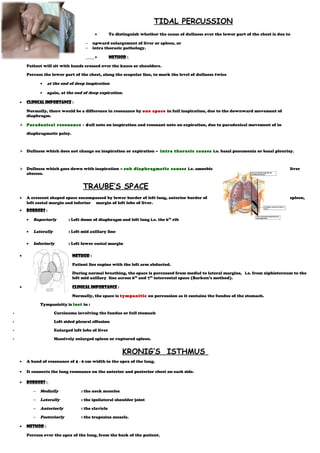
Patient will sit with hands crossed over the knees or shoulders.
Percuss the lower part of the chest, along the scapular line, to mark the level of dullness twice
• at the end of deep inspiration
• again, at the end of deep expiration.
• Clinical Importance :
Normally, there would be a difference in resonance by one space in full inspiration, due to the downward movement of
diaphragm.
 Paradoxical resonance - dull note on inspiration and resonant note on expiration, due to paradoxical movement of in
diaphragmatic palsy.
 Dullness which does not change on inspiration or expiration – intra thoracic causes i.e. basal pneumonia or basal pleurisy.
 Dullness which goes down with inspiration – sub diaphragmatic causes i.e. amoebic liver
abscess.
TRAUBE’S SPACE
• A crescent shaped space encompassed by lower border of left lung, anterior border of spleen,
left costal margin and inferior margin of left lobe of liver.
• Borders :
• Superiorly : Left dome of diaphragm and left lung i.e. the 6th
rib
• Laterally : Left mid axillary line
• Inferiorly : Left lower costal margin
• Method :
Patient lies supine with the left arm abducted.
During normal breathing, the space is percussed from medial to lateral margins, i.e. from xiphisternum to the
left mid axillary line across 6th
and 7th
intercostal space (Barkun’s method).
• Clinical Importance :
Normally, the space is tympanitic on percussion as it contains the fundus of the stomach.
Tympanicity is lost in :
- Carcinoma involving the fundus or full stomach
- Left sided pleural effusion
- Enlarged left lobe of liver
- Massively enlarged spleen or ruptured spleen.
KRONIG’S ISTHMUS
• A band of resonance of 5 - 6 cm width in the apex of the lung.
• It connects the lung resonance on the anterior and posterior chest on each side.
• Borders :
– Medially : the neck muscles
– Laterally : the ipsilateral shoulder joint
– Anteriorly : the clavicle
– Posteriorly : the trapezius muscle.
• Method :
Percuss over the apex of the lung, from the back of the patient.
Pleximeter finger should be placed over the supraclavicular fossa, across the anterior border of trapezius, perpendicular to the
clavicle.
• Clinical importance :
Normally the note is resonant.
Area becomes dull in :
– Apical tuberculosis, Pancoast’s tumour, Apical pneumonia.

Weitere ähnliche Inhalte

Was ist angesagt?

Examination of gastrointestinal system by HX
Examination of gastrointestinal system by HXExamination of gastrointestinal system by HX
Examination of gastrointestinal system by HX
Dr. Rubz
 

Was ist angesagt? (20)

Clubbing
ClubbingClubbing
Clubbing
 
Pleural effusion(X-ray Findings)
Pleural effusion(X-ray Findings)Pleural effusion(X-ray Findings)
Pleural effusion(X-ray Findings)
 
Abdominal Exam
Abdominal ExamAbdominal Exam
Abdominal Exam
 
Approach to Chest X-Ray and Interpretation
Approach to Chest X-Ray and InterpretationApproach to Chest X-Ray and Interpretation
Approach to Chest X-Ray and Interpretation
 
Chest X-rays for Undergraduates
Chest X-rays for UndergraduatesChest X-rays for Undergraduates
Chest X-rays for Undergraduates
 
Types of Arterial pulses
Types of  Arterial  pulsesTypes of  Arterial  pulses
Types of Arterial pulses
 
Clinical Examination of CVS
Clinical Examination of CVSClinical Examination of CVS
Clinical Examination of CVS
 
Thyroid surgical anatomy
Thyroid surgical anatomyThyroid surgical anatomy
Thyroid surgical anatomy
 
Abdomen xray signs
Abdomen xray signsAbdomen xray signs
Abdomen xray signs
 
IriS
IriSIriS
IriS
 
Abnormal Chest xray
Abnormal Chest xray Abnormal Chest xray
Abnormal Chest xray
 
Examination of the respiratory system
Examination of the respiratory systemExamination of the respiratory system
Examination of the respiratory system
 
Abdominal X ray
Abdominal X rayAbdominal X ray
Abdominal X ray
 
Hepatospleenomegaly in children
Hepatospleenomegaly in childrenHepatospleenomegaly in children
Hepatospleenomegaly in children
 
Radiological features of pneumonia
Radiological features of pneumoniaRadiological features of pneumonia
Radiological features of pneumonia
 
Radiographic manifestations of pulmonary tuberculosis
Radiographic manifestations of pulmonary tuberculosisRadiographic manifestations of pulmonary tuberculosis
Radiographic manifestations of pulmonary tuberculosis
 
Lung consolidation
Lung consolidationLung consolidation
Lung consolidation
 
Clubbing
ClubbingClubbing
Clubbing
 
Chest xray
Chest xray  Chest xray
Chest xray
 
Examination of gastrointestinal system by HX
Examination of gastrointestinal system by HXExamination of gastrointestinal system by HX
Examination of gastrointestinal system by HX
 

Ähnlich wie Jerrin's

Percussion in respiratory system
Percussion in respiratory systemPercussion in respiratory system
Percussion in respiratory system
Kurian Joseph
 
14828percussion_and_auscultation_of_the_chest[1].pdf
14828percussion_and_auscultation_of_the_chest[1].pdf14828percussion_and_auscultation_of_the_chest[1].pdf
14828percussion_and_auscultation_of_the_chest[1].pdf
axmedfare138
 
Cardiopulmonary assessment
Cardiopulmonary assessment Cardiopulmonary assessment
Cardiopulmonary assessment
Sakshee Jain
 
Abdomen history and physical examination.pdf
Abdomen history and physical examination.pdfAbdomen history and physical examination.pdf
Abdomen history and physical examination.pdf
angelicocos1
 
Anatomy of gastroesophagial junction with specail reference to hiatus hernia...
Anatomy of gastroesophagial junction  with specail reference to hiatus hernia...Anatomy of gastroesophagial junction  with specail reference to hiatus hernia...
Anatomy of gastroesophagial junction with specail reference to hiatus hernia...
Rana Singh
 

Ähnlich wie Jerrin's (20)

Clinical examination of Respiratory System by Pandian M, tutor, Dept of Physi...
Clinical examination of Respiratory System by Pandian M, tutor, Dept of Physi...Clinical examination of Respiratory System by Pandian M, tutor, Dept of Physi...
Clinical examination of Respiratory System by Pandian M, tutor, Dept of Physi...
 
Chest examination
Chest examinationChest examination
Chest examination
 
Percussion in respiratory system
Percussion in respiratory systemPercussion in respiratory system
Percussion in respiratory system
 
14828percussion_and_auscultation_of_the_chest[1].pdf
14828percussion_and_auscultation_of_the_chest[1].pdf14828percussion_and_auscultation_of_the_chest[1].pdf
14828percussion_and_auscultation_of_the_chest[1].pdf
 
Dr. Adanwali Hassan Physical examination.pptx
Dr. Adanwali Hassan Physical examination.pptxDr. Adanwali Hassan Physical examination.pptx
Dr. Adanwali Hassan Physical examination.pptx
 
Cardiopulmonary assessment
Cardiopulmonary assessment Cardiopulmonary assessment
Cardiopulmonary assessment
 
Surgical Anatomy of Esophagus
Surgical Anatomy of EsophagusSurgical Anatomy of Esophagus
Surgical Anatomy of Esophagus
 
Ivor lewis esophagectomy
Ivor lewis esophagectomyIvor lewis esophagectomy
Ivor lewis esophagectomy
 
CPT
CPTCPT
CPT
 
Diaphragm and mechanism of respiration.pptx
Diaphragm and mechanism of respiration.pptxDiaphragm and mechanism of respiration.pptx
Diaphragm and mechanism of respiration.pptx
 
Learn Chest X-Ray With Its Normal Positioning & Radio-Anatomy
Learn Chest X-Ray With Its Normal Positioning & Radio-AnatomyLearn Chest X-Ray With Its Normal Positioning & Radio-Anatomy
Learn Chest X-Ray With Its Normal Positioning & Radio-Anatomy
 
Chest xray
Chest xrayChest xray
Chest xray
 
Femoral hernia with umbilical hernia
Femoral hernia with umbilical herniaFemoral hernia with umbilical hernia
Femoral hernia with umbilical hernia
 
Abdomen history and physical examination.pdf
Abdomen history and physical examination.pdfAbdomen history and physical examination.pdf
Abdomen history and physical examination.pdf
 
abdominal wall final (1).pptx
abdominal wall final (1).pptxabdominal wall final (1).pptx
abdominal wall final (1).pptx
 
Spleen...
Spleen...Spleen...
Spleen...
 
SURGICAL ANATOMY
SURGICAL ANATOMYSURGICAL ANATOMY
SURGICAL ANATOMY
 
Lecture 1- Esophagus and stomach.ppt
Lecture 1- Esophagus and stomach.pptLecture 1- Esophagus and stomach.ppt
Lecture 1- Esophagus and stomach.ppt
 
Respiratory examination
Respiratory examinationRespiratory examination
Respiratory examination
 
Anatomy of gastroesophagial junction with specail reference to hiatus hernia...
Anatomy of gastroesophagial junction  with specail reference to hiatus hernia...Anatomy of gastroesophagial junction  with specail reference to hiatus hernia...
Anatomy of gastroesophagial junction with specail reference to hiatus hernia...
 

Mehr von Kurian Joseph (20)

Vj tongue
Vj tongueVj tongue
Vj tongue
 
Urine casts &crystals
Urine casts &crystalsUrine casts &crystals
Urine casts &crystals
 
Urinary bladder nerve supply
Urinary bladder nerve supply Urinary bladder nerve supply
Urinary bladder nerve supply
 
Types of portal hypertension
Types of portal hypertensionTypes of portal hypertension
Types of portal hypertension
 
Trigeminal
TrigeminalTrigeminal
Trigeminal
 
Faces in medicine
Faces in medicine Faces in medicine
Faces in medicine
 
Superficial reflexes
Superficial reflexesSuperficial reflexes
Superficial reflexes
 
Signs of aortic regurgitation
Signs of aortic regurgitationSigns of aortic regurgitation
Signs of aortic regurgitation
 
Sensory dermatomes
Sensory dermatomesSensory dermatomes
Sensory dermatomes
 
Second heart sound
Second heart soundSecond heart sound
Second heart sound
 
S3 and s4 heart sounds
S3 and s4 heart soundsS3 and s4 heart sounds
S3 and s4 heart sounds
 
Rheumatic fever
Rheumatic feverRheumatic fever
Rheumatic fever
 
Pupils
PupilsPupils
Pupils
 
Pulse
PulsePulse
Pulse
 
Primitive reflexes
Primitive reflexesPrimitive reflexes
Primitive reflexes
 
Parkinson's disease
Parkinson's diseaseParkinson's disease
Parkinson's disease
 
Para sternal puls (bavya)
Para sternal puls (bavya)Para sternal puls (bavya)
Para sternal puls (bavya)
 
Nystagmus
NystagmusNystagmus
Nystagmus
 
Kidn
KidnKidn
Kidn
 
Internal capsule
Internal capsuleInternal capsule
Internal capsule
 

Jerrin's

  • 1. TIDAL PERCUSSION • To distinguish whether the cause of dullness over the lower part of the chest is due to – upward enlargement of liver or spleen, or – intra thoracic pathology. • Method : Patient will sit with hands crossed over the knees or shoulders. Percuss the lower part of the chest, along the scapular line, to mark the level of dullness twice • at the end of deep inspiration • again, at the end of deep expiration. • Clinical Importance : Normally, there would be a difference in resonance by one space in full inspiration, due to the downward movement of diaphragm.  Paradoxical resonance - dull note on inspiration and resonant note on expiration, due to paradoxical movement of in diaphragmatic palsy.  Dullness which does not change on inspiration or expiration – intra thoracic causes i.e. basal pneumonia or basal pleurisy.  Dullness which goes down with inspiration – sub diaphragmatic causes i.e. amoebic liver abscess. TRAUBE’S SPACE • A crescent shaped space encompassed by lower border of left lung, anterior border of spleen, left costal margin and inferior margin of left lobe of liver. • Borders : • Superiorly : Left dome of diaphragm and left lung i.e. the 6th rib • Laterally : Left mid axillary line • Inferiorly : Left lower costal margin • Method : Patient lies supine with the left arm abducted. During normal breathing, the space is percussed from medial to lateral margins, i.e. from xiphisternum to the left mid axillary line across 6th and 7th intercostal space (Barkun’s method). • Clinical Importance : Normally, the space is tympanitic on percussion as it contains the fundus of the stomach. Tympanicity is lost in : - Carcinoma involving the fundus or full stomach - Left sided pleural effusion - Enlarged left lobe of liver - Massively enlarged spleen or ruptured spleen. KRONIG’S ISTHMUS • A band of resonance of 5 - 6 cm width in the apex of the lung. • It connects the lung resonance on the anterior and posterior chest on each side. • Borders : – Medially : the neck muscles – Laterally : the ipsilateral shoulder joint – Anteriorly : the clavicle – Posteriorly : the trapezius muscle. • Method : Percuss over the apex of the lung, from the back of the patient.
  • 2. Pleximeter finger should be placed over the supraclavicular fossa, across the anterior border of trapezius, perpendicular to the clavicle. • Clinical importance : Normally the note is resonant. Area becomes dull in : – Apical tuberculosis, Pancoast’s tumour, Apical pneumonia.