Models of Human Diseases Conference (2010) Tetrahymena model by Dr. R. Pearl...
publication 3
1. 1
Centre for Cellular and Molecular Biology, Council of Scientific & Industrial Research (CSIR), Uppal Road, Hyderabad 500 007,
India
Neurons are among the most morphologically distinct cells.
Their differentiation involving neurite outgrowth (NOG) and
changes in electrophysiological characteristics is dependent
on environmental cues and multiple cellular functions such
as vesicle trafficking and actin reorganization. A large
number of signalling components are responsible for trans-
mitting growth factor signals to the neuronal cytoskeleton to
induce morphological changes such as axon growth (Kaplan
and Miller 2000; Frebel and Wiese 2006). The neuronal
growth cone guides the extending neurite by dynamic
extension of filopodia which act as sensors (Dent et al.
2007).
We have recently demonstrated a role for the guanine
nucleotide exchange factor, C3G in actin reorganization and
induction of filopodia in epithelial cells (Radha et al. 2007).
C3G is an ubiquitously expressed protein with a catalytic
domain at the C-terminal and a central proline rich domain
through which it interacts with Crk, Hck, Cas and c-Abl
(Knudsen et al. 1994; Tanaka et al. 1994; Kirsch et al. 1998;
Shivakrupa et al. 2003; Radha et al. 2007). C3G stimulates
guanine nucleotide exchange of Ras family GTPases, Rap1,
Rap2, R-Ras and also Rho family GTPase, TC10 (Gotoh
et al. 1995, 1997; Mochizuki et al. 2000; Ohba et al. 2000;
Chiang et al. 2006). It is activated upon tyrosine phosphor-
ylation and membrane recruitment in response to a variety of
exogenous stimuli such as growth factors, neurotrophins,
cytokines, integrin engagement and mechanical force (de
Jong et al. 1998; Ichiba et al. 1999; Alsayed et al. 2000;
Ballif et al. 2004; Radha et al. 2004; Tamada et al. 2004;
Received June 3, 2008; accepted September 23, 2008.
Address correspondence and reprint requests to V. Radha, Centre for
Cellular and Molecular Biology, Uppal Road, Hyderabad – 500 007,
INDIA. E-mail: vradha@ccmb.res.in
1
The present address of Ajumeera Rajanna is the Division of Repro-
ductive Health and Nutrition, Indian Council of Medical Research,
Ansari Nagar, New Delhi – 110 029
Abbreviations used: db-cAMP, dibutyryl cyclic AMP; FCS, fetal calf
serum; GFP, green fluorescent protein; GNEF, guanine nucleotide
exchange factor; MAPK, mitogen-activated protein kinase; NB, neuro-
blastoma; NGF, nerve growth factor; NOG, neurite outgrowth; p-C3G,
phospho-tyr504 C3G; PV, pervanadate; RFP, red fluorescent protein;
SFK, Src family kinase; WT, wild type.
Abstract
Neuronal differentiation involving neurite growth is dependent
on environmental cues which are relayed by signalling path-
ways to actin cytoskeletal remodelling. C3G, the exchange
factor for Rap1, functions in pathways leading to actin reor-
ganization and filopodia formation, processes required during
neurite growth. In the present study, we have analyzed the
function of C3G, in regulating neuronal cell survival and
plasticity. Human neuroblastoma cells, IMR-32 induced to
differentiate by serum starvation or by treatment with nerve
growth factor (NGF) or forskolin showed enhanced C3G pro-
tein levels. Transient over-expression of C3G stimulated
neurite growth and also increased responsiveness to NGF
and serum deprivation induced differentiation. C3G-induced
neurite growth was dependent on both its catalytic and N-
terminal regulatory domains, and on the functions of Cdc42
and Rap1. Knockdown of C3G using small hairpin RNA
inhibited forskolin and NGF-induced morphological differenti-
ation of IMR-32 cells. Forskolin-induced differentiation was
dependent on catalytic activity of C3G. Forskolin and NGF
treatment resulted in phosphorylation of C3G at Tyr504 pre-
dominantly in the Golgi. C3G expression induced the cell cy-
cle inhibitor p21 and C3G knockdown enhanced cell death in
response to serum starvation. These findings demonstrate a
novel function for C3G in regulating survival and differentiation
of human neuroblastoma cells.
Keywords: neurite outgrowth, differentiation, C3G, survival,
neuroblastoma cells, guanine nucleotide exchange factor.
J. Neurochem. (2008) 107, 1424–1435.
JOURNAL OF NEUROCHEMISTRY | 2008 | 107 | 1424–1435 doi: 10.1111/j.1471-4159.2008.05710.x
1424 Journal Compilation Ó 2008 International Society for Neurochemistry, J. Neurochem. (2008) 107, 1424–1435
Ó 2008 The Authors
2. Fukuyama et al. 2005). It is therefore involved in regulating
a variety of cellular functions such as proliferation, adhesion,
migration and apoptosis. C3G is essential for mouse
embryonic development with mutant embryos lacking
C3G, showing defects in nervous system and blood vessel
maturation (Ohba et al. 2001; Voss et al. 2003, 2006). C3G
deficiency causes an increase in cortical neural precursor cell
population, suggesting that C3G may be required for
proliferation arrest and maturation of these cells. Reelin, a
secreted protein required for survival of differentiated
neurons, stimulates tyrosine phosphorylation of C3G and
activates Rap1 in primary cortical culture (Ballif et al. 2004).
NGF stimulates persistent mitogen-activated protein kinase
(MAPK) activation through Rap1 in the endosomal com-
partment and thereby enables long-lived signalling (Wu et al.
2001).
The mechanism of action of neurotrophins and the
signalling components they engage to simultaneously induce
cell survival, cell cycle arrest and actin cytoskeleton-
dependent morphological changes are not yet fully
established. Candidates that mediate transition from an
undifferentiated state to neurite bearing morphology need
to be identified. Earlier work from our laboratory showed
that phosphorylated C3G localizes specifically to the Golgi
and sub-cortical actin cytoskeleton (Radha et al. 2004). Rap1
as well as TC10, which are targets of C3G play an important
role in neurite extension and neuronal differentiation (York
et al. 1998; Abe et al. 2003). Since over-expression of C3G
induces filopodia in epithelial cells through actin cytoskeletal
changes, we were prompted to investigate a role for C3G in
neuronal differentiation. Using a human neuroblastoma cell
line as a model for neuronal differentiation, we show that
C3G protein levels increase when induced to differentiate by
serum withdrawal or treatment with forskolin or NGF. We
analyzed the functional consequence of C3G protein level
changes on neuronal differentiation. IMR-32 cells differen-
tiate upon C3G over-expression, dependent on its catalytic
activity and on the functions of Rap1 and Cdc42. We also
show that NGF as well as forskolin-induced differentiation of
human neuroblastoma cells is dependent on C3G, and that
down-regulation of C3G enhances serum starvation-induced
cell death.
Materials and methods
Cell culture, transfection and differentiation
The human neuroblastoma cell line IMR-32 was grown in
Dulbecco’s modified Eagle’s medium containing 10% fetal calf
serum (FCS) in a humidified CO2 incubator. Transfections were
performed using lipofectamine plus (Invitrogen Carlsbad, CA,
USA). For differentiation, exponentially growing cells were kept in
Dulbecco’s modified Eagle’s medium containing 1% FCS for 16 h
and Forskolin (25 lM) or NGF (50 ng/mL) added for a further
period of 24–96 h. As controls, cells were also serum starved by
growing them in 1% FCS containing medium for an equal length of
time. Images of live cells were captured using Axiovert Inverted
Scope from Carl Zeiss. Pervanadate (PV) treatment was carried out
by exposing cells to a solution of 50 lM freshly prepared solution
of PV for 20 min (Radha et al. 2004). MAPK inhibitors, PD98059
(50 lM) & SB203580 (10 lM), and Src family kinase (SFK)
inhibitor PP2 (1 lM) were added 4 h after transfection.
Plasmids and antibodies
C3G, Y504F mutant, various deletion constructs, vectors expressing
shRNA that target C3G and corresponding mutants, N-Wasp-Crib-
GFP and dnCdc42 have been described earlier (Shivakrupa et al.
2003; Radha et al. 2007). Rap1 plasmids cloned in pCGN with HA
tag were kindly provided by Dr Lawrence Quilliam. TC10/T31N-
HA cloned in pKH3, a dominant negative mutant of TC10, was
provided by Dr Jeffrey Pessin. CrkII expression plasmid cloned in
pEBB was kindly provided by Dr. Bruce Mayor. GFP and red
fluorescent protein (RFP) plasmids were from Clontech (Palo Alto,
CA, USA). C3G, p-C3G, a-tubulin, p-H2B (S14) and Cdk2
antibodies were from Santa Cruz (Santa Cruz, CA, USA).
Polyclonal antibody (C9) that specifically detects all over-expressed
C3G forms was raised in our laboratory (Radha et al. 2007). HA,
c-Myc, p21 and cleaved caspase-3 antibodies were from Roche,
Oncogene Research Products, BD Biosciences (San Jose, CA, USA)
and Cell Signalling Technologies (Danvers, MA, USA) respectively.
RT97 was from Abcam (Cambridge, MA, USA). Acetylated tubulin
antibody was from Sigma (St Louis, MO, USA). Fluorescent tagged
secondary antibodies, Oregon-green phalloidin and Rhodamine
phalloidin were from Molecular Probes (Eugene, OR, USA).
Western blotting, indirect immunofluorescence and quantitation
of neurite growth
Exogenously expressed or endogenous proteins were detected by
indirect immunoflourescence as described (Radha et al. 2004). When
visualization of two proteins was required, they were stained
sequentially using secondary antibodies tagged with either Cy3 or
Alexa 488. Images of cells were captured using a Zeiss Confocal
Microscope (LSM510 META) or on AX10-Imager Z1 from Carl
Zeiss (GmBH, Jena, Germany). Neurite outgrowth was quantitated by
visual observation of cell morphology after staining for F-actin using
a 40· objective of an Olympus microscope. Cells possessing
extensions greater than two cell body diameters (i.e. > 20 lM) were
considered as neurite bearing. Proportion of neurite bearing cells
among cells expressing the exogenous proteins and among those from
non-expressing cells in the same field were quantitated. A minimum
of 200 expressing and 800 non-expressing cells per each coverslip
were scored. Data represents mean ± standard deviation from at least
three experiments performed using duplicate coverslips after
subtracting background. ‘p’ values were determined using Student’s
t-test and values considered significant when p < 0.05. Immuno-
blotting was performed as described earlier (Radha et al. 2004).
Survival assay
IMR-32 cells growing in 24-well plates were transfected with
plasmids encoding shRNA and mshRNA constructs (380 ng) along
with 20 ng of GFP/RFP. One day later cells from each well were
trypsinised and plated on two coverslips and maintained in either
complete medium or in medium containing 0.5% FCS for a further
Ó 2008 The Authors
Journal Compilation Ó 2008 International Society for Neurochemistry, J. Neurochem. (2008) 107, 1424–1435
C3G in neuronal differentiation and survival | 1425
3. period of 96 h. Cells were fixed and stained with diamidinophenyl-
indole (DAPI) and positive cells scored for cell death based on
morphological examination as described earlier (Radha et al. 1999)
or on the basis of pH2B (S14) positivity.
Results
C3G protein increases during differentiation and C3G
over-expression induces neurite growth
Neuroblastomas in culture provide a good model system for
studying neuronal differentiation. IMR-32 is an N-myc over-
expressing, p53 positive cell line that undergoes growth
arrest and extends neurites in response to serum starvation or
treatment with forskolin or NGF, agents which induce
differentiation (Prasad et al. 1973; Reynolds and Perez-Polo,
1981; Chang et al. 2005). Exponentially growing IMR-32
cells were subjected to serum starvation and treatment with
either 25 lM forskolin or 50 ng/mL NGF for 72 h. Serum
starvation inhibits IMR-32 cell proliferation and about 10%
of cells show neurites. A large number of cells also show
flattened and spread out morphology and by 72 h show
enhanced cell death (Fig. 1a). Upon treatment with forskolin
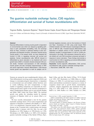
(b)
(a)
(c) (d)
(f)(e)
(g)
Fig. 1 C3G levels increase upon differentiation of neuroblastoma cells
and C3G over-expression induces neurite growth. (a) Exponentially
growing IMR-32 cells (UT), or those subjected to serum starvation
(SS) and treatment with 25 lM forskolin (fsk) or 50 ng/mL NGF for
72 h were observed for morphological changes and images captured
using a live cell imaging system. Bar, 20 lm. (b and c) Whole cell
lysates made from these cells were subjected to immunoblotting using
C3G antibodies and Cdk/Tubulin as loading controls. IMR-32 cells
were transiently transfected with C3G expression vector and after 72 h
subjected to immunoblotting (d) or indirect immunofluorescence to
detect C3G expressing cells (red) and F-actin using Oregon-green
phalloidin (e). Panels shown are confocal images taken at different
magnifications. (f) The growth cone of an extending neurite induced by
C3G expression and cortical area of GFP expressing cells (boxed area
in left panels) are shown at higher magnification. Bar, 10 lm. (g)
Quantitation of neurite growth in C3G expressing and non-expressing
cells grown in complete medium or in 1% serum containing medium or
in medium containing NGF. Data indicate mean ± SD for percentage
of neurite bearing cells averaged from multiple fields on duplicate
coverslips from three different experiments. *p < 0.05;**p < 0.01.
Journal Compilation Ó 2008 International Society for Neurochemistry, J. Neurochem. (2008) 107, 1424–1435
Ó 2008 The Authors
1426 | V. Radha et al.
4. or NGF, a larger proportion (32% and 23% respectively)
change their morphology from polygonal shaped cells to
elongated cells with long neurites (Fig. 1a). C3G protein
levels increase upon serum starvation as well as upon
treatment with NGF and forskolin (Fig. 1b and c) The
multiple bands observed are likely, alternately spliced
variants of C3G, which have earlier been described in other
cell types and tissues (Shivakrupa et al. 1999; Gutierrez-
Berzal et al. 2006), or are the result of altered mobility
caused by post-translational modifications.
Since C3G levels increased during differentiation, we
determined if C3G over-expression induced neurite growth
in IMR-32 cells. Cells transiently expressing C3G for 72 h
were immunostained for C3G and with phalloidin for F-actin
to observe morphological changes. A large number of C3G
expressing cells showed NOG, a hallmark of differentiation.
Cells that did not express C3G retained their polygonal shape
(Fig. 1e). Generally, C3G-induced neurites were linear
extensions with very limited branching. Neurite growth is
enabled by filopodia at the growth cone which act as sensors
for guidance cues. The staining in the growth cone was
visualized by scanning a specific area and an enrichment of
C3G in the growth cone and in filopodia was observed
(Fig. 1f). Expression of green fluorescent protein (GFP) did
not result in NOG or cause any morphological change. GFP
did not show enrichment in the cortical areas and no
filopodia were seen at the cell periphery. Quantitation of the
number of cells with neurites showed that about 22% of C3G
expressing cells showed neurites relative to only about 3% of
non-expressing cells (Fig. 1g). These changes were observed
in the absence of any external stimuli. Over-expression of
C3G also resulted in enhanced responsiveness to serum
starvation and NGF-induced differentiation (Fig. 1g). Com-
pared with non-expressing cells exposed to these conditions,
C3G expressing cells showed a significant increase in the
percentage of neurite bearing cells.
Acetylated tubulin (Ac-Tub), a marker for stable micro-
tubules required for neurite growth, was used to assess
C3G-induced neurites. Shafts of neurites formed in C3G
expressing cells showed enhanced staining (Supporting
information Fig. S1a). Non-expressing cells showed a weak
filamentous microtubule network. Neurofilament (NF)
proteins are important for neuronal process formation and
are expressed and phosphorylated during neuronal differen-
tiation. Using a phospho-dependent anti-NF antibody RT97
(Coleman and Anderton 1990), we determined their expres-
sion by indirect immunofluorescence in cells transfected with
C3G. This antibody did not show staining of exponentially
growing IMR-32 cells, in interphase. 20–40% C3G express-
ing cells with and without neurites showed enhanced
phospho-NF expression, being maximal at 50 h after trans-
fection (Supporting information Fig. S1b). C3G-induced
morphological changes are therefore consistent with neuronal
differentiation.
Cellular C3G is present as a complex with Crk in the
cytosol and this complex associates with membranes in
response to growth factor stimuli. C3G associates with CrkII
in IMR-32 cells (unpublished observations) and CrkII
induces differentiation in PC12 cells (Tanaka et al. 1994).
We determined the consequence of co-expressing C3G with
Crk and observed an additive effect on the number of cells
with NOG when C3G and Crk were co-expressed at 1 : 1
ratio (Supporting information Fig. S2). Neurite growth was
more robust with longer neurites showing extensive branch-
ing, unlike those seen upon expression of C3G alone.
Domain requirements of C3G for induction of neurite
growth
C3G has catalytic activity dependent as well as independent
functions. We characterized the domain requirements of C3G
responsible for neuronal differentiation. Deletion mutants
lacking either the catalytic domain (DC) or the
protein-interaction domain (DN) or possessing only the
protein-interaction domain (CBR) (Fig. 2a) were transiently
expressed and NOG monitored. Constructs lacking the
catalytic domain were unable to promote NOG in high
serum (Fig. 2b and c) or low serum medium (data not
shown). This was not due to expression level differences or
due to differences in their sub-cellular localization, as all
variants showed similar localization. Surprisingly, the DN
construct which was earlier shown to possess enhanced
exchange factor activity, induced NOG, but in fewer cells
than did full length C3G. These results suggested that while
catalytic activity is essential for C3G to induce differentia-
tion, its protein-interaction domain also contributes to its
ability to induce differentiation. Phosphorylation of C3G at
Y504 enhances its catalytic activity (Ichiba et al. 1999). A
phosphorylation site defective mutant Y504F was tested and
found to be partially compromised in its ability to induce
neurites (Fig. 2b and c).
Rap1 mediates C3G-induced neuronal differentiation
Rap1 is the major effector of C3G mediated signalling and
expression of an activated form of Rap1 induces neurites in
human neuroblastoma cells (Uchida et al. 2006). Since the
ability of C3G to induce differentiation was dependent on its
catalytic activity, we tested whether Rap1 was required in
this pathway. C3G was expressed along with control
plasmid, WT Rap1, dominant negative (dn) Rap1 or
constitutively active Rap1 at a ratio of 1 : 1. Quantitation
of NOG showed that dnRap1 reduced the ability of C3G to
induce neurites (Fig. 3a and b). While co-expression of WT
Rap1 showed only a marginal increase in number of cells
with neurites, constitutively active Rap1 enhanced number of
cells with C3G-induced processes. When expressed alone,
only activated Rap1 induced neurites. TC10, a target of C3G
is induced during NGF or, db-cAMP induced differentiation
of PC12 cells (Abe et al. 2003). Co-expression of dn TC10
Ó 2008 The Authors
Journal Compilation Ó 2008 International Society for Neurochemistry, J. Neurochem. (2008) 107, 1424–1435
C3G in neuronal differentiation and survival | 1427
5. did not inhibit C3G-induced neurite formation unlike
dnRap1 indicating that C3G is selective in engaging
downstream effectors in human neuroblastoma cells (data
not shown).
Requirement of Cdc42 for C3G-induced neurite growth
Cdc42, and its effector, N-Wasp are implicated in actin
reorganization that takes place during neurite growth (Banzai
et al. 2000; Rosario et al. 2007). Using dominant-negative
mutants, we examined the involvement of Cdc42 in C3G-
induced neuronal differentiation. Co-expression of dnCdc42
(Fig. 3c and d) or N-Wasp- crib-GFP (Fig. 3e and f) reduced
the ability of C3G to induce neurites suggesting that these
molecules are engaged in the pathway of C3G-induced actin
reorganization during neuronal differentiation. In some
signalling pathways, Rap1 functions to activate Cdc42
(Schwamborn and Puschel 2004; Fukuyama et al. 2005).
We observed that dnCdc42 inhibits NOG induced by C3G
co-expressed with WT Rap1, but had a marginal effect on
NOG induced by C3G and activated Rap1 (Supporting
information Fig. S3). NOG induced by activated Rap1 was
independent of Cdc42.
C3G is required for NGF and forskolin-induced neuronal
cell differentiation
Since C3G levels increased upon differentiation, we exam-
ined the requirement of C3G for neurite growth induced by
NGF and forskolin. ShRNA vectors targeting C3G, ShA and
ShC reduced endogenous C3G protein by 75% and 60%
respectively relative to cells expressing mutant vectors
(mShA and mShC) (Fig. 4a). IMR-32 cells transiently
expressing the WT and mutant shRNAs along with GFP
were treated with NGF and neurite growth monitored among
GFP expressing and non-expressing cells. Expression of
mutant shRNA vectors did not affect the number of cells with
NOG, whereas, very few cells expressing WT shRNA have
neurite extensions and retain morphology similar to undif-
ferentiated cells (Fig. 4b and d). The expression of C3G
shRNA constructs also blocked forskolin-induced neurite
growth by about 75% (Fig. 4c and d).
Since catalytic activity of C3G was required for inducing
neuronal differentiation, we determined whether C3G cata-
lytic activity was required for forskolin-induced differentia-
tion. The deletion construct, CBR functions as a dominant
negative to inhibit signalling as a result of activation of
endogenous C3G (Schmitt and Stork 2002). Cells treated with
forskolin after CBR transfection were compromised in their
ability to differentiate compared to non-expressing cells (8.1%
against 30.2%) indicating that the exchange factor activity of
C3G is required for forskolin-induced differentiation (Fig. 5a
and b). As a target of C3G, we determined the role of Rap1 in
mediating forskolin and NGF-induced NOG in IMR-32 cells.
It was seen that dominant negative Rap1 expression reduced
forskolin-induced NOG from 29.2 ± 1.93% to 6.05 ± 1.1%.
(a)
(b)
(c) (d)
Fig. 2 Requirement of C3G catalytic do-
main for induction of neurite growth. Various
C3G constructs as depicted schematically in
(a) were tested for ability to induce neurites.
(b) Cells were fixed and stained for C3G
expression and representative fields show-
ing expression of various constructs imaged
using confocal microscope. Bar, 10 lm (c).
Immuonblot showing expression in cell ly-
sates (d). Comparison of the ability of vari-
ous constructs to induce neurites depicted
as mean ± SD obtained by quantitation of
neurite bearing cells among C3G express-
ing cells. Non-expressing cells in the same
coverslips were also examined for neurite
growth and subtracted as background prior
to averaging the data. *p < 0.02.**p < 0.01.
Journal Compilation Ó 2008 International Society for Neurochemistry, J. Neurochem. (2008) 107, 1424–1435
Ó 2008 The Authors
1428 | V. Radha et al.
6. Similarly, NGF-induced NOG was reduced from 19.2 ± 4.7%
to 2.7 ± 1.2%. These results suggest that a pathway involving
C3G and Rap1 enables differentiation of IMR-32 cells in
response to forskolin and NGF.
In NIH3T3 cells, forskolin activates Rap1 dependent on
C3G (Schmitt and Stork 2002). Tyrosine kinases contribute
to regulation of neurite growth (Loh et al. 2008) and
forskolin activates c-Src. IMR-32 cells treated with forskolin
for 72 h showed p-C3G staining, predominantly at a
juxtanuclear organelle (Fig. 5c). No staining is seen in
untreated cells or in cells treated with forskolin for short
periods of time (30 min to 1 hr). When tyrosine phospha-
tases were inhibited by treatment with PV, we observed more
prominent staining of p-C3G in the plasma membrane and in
the Golgi region, indicating that forskolin treatment results in
phosphorylation of C3G. The location of the juxtanuclear
staining was confirmed by co-staining for giantin, a golgi
marker indicating that p-C3G is particularly localized at the
Golgi (Fig. 5d). Cells treated with PV alone as control
showed weak staining at the plasma membrane in addition to
staining at the Golgi. p-C3G staining was seen in cells treated
with NGF for either short (30 min) (Fig. 5e) or long (72 h)
(data not shown) periods of time. To determine whether
SFKs were responsible for phosphorylation of C3G, cells
were pre-treated with PP2, a specific inhibitor. p-C3G
staining was significantly reduced in the presence of the
(a) (b)
(c) (d)
(f)(e)
Fig. 3 C3G-induced neurite growth is
dependent on Rap1 and Cdc42 (a) IMR-32
cells transfected with C3G and the indicated
constructs were fixed and stained for C3G
and HA epitope (to visualize Rap con-
structs). Representative confocal images of
expressing cells are shown. Bar, 10 lm. (b)
Extent of neurite growth under the various
conditions described above is depicted.
Neurite growth induced upon expression
of Rap1 constructs alone is also depicted
after background subtraction.**p < 0.01,
*p < 0.05 (c) C3G was expressed along
with either control vector or dnCdc42 and
stained for C3G and myc (tag for dnCdc42).
Images depicting morphology of the
expressing cells captured using a confocal
microscope are shown. Bar, 10 lm. (d)
Quantitation of neurite growth. *p < 0.01 (e)
GFP or N-Wasp Crib-GFP vectors were co-
transfected along with C3G and images of
expressing cells captured using confocal
microscope 72 h after transfection. Bar,
10 lm. (f) Bar diagram represents propor-
tion of neurite bearing cells among the
expressing cells after background subtrac-
tion. *p < 0.01. Expression levels of various
proteins are shown in the corresponding
immunoblots.
Ó 2008 The Authors
Journal Compilation Ó 2008 International Society for Neurochemistry, J. Neurochem. (2008) 107, 1424–1435
C3G in neuronal differentiation and survival | 1429
7. inhibitor (Fig. 5e). These results suggested that endogenous
C3G is phosphorylated on Y504 by SFKs, in response to
forskolin or NGF treatment. We also examined whether SFK
activity was required during NOG induced by C3G. We
found that the presence of 1 lM PP2 during transient
expression, did not affect the ability of C3G to induce
neurites (data not shown).
C3G expression induces the cell cycle inhibitor, p21
Control of cell cycle is important for regulating neuronal
differentiation as precursor cells need to exit the cell cycle.
The cell cycle inhibitor p21 is induced and shows nuclear
localization in cells undergoing proliferation arrest (Sherr
and Roberts 1999). We looked for endogenous p21
expression in IMR-32 cells transiently expressing C3G for
72 h. A small proportion of IMR-32 cells (13.6%) showed
p21 positivity, but among C3G expressing cells, 57%
showed p21 positivity (Fig. 6a). Nuclear p21 was seen in
many C3G expressing cells that did not show neurite
growth suggesting that p21 expression may be an event that
precedes neurite growth. Prolonged MAPK activation is
associated with Rap1 activation and neuronal differentiation
(York et al. 1998). MAPK pathways are also important for
up-regulation of p21. We examined whether C3G-induced
NOG and p21 protein increase were dependent on MAPK
activation. Extracellular signal regulated kinase (ERK)
inhibitor, PD98059 and p38 MAPK inhibitor, SB203580
reduced ability of C3G to induce neurites. (Fig. 6a and b).
The intensity of nuclear p21 fluorescence from expressing
and non-expressing cells in various fields determined using
LSM-FCS software showed a 2.4-fold difference between
C3G expressing and non-expressing cells. ERK inhibitor
significantly decreased the number of cells, as well as
intensity of nuclear p21 staining. (Fig. 6b and c).
(a)
(c) (d)
(b)
Fig. 4 C3G knockdown inhibits NGF and
forskolin-induced neurite growth. (a) Ly-
sates of IMR-32 cells expressing various
hairpin RNA constructs as indicated were
subjected to immunoblotting for expression
of C3G and Cdk2 (loading control). IMR-32
cells were transfected with 380 ng of indi-
cated shRNA constructs and 20 ng of GFP
and subjected to NGF stimulation (b) or
forskolin treatment (c) as described in
Methods. After 72 h of treatment, extent of
neurite growth was compared between
GFP expressing and non-expressing cells.
Representative fields are shown in (b) & (c)
and quantitation of neurite growth in (d).
Bar, 10 lm. *p < 0.01.
Journal Compilation Ó 2008 International Society for Neurochemistry, J. Neurochem. (2008) 107, 1424–1435
Ó 2008 The Authors
1430 | V. Radha et al.
8. C3G knockdown enhances serum starvation-induced cell
death
Since C3G levels increased during serum starvation of IMR-
32 cells, we determined whether C3G played a role in
survival of neuroblastoma cells during serum starvation,
which induces differentiation as well as cell death. IMR-32
cells were transfected with shRNA targeting C3G along with
20 ngs of GFP/RFP plasmids to detect transfected cells. Post-
transfection, cells were cultured in either complete medium
or in medium containing 0.5% FCS for a period of 96 h, and
observed for cell death among expressing cells using
morphological criteria and p-H2B positivity (Cheung et al.
2003) (Fig. 7a and b). While a majority of RFP positive cells
expressing mShC, do not show p-H2B staining, a large
number of cells expressing ShC were p-H2B positive. Down-
regulation of C3G using two different shRNA enhanced
serum starvation-induced cell death relative to cells express-
ing mshRNA (Fig. 7d). No significant difference in cell
death was observed because of reduction in C3G levels when
cells were grown in complete medium. Down-regulation of
C3G during serum starvation also resulted in activation of
caspase-3 (Fig. 7c).
Discussion
Several reports have indicated a negative role for C3G in cell
proliferation (Schmitt and Stork 2002; Stork 2003; Guerrero
et al. 2004). This is the first study implying a role for C3G in
differentiation, a process closely associated with cell cycle
arrest. Cells over-expressing C3G show morphological
changes, assuming a terminally differentiated neuronal
phenotype even in the absence of any external stimuli.
Presence of acetylated tubulin and phosphorylation of NF
proteins that was found to accompany C3G expression
provided further evidence for neuron specific differentiation
induced by C3G. C3G also enhanced neurite growth induced
by serum starvation and NGF treatment implying its ability
to potentiate signalling mediated by these inducers. The
ability of C3G to induce neurites was dependent on its
catalytic activity as well as its protein interaction domain.
(a)
(c)
(d)
(e)
(b)
Fig. 5 Catalytic activity of C3G is required
for neurite growth. IMR-32 cells were
transfected with control or CBR vector prior
to treatment of cells with forskolin. 72 h la-
ter, cells were stained for C3G expression
and neurite growth compared between
expressing (CBR) and non-expressing cells
(NE). Representative fields are shown in
(a) and quantitation in (b). Bar, 10 lm.
*p < 0.01. CBR expression is shown in
Western blot. (c) Forskolin induces C3G
phosphorylation. IMR-32 cells were treated
with forskolin (25 lM) for 72 h and sub-
jected to pervanadate (PV) treatment or left
untreated. Serum starved cells were also
treated with PV as controls. Cells were
stained using an antibody that detects C3G
phosphorylated on Y504. (d) The same
coverslips were also subjected to giantin
staining as a marker for Golgi localization.
(e) SFK inhibition represses forskolin and
NGF-induced C3G phosphorylation. Cells
were pre-treated with PP2 prior to forskolin
(72 h) or NGF (30 min) stimulation and
stained for p-C3G.
Ó 2008 The Authors
Journal Compilation Ó 2008 International Society for Neurochemistry, J. Neurochem. (2008) 107, 1424–1435
C3G in neuronal differentiation and survival | 1431
9. This is in concordance with the requirement of Rap1
activation in this pathway. Rap1 is a direct target for the
exchange factor activity of C3G and it is possible that the N-
terminal sequences of C3G may be required for proper
localization and activation of Rap1 or other additional
downstream effectors.
Forskolin and NGF engage common as well as divergent
pathways to induce neurite growth (Hoshino and Nakamura
2003). Our study shows that C3G and its effector, Rap1 are
major players in human cells in transmitting signals from both
NGF and forskolin to induce differentiation, suggesting their
role in a common effector function during the differentiation
(a)
(b) (c) Fig. 6 C3G expression induces neurite
growth and p21 protein dependent on
MAPK pathway. (a) IMR-32 cells transfect-
ed with C3G were grown in the presence or
absence of MAPK inhibitors and stained for
p21 and C3G after 72 h. Representative
fields are depicted. Bar, 10 lm. (b) Quan-
titation of NOG in C3G expressing cells in
the presence of inhibitors. (c) p21 fluores-
cence intensity was quantitated from a large
number of C3G expressing and non-
expressing cells in various fields using the
LSM software. Values depict mean ± s.d.
of nuclear p21 fluorescence intensity.
*p < 0.01.
(a) (c)
(b) (d)
Fig. 7 Effect of C3G knockdown on sur-
vival of IMR-32 cells subjected to serum
starvation. IMR-32 cells were transfected
with two shRNA vectors and corresponding
mutants along with GFP or RFP. Cells were
replated on coverslips and maintained ei-
ther in 10% serum medium (CM) or 0.5%
serum medium (SS). 96 h later, cells were
fixed, stained with DAPI and examined for
cell death using morphological criteria (a),p-
H2B (b) or cleaved caspase-3 staining (c).
Scale bar, 10 lm. (d) Proportion of GFP
expressing cells showing morphological
features of apoptosis was quantitated and
average from multiple experiments depicted
as mean ± SD. *p < 0.01.
Journal Compilation Ó 2008 International Society for Neurochemistry, J. Neurochem. (2008) 107, 1424–1435
Ó 2008 The Authors
1432 | V. Radha et al.
10. process. In the mouse model, absence of C3G resulted in
enhanced growth of certain neuronal cell types indicating that
C3G is required during brain development to arrest cell cycle
(Voss et al. 2006). Our findings of the ability of C3G to
induce neuronal differentiation can explain the brain abnor-
malities seen in mutant mice. Unpublished observations from
our laboratory have also shown that C3G levels increase
during differentiation of human myelomonocytic cells to a
macrophage lineage, suggesting that a role for C3G in
differentiation may not be restricted to specific cell types.
In response to NGF, Rap1 is activated only in the
endosomal compartment, thereby enabling persistent signal-
ling (Arevalo et al. 2004; Hisata et al. 2007). Our results
show localization of p-C3G to the Golgi region in cells
differentiated by forskolin or NGF treatment, suggesting that
localized activation of C3G in this compartment contributes
to Rap1 activation. Forskolin as well as NGF activate SFKs
(Schmitt and Stork 2002; Loh et al. 2008), and our results
imply that in response to these treatments, SFKs target C3G.
This modification may be one of the means of activating
C3G. But neurite growth induced by over-expression of C3G
is not dependent on activity of SFKs. When over-expressed,
tyrosine phosphorylation defective mutant of C3G is only
partially compromised in its ability to induce neurites
indicating that phosphorylation alone is insufficient for
triggering downstream events. Our results using a dominant
negative approach also substantiate the findings that C3G
catalytic activity is required for forskolin-induced differen-
tiation. A recent study in PCl2 cells showed a major role for
PDZ-GEF1 in activating Rap1 and in inducing NOG in
response to NGF (Hisata et al. 2007). In these cells they
show that C3G knockdown by 60–70% reduces NGF-
induced NOG by only 15% suggesting that C3G plays a
minor role in signalling neurite growth in murine cells.
PC12 cells over-expressing gut tyrosine kinase (GTK), a
tyrosine kinase, undergo differentiation and showed an
increased association of p130 Cas and FAK with Crk II
and C3G, resulting in Rap1 activation (Anneren et al. 2000).
NGF induces complex formation of TrkA, Grb2, Shp2, gut
tyrosine kinase (GTK) and C3G to activate Rap1(Kao et al.
2001). C3G over-expression may be enabling such complex
formation to induce differentiation even in the absence of
receptor activation by NGF. Our observation that C3G can
further increase the extent of neurite growth induced by
activated Rap1 suggests that C3G may be activating
pathways in addition to Rap1 activation, or C3G activates
endogenous Rap1, the effect of which becomes additive to
that of activated Rap1. Exogenously expressed activated
Rap1 may engage alternate pathways leading to the differ-
entiation process. C3G-induced neurite growth is dependent
on Cdc42, but expression of activated Rap1 engages a
pathway independent of Cdc42. During axon specification
and nectin-induced signalling, activation of Cdc42 is a
function of Rap1 (Schwamborn and Puschel 2004; Fukuy-
ama et al. 2005). It is possible that Cdc42 is bypassed when
constitutively active Rap1 is expressed. In epithelial cells, we
had earlier shown that C3G induces actin reorganization and
filopodia formation independent of Cdc42 (Radha et al.
2007). Another difference was that filopodia formation did
not require catalytic activity of C3G, whereas neurite growth
was dependent on both catalytic activity as well as its protein
interaction domain. These observations indicate that in a
cell type dependent manner, C3G engages distinct effector
pathways to bring about actin cytoskeletal reorganization.
Though C3G is capable of activating the Rho family GTPase
TC10, we observed that in IMR-32 cells C3G over-
expression induced signalling leading to neurite growth does
not engage TC10.
The induction of NF phosphorylation and cell cycle
inhibitor, p21 in C3G expressing cells suggested that C3G
induces differentiation while simultaneously repressing the
cell cycle. Neuronal differentiation is often coupled with cell
cycle exit and therefore molecules that induce differentiation
are responsible for halting cell cycle progression (Nguyen
et al. 2006). C3G may also be acting bifunctionally to cause
cell cycle exit as well as differentiation by engaging the
MAPK pathways. The role of C3G in causing cell cycle exit
is also corroborated by studies in C3G knockout mice which
show increase in cortical cell population (Voss et al. 2006).
Our study shows that induction of p21 may be one of the
mechanisms by which C3G induces cell cycle arrest. Earlier,
the ability of C3G to repress anchorage dependent growth of
transformed cells has been described. They showed changes
in expression of cyclinA in response to C3G expression,
indicating that C3G can signal to cell cycle regulatory
molecules (Guerrero et al. 2004). Molecules that trigger a
differentiation programme also enable activation of survival
pathways to prevent cell death (Poluha et al. 1996; Frebel
and Wiese 2006). Our results show that higher levels of cell
death are induced in serum starved cells where C3G has been
knocked down. Therefore, enhanced C3G protein seen
during serum starvation may aid in cell survival. p21 is
required for survival of differentiating neuroblastoma cells
(Poluha et al. 1996). The increase in cell death observed in
C3G knockdown cells may therefore be due to inability of
these cells to express p21 in response to serum starvation.
Taken together, our observations implicate a function for
C3G in neuronal differentiation. This property may be
mediated through its ability to influence actin reorganization,
induce cell cycle inhibitors and suppress cell death. Through
induction of cell cycle arrest and terminal maturation, C3G
over-expression could help in reverting the neoplastic
phenotype of human neuroblastomas.
Acknowledgements
We thank Dr. G. Swarup for critical reading of the manuscript and
Nandini Rangaraj for help in using the confocal microscope. We
Ó 2008 The Authors
Journal Compilation Ó 2008 International Society for Neurochemistry, J. Neurochem. (2008) 107, 1424–1435
C3G in neuronal differentiation and survival | 1433
11. also thank Dr Pessini, SUNY, New York, Dr Quilliam, Indiana
University School of Medicine, Dr Takenawa, University of
Tokyo, Dr Bruce Mayer, University of Connecticut Health Center,
and Dr Philip Stork, Oregon Health and Science University,
Portland for generous gift of expression vectors. This work was
supported by funding from the Department of Science &
Technology, Govt. of India. KD acknowledges receipt of Junior
Research Fellowship from Council of Scientific and Industrial
Research, India.
Supporting information
Additional Supporting Information may be found in the online
version of this article:
Fig S1. C3G expressing cells were stained to detect presence of
C3G and acetylated tubulin (Ac-Tub) (a) or phospho-neurofilament
(p-NF) (b). Representative fields of cells captured using a confocal
microscope are shown.
Fig S2. Effect of CrkII co-expression on C3G-induced neurite
growth.
Fig S3. Role of Cdc42 in mediating neurite growth induced upon
co-expression of C3G with Rap1 and activated Rap1. IMR-32 cells
were transfected as indicated and NOG quantitated.
Please note: Wiley-Blackwell are not responsible for the content
or functionality of any supporting materials supplied by the authors.
Any queries (other than missing material) should be directed to the
corresponding author for the article.
References
Abe T., Kato M., Miki H., Takenawa T. and Endo T. (2003) Small
GTPase TC10 and its homologue Rho T induce N-Wasp mediated
long process formation and neurite outgrowth. J. Cell Sci. 116,
155–168.
Alsayed Y., Uddin S., Ahmad S., Majchrzak B., Druker B. J., Fish E. N.
and Platanias L. C. (2000) IFN-gamma activates the C3G/Rap1
signaling pathway. J. Immunol. 164, 1800–1806.
Anneren C., Reedquist K. A., Bos J. L. and Welsh M. (2000) GTK, a
Src-related tyrosine kinase induces NGF-independent neurite
growth in PC12 cells through activation of the Rap1 pathway.
J. Biol. Chem. 275, 29153–29161.
Arevalo J. C., Yano H., Teng K. K. and Chao M. V. (2004) A unique
pathway for sustained neurotrophin signalling through an ankyrin-
rich membrane spanning protein. EMBO J. 23, 2358–2368.
Ballif B. A., Arnaud L., Arthur W. T., Guris D., Imamoto A. and Cooper
J. A. (2004) Activation of a Dab1/CrkL/C3G/Rap1 pathway in
Reelin-stimulated neurons. Curr. Biol. 14, 606–610.
Banzai Y., Miki H., Yamaguchi H. and Takenawa T. (2000) Essential
role of N-Wasp in neurite extension in PC12 cells and rat
hippocampal primary culture cells. J. Biol. Chem. 275, 11987–
11992.
Chang W., Chen H., Chiou R., Chen C. and Huang G. (2005) A novel
function of transcription factor a-Pal/NRF-1 : increasing neurite
outgrowth. Biochem. Biophys. Res. Comm. 334, 199–206.
Cheung W., Ajiro K., Samejima K., Kloc M., Cheung P., Mizzen C.,
Beeser A., Etkin L., Chernoff J. and Earnshah W. (2003) Apoptotic
phosphorylation of histone H2B is mediated by mammalian STE20
kinase. Cell 113, 507–517.
Chiang S. H., Chang L. and Saltiel A. R. (2006) TC10 and insulin-
stimulated glucose transport. Methods Enzymol. 406, 701–714.
Coleman M. P. and Anderton B. H. (1990) Phosphate-dependent
monoclonal antibodies to neurofilaments and Alzheimer neurofi-
brillary tangles recognize a synthetic phosphopeptides. J. Neuro-
chem. 54, 1548–1555.
Dent E. W., Kwiatkowski A. V., Mebane L. M. et al. (2007) Filopodia
are required for cortical neurite formation. Nat. Cell Biol. 9, 1347–
1359.
Frebel K. and Wiese S. (2006) Signalling molecules essential for neu-
ronal survival and differentiation. Biochem. Soc. Trans. 34, 1287–
1290.
Fukuyama T., Ogita H., Kawakatsu T., Fukuhara T., Yamada T., Sato T.,
Shimizu K., Nakamura T., Matsuda M. and Takai Y. (2005)
Involvement of the c-Src-Crk-C3G-Rap1 signaling in the nectin-
induced activation of Cdc42 and formation of adherens junctions.
J. Biol. Chem. 280, 815–825.
Gotoh T., Hattori S., Nakamura S. et al. (1995) Identification of Rap1 as
a target for the Crk SH3 domain-binding guanine nucleotide-
releasing factor C3G. Mol. Cell. Biol. 15, 6746–6753.
Gotoh T., Niino Y., Tokuda M., Hatase O., Nakamura S., Matsuda M.
and Hattori S. (1997) Activation of R-Ras by Ras-guanine nucle-
otide-releasing factor. J. Biol. Chem. 272, 18602–18607.
Guerrero C., Martan-Encabo S., Fernandez-Medarde A. and Santos E.
(2004) C3G-mediated suppression of oncogene-induced focus
formation in fibroblasts involves inhibition of ERK activation,
cyclin A expression and alterations of anchorage-independent
growth. Oncogene 23, 4885–4893.
Gutierrez-Berzal J., Castellano E., Martin-Encabo S., Guitierrez-Cianca
N., Hernandez J. M., Santos E. and Guerrero C. (2006) Charac-
terization of p87 C3G, a novel, truncated C3G isoform that is
overexpressed in chronic myeloid leukemia and interacts with Bcr-
Abl. Exp. Cell Res. 312, 938–948.
Hisata S., Sakisaka T., Baba T., Yamada T., Aoki K., Matsuda M. and
Takai Y. (2007) Rap1-PDZ-GEF1 interacts with a neurotrophin
receptor at late endosomes, leading to sustained activation of Rap1
and ERK and neurite outgrowth. J. Cell Biol. 178, 843–860.
Hoshino M. and Nakamura S. (2003) Small GTPase Rin induces neurite
outgrowth through Rac/Cdc42 and calmodulin in PC12 cells.
J. Cell Biol. 163, 1067–1076.
Ichiba T., Hashimoto Y., Nakaya M., Kuraishi Y., Tanaka S., Kurata T.,
Mochizuki N. and Matsuda M. (1999) Activation of C3G guanine
nucleotide exchange factor for Rap1 by phosphorylation of tyro-
sine 504. J. Biol. Chem. 274, 14376–14381.
de Jong R., van Wijk A., Heisterkamp N. and Groffen J. (1998) C3G is
tyrosine-phosphorylated after integrin-mediated cell adhesion in
normal but not in Bcr/Abl expressing cells. Oncogene. 17, 2805–
2810.
Kao S., Jaiswal R. K., Kolch W. and Landreth G. E. (2001) Identification
of the mechanisms regulating the differential activation of the
mapk cascade by epidermal growth factor and nerve growth factor
in PC12 cells. J. Biol. Chem. 276, 18169–18177.
Kaplan D. R. and Miller F. D. (2000) Neurotrophin signal transduction in
the nervous system. Curr. Op. Neurobiol. 10, 381–391.
Kirsch K. H., Georgescu M. M. and Hanafusa H. (1998) Direct binding
of p130(Cas) to the guanine nucleotide exchange factor C3G.
J. Biol. Chem. 273, 25673–25679.
Knudsen B. S., Feller S. M. and Hanafusa H. (1994) Four proline-rich
sequences of the guanine-nucleotide exchange factor C3G bind
with unique specificity to the first Src homology 3 domain of Crk.
J. Biol. Chem. 269, 32781–32787.
Loh S. H., Francescut L., Lingor P., Bahr M. and Nicotera P. (2008)
Identification of new kinase clusters required for neurite outgrowth
and retraction by a loss of function RNAi. Cell Death Differen. 15,
283–298.
Journal Compilation Ó 2008 International Society for Neurochemistry, J. Neurochem. (2008) 107, 1424–1435
Ó 2008 The Authors
1434 | V. Radha et al.
12. Mochizuki N., Ohba Y., Kobayashi S., Otsuka N., Graybiel A. M.,
Tanaka S. and Matsuda M. (2000) Crk activation of JNK via C3G
and R-Ras. J. Biol. Chem. 275, 12667–12671.
Nguyen L., Besson A., Roberts J. M. and Guillemot F. (2006) Coupling
cell cycle exit neuronal differentiation and migration in cortical
neurogenesis. Cell Cycle 5, 2314–2318.
Ohba Y., Mochizuki N., Matsuo K., Yamashita S., Nakaya M.,
Hashimoto Y., Hamaguchi M., Kurata T., Nagashima K. and
Matsuda M. (2000) Rap2 as a slowly responding molecular switch
in the Rap1 signaling cascade. Mol. Cell. Biol. 20, 6074–6083.
Ohba Y., Ikuta K., Ogura A. et al. (2001) Requirement for C3G-
dependent Rap1 activation for cell adhesion and embryogenesis.
EMBO J. 20, 3333–3341.
Poluha W., Poluha D. K., Chang B., Crosbie N. E., Schonhoff C. M.,
Kilpatrick D. L. and Ross A. H. (1996) The CDKI p21WAF
is
required for survival of differentiating neuroblastoma cells. Mol.
Cell. Biol. 16, 1335–1341.
Prasad K. N., Mandal B. and Kumar S. (1973) Human neuroblastoma
cell culture: Effect of 5 BrdU on morphological differentiation and
levels of neural enzymes. Proc. Soc. Exp. Biol. Med. 144, 38–42.
Radha V., Sudhakar C. and Swarup G. (1999) Induction of p53 depen-
dent apoptosis upon overexpression of a nuclear protein tyrosine
phosphatase. FEBS Lett. 453, 308–312.
Radha V., Rajanna A. and Swarup G. (2004) Phosphorylated guanine
nucleotide exchange factor C3G, induced by pervanadate and Src
family kinases localizes to the Golgi and subcortical actin cyto-
skeleton. BMC Cell Biol. 5, 31.
Radha V., Rajanna A., Mitra A., Rangaraj N. and Swarup G. (2007) C3G
is required for c-Abl-induced filopodia and its overexpression
promotes filopodia formation. Exp. Cell Res. 313, 2476–2492.
Reynolds C. P. and Perez-Polo J. R. (1981) Induction of neurite out-
growth in IMR-32 human neuroblastoma cell line by NGF.
J. Neurosci. Res. 6, 319–325.
Rosario M., Franke R., Bednarski C. and Birchmeier W. (2007) The
neurite outgrowth multiadaptor RhoGAP, NOMA-GAP, regulates
neurite extension through SHP-2 and Cdc42. J. Cell Biol. 178,
503–516.
Schmitt J. M. and Stork P. J. (2002) PKA phosphorylation of Src mediates
cAMP’s inhibition of cell growth via Rap1. Mol. Cell. 9, 85–94.
Schwamborn J. C. and Puschel A. W. (2004) The sequential activity of
GTPases Rap1B and Cdc42 determines neuronal polarity. Nat.
Neurosci. 7, 923–929.
Sherr C. J. and Roberts J. M. (1999) CDK inhibitors: positive and negative
regulators of G1-phase progression. Genes Dev. 13, 1501–1512.
Shivakrupa R., Singh R. and Swarup G. (1999) Identification of a novel
splice variant of C3G which shows tissue-specific expression. DNA
Cell Biol. 18, 701–708.
Shivakrupa R., Radha V., Sudhakar Ch. and Swarup G. (2003) Physical
and functional interaction between Hck tyrosine kinase and
guanine nucleotide exchange factor C3G results in apoptosis,
which is independent of C3G catalytic domain. J. Biol. Chem. 278,
52188–52194.
Stork P. J. (2003) Does Rap1 deserve a bad Rap? Trends Biochem. Sci.
28, 267.
Tamada M., Sheetz M. P. and Sawada Y. (2004) Activation of a signaling
cascade by cytoskeleton stretch. Dev. Cell 7, 709–718.
Tanaka S., Morishita T., Hashimoto Y. et al. (1994) C3G, a guanine
nucleotide-releasing protein expressed ubiquitously, binds to the
Src homology 3 domains of CRK and GRB2/ASH proteins. Proc.
Natl. Acad. Sci. USA 91, 3443–3447.
Uchida M., Enomoto A., Fukuda T. et al. (2006) Dsk-4 regulates GDNF-
dependent neurite outgrowth through downstream activation of
Rap1 and MAPK. J. Cell Sci. 119, 3067–3077.
Voss A. K., Gruss P. and Thomas T. (2003) The guanine nucleotide
exchange factor C3G is necessary for the formation of focal
adhesions and vascular maturation. Development 130, 355–367.
Voss A. K., Krebs D. L. and Thomas T. (2006) C3G regulates the size of
the cerebral cortex neural precursor population. EMBO J. 25,
3652–3663.
Wu C., Lai C. F. and Mobley W. C. (2001) Nerve growth factor activates
persistent Rap1 signalingin endosomes. J. Neurosci.21, 5406–5416.
York R. D., Yao H., Dillon T., Ellig C. L., Eckert S. P., Mc Claskey E. W.
and Stork P. J. S. (1998) Rap1 mediates sustained MAPK activa-
tion induced by NGF. Nature 392, 622–626.
Ó 2008 The Authors
Journal Compilation Ó 2008 International Society for Neurochemistry, J. Neurochem. (2008) 107, 1424–1435
C3G in neuronal differentiation and survival | 1435