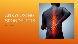
Ankylosing spondylitis
•Als PPTX, PDF herunterladen•
19 gefällt mir•8,370 views

AS

Empfohlen
Weitere ähnliche Inhalte
Was ist angesagt?
Was ist angesagt? (20)
Ähnlich wie Ankylosing spondylitis
Ähnlich wie Ankylosing spondylitis (20)
Mehr von Kristine Faith Tablizo
Mehr von Kristine Faith Tablizo (18)
Kürzlich hochgeladen
Kürzlich hochgeladen (20)
Ankylosing spondylitis
- 1. Tabz, 2015
- 2. SERONEGATIVE SPA’S Pathologic changes in the ligamentous attachments rather than synovium Involvement of SI joints with or without other joints Absence of RF– Thus, seronegative Association with HLA-B27 Immune-mediated manifestations, triggered by a T-cell response presumably directed against an undefined antigen that may cross-react with native molecules of the MS system
- 3. ANKYLOSIS?
- 7. ANKYLOSING SPONDYLITIS (AS) A seronegative spondyloarthropathy chronic, multisystem inflammatory disorder involves primarily the sacroiliac (SI) joints and the axial skeleton. outcome is generally good compared with that in patients with a disease such as rheumatoid arthritis
- 8. Etiology and Pathogenesis: Not fully established Environmental and genetic factors HLA-B27 antigen- associated hereditary marker 90% of people with AS express the HLA-B27 genotype association of AS with HLA-B27 suggests the condition involves CD8 T cells, which interact with HLA-B. possibility that CD4+ T lymphocytes are involved since HLA-B27 possibly with an ability to interact with T cell receptors in association with CD4 (usually CD8+ cytotoxic T cell with HLAB antigen as it is a MHC class 1 antigen) IL-23 receptor gene
- 9. Hx: (Key components of the patient history that suggest AS include the following): Insidious onset of low back pain - The most common symptom Onset of symptoms before age 40 years Presence of symptoms for more than 3 months Symptoms worse in the morning or with inactivity Improvement of symptoms with exercise
- 10. General SSx: Those related to inflammatory back pain - Stiffness of the spine and kyphosis resulting in a stooped posture are characteristic of advanced- stage AS. Sacroiliitis- pathologic hallmark of Sero(-) SAp’s Peripheral enthesitis and arthritis Constitutional and organ-specific extra-articular manifestations
- 11. Entheses are any point of attachment of skeletal muscles to the bone, where recurring stress or inflammatory autoimmune disease can cause inflammation or occasionally fibrosis andcalcification. Enthesitis inflammation of the entheses, the sites where tendons or ligaments insert into the bone. Vs. synovium** It is associated with HLA B27 arthropathies like AS, PS, and ReAs. also called enthesopathy
- 12. Extra-articular manifestations: Uveitis Cardiovascular disease Pulmonary disease Renal disease Neurologic disease Gastrointestinal (GI) disease Metabolic bone disease
- 13. Dx: combining the clinical criteria of inflammatory back pain (Hx) and enthesitis or arthritis (PE) with radiologic findings. Serologic Tests (ESR, CRP) Radiography Power Doppler ultrasonography MRI and CT scanning Genetic Testing Bath Ankylosing Spondylitis Disease Activity Index (BASDAI)
- 14. Dx: combining the clinical criteria of inflammatory back pain and enthesitis or arthritis with radiologic findings. Radiography inflammatory changes in the SI joints and spine earliest changes in SI shows erosions and sclerosis. inflammatory lesions at vertebral entheses may result in sclerosis of the superior and inferior margins of the vertebral bodies, called shiny corners (Romanus lesion). Progression of the erosions pseudo widening of the joint space and bony ankylosis squaring of the vertebral bodies caused by erosions of the superior and inferior margins loss of the normal concave contour of the bodies’ anterior surface. Syndesmophyte-bony growth originating inside a ligament, leading to fusion of vertebrae bamboo spine appearance.
- 15. Dx: combining the clinical criteria of inflammatory back pain and enthesitis or arthritis with radiologic findings. Power Doppler ultrasonography can be used to document active enthesitis. In addition, this technology may be useful in the assessment of changes in inflammatory activity at entheses during the institution of new therapies.
- 16. Dx: combining the clinical criteria of inflammatory back pain and enthesitis or arthritis with radiologic findings. MRI and CT scanning Magnetic resonance imaging (MRI) or computed tomography (CT) scanning of the SI joints, spine, and peripheral joints may reveal evidence of early sacroiliitis, erosions, and enthesitis that are not apparent on standard radiographs.
- 17. Schober's test - a test used in rheumatology to measure the ability of a patient to flex his/her lower back. if the distance increases less than 5 cm, then there is an indication that the flexion of the lower back is limited. diagnostically useful as part of a clinical diagnosis of syndromes such as ankylosing spondylitis.
- 18. none
- 19. Tx: Pharmacologic therapy Nonsteroidal anti-inflammatory drugs (NSAIDs)-mainstay tx ibuprofen, phenylbutazone, diclofenac, indomethacin, naproxen an d COX-2 inhibitors. Indomethacin is a drug of choice. Opioid painkillers Medications used to treat the progression of the disease include the following: Disease-modifying antirheumatic drugs (DMARDs) sulfasalazine can be used in people with peripheral arthritis. Tumor necrosis factor-alpha (TNFα) blockers (antagonists), biologics etanercept, infliximab, golimumab and adalimumab Anti-interleukin-6 inhibitors tocilizumab and rituximab, undergoing trials.
- 20. Surgical therapy Vertebral osteotomy - Patients with fusion of the cervical or upper thoracic spine may benefit from extension osteotomy of the cervical spine [11] Fracture stabilization Joint replacement - Patients with significant involvement of the hips may benefit from total hip arthroplasty Tx:
- 21. Physical therapy Though physical therapy remedies have been scarcely documented, some therapeutic exercises are used to help manage lower back, neck, knee, and shoulder pain. Low intensity aerobic exercise Transcutaneous electrical nerve stimulation (TENS) Thermotherapy Proprioceptive neuromuscular facilitation (PNF) Tx:
- 22. HISTORY: A 72 year old woman was brought to the mergency room by her son-in-law after falling in her bath tub. She was previously in good health despite leading a relatively a sedentary lifestyle and having a 30-pack-year history of cigarette smoking. The only medication she currently takes is Inderal (propranolol)for mild hypertension. She fell upon entering the bath tub when her right leg slip out from under her; she lander on her right hip. There was no trauma to her hear nor does she complain of right or left wrist pain. However, she reports severe pain in the right hip and upper thigh and was unable to get up after her fall. An injection oxymorphone HCl (Numorphan) helped relieved her pain and she was taken to the radiology department for an xray of her right leg and hip