Updated1.interaction of-radiation-with-biological-matter
•Als PPT, PDF herunterladen•
0 gefällt mir•99 views

This document discusses the interaction of radiation with biological matter and provides context on the history and use of radiation therapy. It notes that approximately 50% of cancer patients receive radiation therapy with curative intent. The document reviews the early history of radiation therapy using X-rays and radioisotopes beginning in the late 19th century. It discusses the importance of dose fractionation in achieving a therapeutic benefit from radiation therapy. It also explains the difference between physical and biological radiation dose.

Empfohlen
Empfohlen
Weitere ähnliche Inhalte
Was ist angesagt?
Was ist angesagt? (6)
Ähnlich wie Updated1.interaction of-radiation-with-biological-matter
Ähnlich wie Updated1.interaction of-radiation-with-biological-matter (20)
Mehr von استاذ الفيزياء النووية
Mehr von استاذ الفيزياء النووية (19)
Kürzlich hochgeladen
Kürzlich hochgeladen (20)
Updated1.interaction of-radiation-with-biological-matter
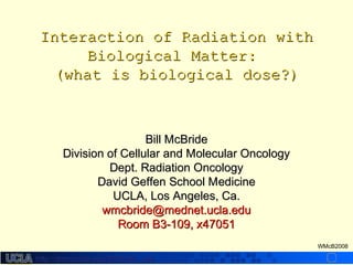
- 1. http://dmco.ucla.edu/McBride_Labhttp://dmco.ucla.edu/McBride_Lab WMcB2008 Interaction of Radiation withInteraction of Radiation with Biological Matter:Biological Matter: (what is biological dose?)(what is biological dose?) Bill McBrideBill McBride Division of Cellular and Molecular OncologyDivision of Cellular and Molecular Oncology Dept. Radiation OncologyDept. Radiation Oncology David Geffen School MedicineDavid Geffen School Medicine UCLA, Los Angeles, Ca.UCLA, Los Angeles, Ca. wmcbride@mednet.ucla.eduwmcbride@mednet.ucla.edu Room B3-109, x47051Room B3-109, x47051
- 2. http://dmco.ucla.edu/McBride_Labhttp://dmco.ucla.edu/McBride_Lab WMcB2008 Objectives:Objectives: – Know the characteristics of ionizingKnow the characteristics of ionizing radiation that make it useful for RTradiation that make it useful for RT – Define LET and RBE and what is meant byDefine LET and RBE and what is meant by quality of radiationquality of radiation – Know the difference between direct andKnow the difference between direct and indirect action of radiation and the role ofindirect action of radiation and the role of free radicalsfree radicals – Recognize the impact of oxygen on initialRecognize the impact of oxygen on initial radiation damage and of hypoxia in tumorradiation damage and of hypoxia in tumor RTRT – Understand how biological radiation doseUnderstand how biological radiation dose and physical radiation dose differand physical radiation dose differ
- 3. http://dmco.ucla.edu/McBride_Labhttp://dmco.ucla.edu/McBride_Lab WMcB2008 Radiation TherapyRadiation Therapy • Approximately 50% of cancer patientsApproximately 50% of cancer patients receive RT with curative intentreceive RT with curative intent • Approximately half of these are curedApproximately half of these are cured Radiation Therapy has a long history!
- 4. http://dmco.ucla.edu/McBride_Labhttp://dmco.ucla.edu/McBride_Lab WMcB2008 Roentgen with his wifeRoentgen with his wife’s’s hand, 1895hand, 1895 X-rays were rapidly adapted for use as a clinical treatment, initially for non- cancerous conditions, but soon for cancer, as well.
- 5. http://dmco.ucla.edu/McBride_Labhttp://dmco.ucla.edu/McBride_Lab WMcB2008 FIRST CURE OF CANCER BY X-RAYSFIRST CURE OF CANCER BY X-RAYS 1899 - BASAL CELL CARCINOMA1899 - BASAL CELL CARCINOMA X-rays were used to cure cancer very soon after their discovery
- 6. http://dmco.ucla.edu/McBride_Labhttp://dmco.ucla.edu/McBride_Lab WMcB2008 And rapidly became a standardAnd rapidly became a standard treatmenttreatment Hammersmith Hospital, London, 1905
- 7. http://dmco.ucla.edu/McBride_Labhttp://dmco.ucla.edu/McBride_Lab WMcB2008 Although side-effects wereAlthough side-effects were encountered!encountered! This is a picture of a 70 year oldThis is a picture of a 70 year old person who was irradiated byperson who was irradiated by Freund at the of age 5 in AustriaFreund at the of age 5 in Austria 1896 for nevus pigmentosus1896 for nevus pigmentosus piliferus.piliferus. L. Freund, Ein mit Rontgenstrahlen behandelterL. Freund, Ein mit Rontgenstrahlen behandelter fall von nevus pigmentosus piliferus. Wein. Med.fall von nevus pigmentosus piliferus. Wein. Med. Wochschr. 47, 428-434 (1987).Wochschr. 47, 428-434 (1987).
- 8. http://dmco.ucla.edu/McBride_Labhttp://dmco.ucla.edu/McBride_Lab WMcB2008 EpilepsyEpilepsy LupusLupus Initially more non-cancerous diseases were treated that cancer (still popular in Europe)
- 9. http://dmco.ucla.edu/McBride_Labhttp://dmco.ucla.edu/McBride_Lab WMcB2008 The Nobel Prize in Physiology or Medicine 1946The Nobel Prize in Physiology or Medicine 1946 "for the discovery of the production of mutations by means of X-ray irradiation Hermann J. Muller However, its use for benign conditions has been limited in most countries for fear of radiation-induced cancer. The carcinogenic effects of X-rays was discovered using fruit flies by Muller in 1946.
- 10. http://dmco.ucla.edu/McBride_Labhttp://dmco.ucla.edu/McBride_Lab WMcB2008 Natural radioactivity was discovered by Becquerel, who was awardedNatural radioactivity was discovered by Becquerel, who was awarded the Nobel Prize in Physics in 1903 along with Marie and Pierrethe Nobel Prize in Physics in 1903 along with Marie and Pierre Curie "in recognition of the extraordinary services they haveCurie "in recognition of the extraordinary services they have rendered by their joint researches on therendered by their joint researches on the radiationradiation phenomena"phenomena" “One wraps a Lumiere photographic plate with a bromide emulsion in two sheets of very thick black paper, such that the plate does not become clouded upon being exposed to the sun for a day. One places on the sheet of paper, on the outside, a slab of the phosphorescent substance, and one exposes the whole to the sun for several hours. When one then develops the photographic plate, one recognizes that the silhouette of the phosphorescent substance appears in black on the negative. If one places between the phosphorescent substance and the paper a piece of money or a metal screen pierced with a cut-out design, one sees the image of these objects appear on the negative. One must conclude from these experiments that the phosphorescent substance in question emits rays which pass through the opaque paper and reduces silver salts.” Paris 1896 Maltese crossHenri BecquerelHenri Becquerel Marie CurieMarie Curie
- 11. http://dmco.ucla.edu/McBride_Labhttp://dmco.ucla.edu/McBride_Lab WMcB2008 Natural RadioactivityNatural Radioactivity ∀ αα particlesparticles – Positively charged, helium nucleusPositively charged, helium nucleus ∀ ββ particlesparticles – Negatively charged, electronsNegatively charged, electrons ∀ γγ-rays-rays – No charge, EMRNo charge, EMR
- 12. http://dmco.ucla.edu/McBride_Labhttp://dmco.ucla.edu/McBride_Lab WMcB2008 Radioisotopes also were soon beingRadioisotopes also were soon being used to treat and cure cancer.used to treat and cure cancer. Radium applicators were used for many other conditions! Radioactive plaques and implants are still in common use, for example in prostate implant seeds. First cure ofFirst cure of cancer bycancer by radium plaque -radium plaque - 19221922
- 13. http://dmco.ucla.edu/McBride_Labhttp://dmco.ucla.edu/McBride_Lab WMcB2008 Therapeutic Benefit and R.T.Therapeutic Benefit and R.T. There is always a need to derive a therapeuticThere is always a need to derive a therapeutic benefit from RT. There are 2 main ways bybenefit from RT. There are 2 main ways by which this is achieved:which this is achieved: 1. Physical means1. Physical means – distributing dose by treatment planningdistributing dose by treatment planning 2. Biological means2. Biological means – dose fractionationdose fractionation
- 14. http://dmco.ucla.edu/McBride_Labhttp://dmco.ucla.edu/McBride_Lab WMcB2008 Grenz Rays Megavoltag e Orthovoltag e Superficial Therapy Contact Therapy 20 KeV 50 KeV 150 KeV 500 KeV 1-25 MeV Major improvements in RTMajor improvements in RT during the mid-1900s cameduring the mid-1900s came from improved penumbrafrom improved penumbra and decreased skin doseand decreased skin dose associated with higherassociated with higher energy x-rays, cobalt, andenergy x-rays, cobalt, and high energy photons.high energy photons. More recently conformalMore recently conformal RT, IMRT, IGRT,RT, IMRT, IGRT, Gammaknife, Cyberknife,Gammaknife, Cyberknife, tomotherapy, SRS, SRT,tomotherapy, SRS, SRT, protons, heavy ions, etc.protons, heavy ions, etc. have added considerablehave added considerable variety to the choices forvariety to the choices for physical radiation deliveryphysical radiation delivery and present radiobiologicaland present radiobiological challenges.challenges.
- 15. http://dmco.ucla.edu/McBride_Labhttp://dmco.ucla.edu/McBride_Lab WMcB2008 18961896 FreundFreund - treated hairy nevus with fractionated doses- treated hairy nevus with fractionated doses 19001900 Stenbeck - cured skin cancer with single dosesStenbeck - cured skin cancer with single doses 19061906 Bergonie and Trubandeau introduced theBergonie and Trubandeau introduced the “Law” that radiosensitivity is related“Law” that radiosensitivity is related to cell proliferation (NOT TRUE!) to explain why fractionated doses sterilizedto cell proliferation (NOT TRUE!) to explain why fractionated doses sterilized rams without skin reactionsrams without skin reactions Regaud - treated uterine cancer with fractionated dosesRegaud - treated uterine cancer with fractionated doses 19141914 SchwartzSchwartz - Fractionation is superior because of cell cycle redistribution- Fractionation is superior because of cell cycle redistribution 19191919 Coutard cures deep-seated H&N tumorsCoutard cures deep-seated H&N tumors 19321932 Coutard shows fractionation superior to single doseCoutard shows fractionation superior to single dose 19441944 Strandquist - empirical laws for changing dose per fractionStrandquist - empirical laws for changing dose per fraction 19671967 Ellis - Nominal Standard Dose (NSD) formulaEllis - Nominal Standard Dose (NSD) formula 1980s Linear Quadratic formula gains favor1980s Linear Quadratic formula gains favor History of FractionationHistory of Fractionation
- 16. http://dmco.ucla.edu/McBride_Labhttp://dmco.ucla.edu/McBride_Lab WMcB2008 The First Radiation Dosimeter!The First Radiation Dosimeter! Early x-ray machines took a long time to deliver effective dose and gave skin reactions that could be circumvented by dose fractionation.
- 17. http://dmco.ucla.edu/McBride_Labhttp://dmco.ucla.edu/McBride_Lab WMcB2008 From Amaldi and Kraft, “Radiotherapy with beams of carbon ions, Reports on Progress in Physics, 68, (2005)
- 18. http://dmco.ucla.edu/McBride_Labhttp://dmco.ucla.edu/McBride_Lab WMcB2008 ““In order to save machine time, a 3-day-a-week schedule wasIn order to save machine time, a 3-day-a-week schedule was initiated in 1962. This schedule was quickly abandoned in pre-initiated in 1962. This schedule was quickly abandoned in pre- operative irradiation because of increased wound healing problems.operative irradiation because of increased wound healing problems. Although acute reactions in the 3-day-a-week schedule for protractedAlthough acute reactions in the 3-day-a-week schedule for protracted radical irradiation were not excessive,radical irradiation were not excessive, late radiation sequelaelate radiation sequelae areare probably more pronounced as observed 2 or more years later.probably more pronounced as observed 2 or more years later.”” Fletcher, 1966.Fletcher, 1966. 3 x 3.3 Gy3 x 3.3 Gy 5 x 2 Gy5 x 2 Gy History has repeatedly shown that dose fractionationHistory has repeatedly shown that dose fractionation results in a therapeutic advantageresults in a therapeutic advantage
- 19. http://dmco.ucla.edu/McBride_Labhttp://dmco.ucla.edu/McBride_Lab WMcB2008 Clinical RT is Changing, which PresentsClinical RT is Changing, which Presents Challenges and Opportunities forChallenges and Opportunities for RadiobiologyRadiobiology Conventional treatment:Conventional treatment: Tumors are irradiated to a specified dose with 2Gy fractions delivered, more or lessTumors are irradiated to a specified dose with 2Gy fractions delivered, more or less homogeneously, in a 6 week time periodhomogeneously, in a 6 week time period • Varying this schedule impacts outcomeVarying this schedule impacts outcome • Radiobiological modeling attempts to provide guidelines for customization of RT usingRadiobiological modeling attempts to provide guidelines for customization of RT using – Radiobiological principlesRadiobiological principles derived from preclinical dataderived from preclinical data – Radiobiological parameters derived from clinical altered fractionation protocolsRadiobiological parameters derived from clinical altered fractionation protocols Modern treatment:Modern treatment: IMRT etc allows optimized non-homogeneous dose distributions, concomitant boosts,IMRT etc allows optimized non-homogeneous dose distributions, concomitant boosts, dose painting -dose painting - dose heterogeneitydose heterogeneity SRS, SRT, HDR, Protons, Heavy Ions -SRS, SRT, HDR, Protons, Heavy Ions - high dose/fx issueshigh dose/fx issues Molecular and chemical targeting -Molecular and chemical targeting - dose adjustmentdose adjustment Molecular prognosis and diagnosis promise individualized treatment plans andMolecular prognosis and diagnosis promise individualized treatment plans and biologicalbiological treatment planningtreatment planning
- 20. http://dmco.ucla.edu/McBride_Labhttp://dmco.ucla.edu/McBride_Lab WMcB2008 • Radiobiology has derived means ofRadiobiology has derived means of understanding why dose fractionation gives aunderstanding why dose fractionation gives a therapeutic benefit.therapeutic benefit. • New physical delivery methods need toNew physical delivery methods need to incorporate and/or modify these concepts.incorporate and/or modify these concepts. • In order to understand either conventional orIn order to understand either conventional or newer treatment effects, one needs to knownewer treatment effects, one needs to know the differences between physical andthe differences between physical and biological radiation dosebiological radiation dose
- 21. http://dmco.ucla.edu/McBride_Labhttp://dmco.ucla.edu/McBride_Lab WMcB2008 What is Radiation?What is Radiation? • Radiation is classified into two main categories:Radiation is classified into two main categories: - Non-ionizing radiationNon-ionizing radiation - Ionizing radiationIonizing radiation
- 22. http://dmco.ucla.edu/McBride_Labhttp://dmco.ucla.edu/McBride_Lab WMcB2008 ELECTROMAGNETIC RADIATIONSELECTROMAGNETIC RADIATIONS Photon E = hν (energy = Planck’s const x frequency) = hc/λ (c = speed of light, λ = wave length) 1010-9-9 1010-8-8 1010-7-7 1010-6-6 1010-5-5 1010-4-4 1010-3-3 1010-2-2 1010-1-1 11 1010 101022 101033 101044 γγ raysrays X-raysX-rays U.V.U.V. vv ii ss ii bb ll ee Infra RedInfra Red Radio WavesRadio Waves MicrowavesMicrowaves Short WavesShort Waves T.V.T.V. RadioRadio RadarRadar IONIZINGIONIZING RADIATIONRADIATION NON-IONIZING RADIATIONNON-IONIZING RADIATION λ (cms) E (eV) 1.24x10⁷ 1.24x10² 1.24x10⁻¹³
- 23. http://dmco.ucla.edu/McBride_Labhttp://dmco.ucla.edu/McBride_Lab WMcB2008 • Non-ionizing radiationNon-ionizing radiation – Is a particle or wave that has enough kinetic energy to raise theIs a particle or wave that has enough kinetic energy to raise the thermal energy of an outer shell electron and causethermal energy of an outer shell electron and cause excitationexcitation with emission ofwith emission of low energy EMR (infrared)low energy EMR (infrared) • Ionizing radiationIonizing radiation – Ionizing radiation has enough kinetic energy to detach at least one electron from anIonizing radiation has enough kinetic energy to detach at least one electron from an atom or molecule, creating ionsatom or molecule, creating ions – Charged particles such as electrons, protons, heavy ions, alpha and beta particles areCharged particles such as electrons, protons, heavy ions, alpha and beta particles are directlydirectly ionizing because they can interact directly with atomic electrons throughionizing because they can interact directly with atomic electrons through coulombic forces and transfer a major part of their kinetic energy directlycoulombic forces and transfer a major part of their kinetic energy directly – In contrast, photons (x rays,In contrast, photons (x rays, γγ rays) and neutrons are chargeless and therefore morerays) and neutrons are chargeless and therefore more penetrating. They arepenetrating. They are indirectlyindirectly ionizing. They have sufficient kinetic energy to free anionizing. They have sufficient kinetic energy to free an orbital electron producing aorbital electron producing a ‘fast’ recoil or Compton electron that is, in turn, directly‘fast’ recoil or Compton electron that is, in turn, directly ionizingionizing • Energy is deposited inEnergy is deposited in “packets”,“packets”, which is why, when it is deposited in DNA, ionizing radiation iswhich is why, when it is deposited in DNA, ionizing radiation is an efficient cytotoxic agentan efficient cytotoxic agent • Ionizing radiation has an energy in excess of 124 eV, which corresponds to aIonizing radiation has an energy in excess of 124 eV, which corresponds to a λλ < about 10< about 10-6-6 cm.cm. γ-ray γ’- ray excitation ionization α particle excitation and ionization
- 24. http://dmco.ucla.edu/McBride_Labhttp://dmco.ucla.edu/McBride_Lab WMcB2008 Ion formation – H2OIon formation – H2O++ and e-and e- Excitation and H and OH radical formationExcitation and H and OH radical formation ION RADICAL LIFETIMEION RADICAL LIFETIME FREE RADICAL LIFETIMEFREE RADICAL LIFETIME BREAKAGE OF BONDSBREAKAGE OF BONDS CHEMICAL REPAIR / MISREPAIRCHEMICAL REPAIR / MISREPAIR ENZYMIC REPAIR / MISREPAIRENZYMIC REPAIR / MISREPAIR EARLY BIOLOGICAL EFFECTSEARLY BIOLOGICAL EFFECTS LATE BIOLOGICAL EFFECTSLATE BIOLOGICAL EFFECTS 1010-18-18 1010-12-12 1010-6-6 101000 101066 SECSSECS Absorption of energyAbsorption of energy Physical effectsPhysical effects Chemical lesionsChemical lesions Chemical repairChemical repair Enzyme repair/lesionEnzyme repair/lesion Cellular effectsCellular effects Tissue effectsTissue effects Systemic effectsSystemic effects Days-YearsDays-Years Hrs-DaysHrs-Days Mins-HrsMins-Hrs Ionization produces ions, ion radicals, and freeIonization produces ions, ion radicals, and free radicals concentrated along tracks and especially atradicals concentrated along tracks and especially at Bragg peak of primary and secondary electrons. TheyBragg peak of primary and secondary electrons. They are highly reactive and cause damage to biologicalare highly reactive and cause damage to biological mattermatter • IonIon - atom or molecule that has lost an electron and is charged.- atom or molecule that has lost an electron and is charged. • Free radicalFree radical - atom or group of atoms that contains an unpaired electron and is highly reactive- atom or group of atoms that contains an unpaired electron and is highly reactive • Aqueous electronAqueous electron - has lost kinetic energy and has been captured by water - a powerful reducing- has lost kinetic energy and has been captured by water - a powerful reducing agent.agent. 1010-16-16 1010-14-14
- 25. http://dmco.ucla.edu/McBride_Labhttp://dmco.ucla.edu/McBride_Lab WMcB2008 The Gray is theThe Gray is the PhysicalPhysical Unit of RadiationUnit of Radiation • 1 GRAY, the unit of absorbed dose (1 joule / Kg),1 GRAY, the unit of absorbed dose (1 joule / Kg), – Causes 1-2 x 10Causes 1-2 x 1055 ionization events / cellionization events / cell – 1% in DNA1% in DNA – A single cobalt 60 ray will deposit about 1mGy in a cellA single cobalt 60 ray will deposit about 1mGy in a cell • Rad (Radiation Absorbed Dose) is the old unit = cGyRad (Radiation Absorbed Dose) is the old unit = cGy
- 26. http://dmco.ucla.edu/McBride_Labhttp://dmco.ucla.edu/McBride_Lab WMcB2008 Direct and IndirectDirect and Indirect ActionAction ofof RadiationRadiation • Indirectly ionizing radiation canIndirectly ionizing radiation can actact directly ordirectly or indirectlyindirectly on biological targetson biological targets • If the ion pairs and free radicalsIf the ion pairs and free radicals are produced inare produced in aa biologic targetbiologic target (DNA) then this is(DNA) then this is direct actiondirect action • If water or other atoms or molecules are ionized,If water or other atoms or molecules are ionized, diffusible free radicals can act as intermediaries todiffusible free radicals can act as intermediaries to cause damage - this iscause damage - this is indirect actionindirect action
- 27. http://dmco.ucla.edu/McBride_Labhttp://dmco.ucla.edu/McBride_Lab WMcB2008 p+ e- photon p+ photon INDIRECT ACTION DIRECT ACTION Direct and Indirect Action of Ionizing Radiation on DNA 4 nm 2 nm e- R. H2O OH .
- 28. http://dmco.ucla.edu/McBride_Labhttp://dmco.ucla.edu/McBride_Lab WMcB2008 • Since HSince H22O is the major component in cells, the most commonO is the major component in cells, the most common ionization event is radiolysis of water, producing reactive oxygenionization event is radiolysis of water, producing reactive oxygen species (ROS)species (ROS) • The most relevant water is within 2nm of the DNA and tightlyThe most relevant water is within 2nm of the DNA and tightly boundbound • ROS produced include: HROS produced include: H.. - reducing; OH- reducing; OH.. - oxidizing; HO- oxidizing; HO22 .. - oxidizing- oxidizing (O(O22 + H+ H.. ); H); H22OO22 - oxidizing- oxidizing • The net effect is oxidation of cellular constituentsThe net effect is oxidation of cellular constituents • About 60% of DNA damage caused by x-rays is due to ROSAbout 60% of DNA damage caused by x-rays is due to ROS • About 75% of the indirect action of radiation is due to hydroxyl radicalsAbout 75% of the indirect action of radiation is due to hydroxyl radicals (OH(OH.. )) Reactive Oxygen Species (ROS)Reactive Oxygen Species (ROS)
- 29. http://dmco.ucla.edu/McBride_Labhttp://dmco.ucla.edu/McBride_Lab WMcB2008 Free OHFree OH.. radicals generateradicals generate organic radicalsorganic radicals by:by: – AdditionAddition R + OHR + OH.. .. ROHROH – Hydrogen abstractionHydrogen abstraction RH + OHRH + OH.. RR.. + H+ H22OO – Electron transferElectron transfer R + OHR + OH.. RR.. + OH+ OH -- Where R is the organic moietyWhere R is the organic moiety
- 30. http://dmco.ucla.edu/McBride_Labhttp://dmco.ucla.edu/McBride_Lab WMcB2008 Free Radicals and theirFree Radicals and their Scavengers MatterScavengers Matter • Biological effects of ionizing radiation are determined in large part by freeBiological effects of ionizing radiation are determined in large part by free radicalsradicals • Free radicals are involved in many biological processes, including cellularFree radicals are involved in many biological processes, including cellular respirationrespiration • We have defenses against free radicalsWe have defenses against free radicals – Endogenous free radical scavengers - most relevant within 2nm of the DNAEndogenous free radical scavengers - most relevant within 2nm of the DNA – Anti-oxidantsAnti-oxidants • eg superoxide dismutase, especially in mitochondria, and catalaseeg superoxide dismutase, especially in mitochondria, and catalase • Free radical scavengers can protect normal tissue from radiationFree radical scavengers can protect normal tissue from radiation – eg Amifostineeg Amifostine • Depleting free radical scavengers will radiosensitizeDepleting free radical scavengers will radiosensitize • What interacts with free radicals, in particular radicals in biologicalWhat interacts with free radicals, in particular radicals in biological materials will be important in determining outcome at this levelmaterials will be important in determining outcome at this level • Oxygen interacts with free radicalsOxygen interacts with free radicals
- 31. http://dmco.ucla.edu/McBride_Labhttp://dmco.ucla.edu/McBride_Lab WMcB2008 Oxygen MattersOxygen Matters • Binds H radicals forming hydrogen peroxideBinds H radicals forming hydrogen peroxide HH.. + O+ O22 HOHO22 .. (+HO(+HO22 .. ) H) H22OO22 (+O(+O22)) • Binds electrons to give superoxideBinds electrons to give superoxide ee-- + O+ O22 OO22 -- + (H+ (H22O) HOO) HO22 .. + OH+ OH-- • Binds organic radicals to form peroxidesBinds organic radicals to form peroxides RR.. + O+ O22 RORO22 .. (radical peroxide)(radical peroxide) RORO22 .. + R+ R’ H ROOH + R’ (hydroperoxide)’ H ROOH + R’ (hydroperoxide) RORO22 .. + R+ R’’.. ROOR’ (peroxide)ROOR’ (peroxide) OxygenOxygen “fixes”“fixes” the radical lesions in DNA in a form that can notthe radical lesions in DNA in a form that can not be easily chemically repaired and therefore is abe easily chemically repaired and therefore is a very powerfulvery powerful radiosensitizer.radiosensitizer.
- 32. http://dmco.ucla.edu/McBride_Labhttp://dmco.ucla.edu/McBride_Lab WMcB2008 Oxygen Enhancement RatioOxygen Enhancement Ratio (OER)(OER) Dose required to produce a specific biological effect in the absence of oxygenDose required to produce a specific biological effect in the absence of oxygen Dose required for the same effect in its presenceDose required for the same effect in its presence== OER varies with level of effect but can be 2.5 - 3 foldOER varies with level of effect but can be 2.5 - 3 fold 1) Culture Cells1) Culture Cells (( 3) Count cells in hemocytometer3) Count cells in hemocytometer 4) irradiate under oxic or hypoxic conditions4) irradiate under oxic or hypoxic conditions 0 Gy 2Gy 4Gy 6Gy0 Gy 2Gy 4Gy 6Gy 5) Plate cells and5) Plate cells and grow for about 12 daysgrow for about 12 days .. .. .. .. .. .... .. 6) Count colonies6) Count colonies Dose (Gy)Dose (Gy) S.F.S.F. 0 2 4 6 8 100 2 4 6 8 10 1.01.0 0.10.1 0.010.01 oxicoxic hypoxichypoxic Physical Dose = Biological DosePhysical Dose = Biological Dose 2) Suspend Cells2) Suspend Cells trysinization)trysinization)
- 33. http://dmco.ucla.edu/McBride_Labhttp://dmco.ucla.edu/McBride_Lab WMcB2008 • Hypoxic areasHypoxic areas occur almost solely in tumorsoccur almost solely in tumors and are moreand are more radioresistant than oxic areas.radioresistant than oxic areas. • Hypoxia contributes to treatment failureHypoxia contributes to treatment failure • Reoxygenation occurs between radiation dose fractions giving aReoxygenation occurs between radiation dose fractions giving a rationale for dose fractionationrationale for dose fractionation • The oxygen effect is greater for low LET than high LET radiationThe oxygen effect is greater for low LET than high LET radiation Giacca and Brown Pimonizadole (oxygen mimetic) staining colorectal carcinoma The effects of hypoxia were first discovered in 1909 by Schwarz who showed that strapping a radium source on the arm gave less of a skin reaction than just placing it there. This was used to give higher doses to deep seated tumors. Clinical Relevance of HypoxiaClinical Relevance of Hypoxia
- 34. http://dmco.ucla.edu/McBride_Labhttp://dmco.ucla.edu/McBride_Lab WMcB2008 RADIATION QUALITY ANDRADIATION QUALITY AND BIOLOGICAL EFFECTIVENESSBIOLOGICAL EFFECTIVENESS
- 35. http://dmco.ucla.edu/McBride_Labhttp://dmco.ucla.edu/McBride_Lab WMcB2008 gamma raysgamma rays deep therapydeep therapy X-raysX-rays soft X-rayssoft X-rays alpha-particlealpha-particle HIGH LETHIGH LET RadiationRadiation LOW LETLOW LET RadiationRadiation Separation of ion clusters in relation toSeparation of ion clusters in relation to size of biological targetsize of biological target LINEAR ENERGY TRANSFERLINEAR ENERGY TRANSFER LET is average energy (dE) imparted by excitationLET is average energy (dE) imparted by excitation and Ionization events caused by a charged particleand Ionization events caused by a charged particle traveling a set distance (dl) -traveling a set distance (dl) - LET = dE/dl (keV/LET = dE/dl (keV/ µµm)m)
- 36. http://dmco.ucla.edu/McBride_Labhttp://dmco.ucla.edu/McBride_Lab WMcB2008 • A dose of 1 Gy will give 2x10A dose of 1 Gy will give 2x1033 ionization events in 10ionization events in 10-10-10 g (the sizeg (the size of a cell nucleus). This can beof a cell nucleus). This can be achieved by:achieved by: – 1MeV electrons1MeV electrons •700 electrons which give 6700 electrons which give 6 ionization events perionization events per µµm.m. – 30 keV electrons30 keV electrons •140 electrons which give 30140 electrons which give 30 ionization events perionization events per µµm.m. – 4 MeV protons4 MeV protons •14 protons which give 30014 protons which give 300 ionization events perionization events per µµm.m. • The biological effectiveness ofThe biological effectiveness of these different radiationsthese different radiations vary!vary! γ-ray γ’-ray excitation ionization α particle excitation and ionization
- 37. http://dmco.ucla.edu/McBride_Labhttp://dmco.ucla.edu/McBride_Lab WMcB2008 Relative BiologicalRelative Biological EffectivenessEffectiveness (RBE) of the(RBE) of the Radiation MattersRadiation Matters Dose of 250 kVp x-rays required to produce an effectDose of 250 kVp x-rays required to produce an effect Dose of test radiation required for the same effectDose of test radiation required for the same effect == S.F.S.F. 1.01.0 0.10.1 0.010.01 0.0010.001 DOSE GyDOSE Gy High LETHigh LET Low LET, HDRLow LET, HDR Physical Dose = Biological DosePhysical Dose = Biological Dose
- 38. http://dmco.ucla.edu/McBride_Labhttp://dmco.ucla.edu/McBride_Lab WMcB2008 Linear Energy Transfer (LET keV/Linear Energy Transfer (LET keV/µµm)m) RBERBE (for cell kill)(for cell kill) 10001000100100101011 00 22 44 66 88 RBERBE DiagnosticDiagnostic X-raysX-rays FastFast NeutronsNeutrons AlphaAlpha ParticlesParticles overkilloverkill 0.10.1 Co-60Co-60 gamma raysgamma rays 00 11 22 33 44 OEROER OEROER OER is the inverse of RBE because OER depends considerably on theOER is the inverse of RBE because OER depends considerably on the indirect action of ionizing radiationindirect action of ionizing radiation RBE is maximal when the average distance between ionization events =RBE is maximal when the average distance between ionization events = distance between DNA strands = 2nmdistance between DNA strands = 2nm RBE and OER as a function of LETRBE and OER as a function of LET
- 39. http://dmco.ucla.edu/McBride_Labhttp://dmco.ucla.edu/McBride_Lab WMcB2008 DNA is the Primary, but not the only,DNA is the Primary, but not the only, Cellular Target for RadiationCellular Target for Radiation • Microbeam irradiation of cell cytoplasmMicrobeam irradiation of cell cytoplasm does not generally cause cell death, butdoes not generally cause cell death, but irradiation of the nucleus doesirradiation of the nucleus does • Tritiated thymidine incorporated into cellsTritiated thymidine incorporated into cells can kill themcan kill them • Radiation-induced chromosomalRadiation-induced chromosomal abnormalities correlate with cell deathabnormalities correlate with cell death and carcinogenesisand carcinogenesis • However, irradiation of the cytoplasm isHowever, irradiation of the cytoplasm is not without biological consequencesnot without biological consequences
- 40. http://dmco.ucla.edu/McBride_Labhttp://dmco.ucla.edu/McBride_Lab WMcB2008 Lesion sizeLesion size about 15-20about 15-20 nucleotidesnucleotides BlobBlob 7 nm diam.7 nm diam. 12 ion pairs12 ion pairs OHOH .. eeaquaqu OHOH .. eeaquaqu OHOH .. eeaquaqu OHOH .. eeaquaqu OHOH .. eeaquaquOHOH .. eeaquaqu OHOH .. eeaquaqu OHOH .. eeaquaqu OHOH .. eeaquaqu OHOH .. eeaquvaquv OHOH .. eeaquaqu OHOH .. eeaquaqu OHOH .. eeaquaqu SpurSpur 4 nm diam4 nm diam 3 ion pairs3 ion pairs 100 eV energy100 eV energy 95% of energy deposition events95% of energy deposition events The lesions in DNA that are associated withThe lesions in DNA that are associated with cell death and carcinogenesis aftercell death and carcinogenesis after radiation exposure areradiation exposure are largelarge The high cytotoxic efficiency of ionizing radiation can be ascribedThe high cytotoxic efficiency of ionizing radiation can be ascribed to the deposition of low levels of energy in small packets withinto the deposition of low levels of energy in small packets within the DNA that cause lesions large enough to be fatalthe DNA that cause lesions large enough to be fatal
- 41. http://dmco.ucla.edu/McBride_Labhttp://dmco.ucla.edu/McBride_Lab WMcB2008 SINGLE STRAND BREAK 1000 / CELL / GRAY BASE CHANGE (eg C - U) BASE LOSS 1000 / CELL / GRAY BASE MODIFICATION (eg thymine/cytosine glycol) SUGAR DAMAGE (abstraction of hydrogen atom) INTRASTRAND CROSSLINK 0.5 / CELL / GRAY INTERSTRAND CROSSLINK DNA-PROTEIN CROSSLINK 1 / CELL / GRAY * DOUBLE STRAND BREAK 30/ CELL / GRAY
- 42. http://dmco.ucla.edu/McBride_Labhttp://dmco.ucla.edu/McBride_Lab WMcB2008 • Not all ionization events are lethal!!Not all ionization events are lethal!! • As a rough guide the fraction of cells surviving 2Gy (SFAs a rough guide the fraction of cells surviving 2Gy (SF2Gy2Gy)) is about 0.5is about 0.5 • If the S.F. 2Gy is 0.5, what is the S.F. after 60Gy?If the S.F. 2Gy is 0.5, what is the S.F. after 60Gy? = 0.5= 0.53030 = 0.9x10= 0.9x10⁻⁹-9 • If the S.F. 2Gy is 0.7, what is the S.F. after 60Gy?If the S.F. 2Gy is 0.7, what is the S.F. after 60Gy? = 0.7= 0.73030 = 2.2x10= 2.2x10⁻⁵-5
- 43. http://dmco.ucla.edu/McBride_Labhttp://dmco.ucla.edu/McBride_Lab WMcB2008 What is the Lethal Lesion?What is the Lethal Lesion?
- 44. http://dmco.ucla.edu/McBride_Labhttp://dmco.ucla.edu/McBride_Lab WMcB2008 Repairable Sublethal DamageRepairable Sublethal Damage X- orX- or γγ-radiation is sparsely ionizing; most-radiation is sparsely ionizing; most damage can be repaireddamage can be repaired 4 nm4 nm 2 nm2 nm
- 45. http://dmco.ucla.edu/McBride_Labhttp://dmco.ucla.edu/McBride_Lab WMcB2008 SingleSingle lethal hit Also known as α - type killing 4 nm4 nm 2 nm2 nm Unrepairable Multiply Damaged Site It is hypothesized that the lethal lesions are large double strand breaks with Multiply Damaged Sites (MDS) that can not be repaired. They are more likely to occur at the end of a track
- 46. http://dmco.ucla.edu/McBride_Labhttp://dmco.ucla.edu/McBride_Lab WMcB2008 At high dose, intertrack repairable Sublethal Damage may Accumulate forming unrepairable, lethal MDS Also known asAlso known as ββ - type killing- type killing
- 47. http://dmco.ucla.edu/McBride_Labhttp://dmco.ucla.edu/McBride_Lab WMcB2008 S.F.S.F. 1.01.0 0.10.1 0.010.01 0.0010.001 DOSE GyDOSE Gy Low LET, HDRLow LET, HDR Low Dose RateLow Dose Rate allows continuous SLDRallows continuous SLDR Dose Rate MattersDose Rate Matters Physical Dose = Biological DosePhysical Dose = Biological Dose
- 48. http://dmco.ucla.edu/McBride_Labhttp://dmco.ucla.edu/McBride_Lab WMcB2008 Chromatin Structure MattersChromatin Structure Matters • Each cell contains about 2m of DNAEach cell contains about 2m of DNA • The basic structure is theThe basic structure is the nucleosome,nucleosome, which is 146 base pairs of DNA wrapped around 2 copies of histones H2A,H2A, H2B, H3, and H4H2B, H3, and H4 • Nucleosomes are in turn wrapped around other proteins toNucleosomes are in turn wrapped around other proteins to formform compacted chromatincompacted chromatin • Chromatin is maximally compacted during mitosisChromatin is maximally compacted during mitosis • Transcription requires decompaction to facilitate initiationTranscription requires decompaction to facilitate initiation (binding of transcription factors and RNAP II) and(binding of transcription factors and RNAP II) and elongationelongation 840nm840nm minibandminiband - 30nm- 30nm
- 49. http://dmco.ucla.edu/McBride_Labhttp://dmco.ucla.edu/McBride_Lab WMcB2008 Chromatin Structure andChromatin Structure and Radiation ResponsesRadiation Responses • Compact chromatin is more radiosensitive than non-compactedCompact chromatin is more radiosensitive than non-compacted –Mitotic cellsMitotic cells • are 2.8 times more sensitive to DNA breaks than interphase cellsare 2.8 times more sensitive to DNA breaks than interphase cells • have a lower OER (eg 2.0 compared with 2.8)have a lower OER (eg 2.0 compared with 2.8) • do not have much of ado not have much of a “shoulder” on their survival curve“shoulder” on their survival curve –Actively transcribing genes are less sensitive to damageActively transcribing genes are less sensitive to damage • Decompaction and compaction require acetylation and deacetylation ofDecompaction and compaction require acetylation and deacetylation of histones by acetyltransferases (HAT) and deacetylases (HDAC)histones by acetyltransferases (HAT) and deacetylases (HDAC) • HDAC inhibitors are entering the clinic as anti-cancer agents and can radiosensitizeHDAC inhibitors are entering the clinic as anti-cancer agents and can radiosensitize • Radiation Damage to DNA is not randomly distributed.Radiation Damage to DNA is not randomly distributed. • It varies with cell cycle phase and level of gene expressionIt varies with cell cycle phase and level of gene expression Physical Dose = Biological DosePhysical Dose = Biological Dose S.F.S.F. 20201616121288440000 .01.01 .1.1 11 Dose (Gy)Dose (Gy) LATE SLATE S EARLY SEARLY S G1 PHASEG1 PHASE G2/M PHASEG2/M PHASE
- 50. http://dmco.ucla.edu/McBride_Labhttp://dmco.ucla.edu/McBride_Lab WMcB2008 12.5Gy 14.0Gy 15.5Gy 17.0Gy Withers, H. R. and Elkind, M. M. Radiology 91:998, 1968Withers, H. R. and Elkind, M. M. Radiology 91:998, 1968 Used the macrocolony assay in mouseUsed the macrocolony assay in mouse jejunum to assessed the effects of 2jejunum to assessed the effects of 2 radiation doses given varying times apartradiation doses given varying times apart to measure the time to and extent of repair,to measure the time to and extent of repair, redistribution, and repopulationredistribution, and repopulation (regeneration) between dose fractions.(regeneration) between dose fractions. RedistributionRedistribution RepairRepair RepopulationRepopulation 700R 1500R Colony derived from a single surviving clonogen
- 51. http://dmco.ucla.edu/McBride_Labhttp://dmco.ucla.edu/McBride_Lab WMcB2008 ACUTE RESPONDING TISSUESACUTE RESPONDING TISSUES (responses seen during standard therapy)(responses seen during standard therapy) GutGut SkinSkin Bone MarrowBone Marrow MucosaMucosa LATE RESPONDING TISSUESLATE RESPONDING TISSUES (responses seen after end of therapy)(responses seen after end of therapy) BrainBrain Spinal CordSpinal Cord KidneyKidney LungLung BladderBladder Tissue Type MattersTissue Type Matters Dose (Gy)Dose (Gy) SurvivingSurviving FractionFraction 20201616121288440000 .01.01 .1.1 11 Late RespondingLate Responding TissuesTissues Acute RespondingAcute Responding Tissues andTissues and Many TumorsMany Tumors Physical Dose = Biological DosePhysical Dose = Biological Dose
- 52. http://dmco.ucla.edu/McBride_Labhttp://dmco.ucla.edu/McBride_Lab WMcB2008 Dose (Gy)Dose (Gy) Dose FractionationDose Fractionation 242420201616121288440000 .01.01 .1.1 11 SurvivingSurviving FractionFraction Single doseSingle dose Late responding tissuesLate responding tissues Single doseSingle dose Acute responding tissuesAcute responding tissues Fractionated doseFractionated dose Acute responding tissuesAcute responding tissues Fractionated doseFractionated dose Late responding tissuesLate responding tissues Dose fractionation spares late responding tissues more than acuteDose fractionation spares late responding tissues more than acute responding tissues and many tumorsresponding tissues and many tumors
- 53. http://dmco.ucla.edu/McBride_Labhttp://dmco.ucla.edu/McBride_Lab WMcB2008 The Aim is to IncreaseThe Aim is to Increase Therapeutic Benefit!Therapeutic Benefit! ProbabilityProbability of tumorof tumor control/control/ of normalof normal tissuetissue damagedamage Dose (Gy)Dose (Gy) A B CA B C 1.01.0 00 therapeutic benefittherapeutic benefit Normal tissue complication dose-response curves are steep!Normal tissue complication dose-response curves are steep!
- 54. http://dmco.ucla.edu/McBride_Labhttp://dmco.ucla.edu/McBride_Lab WMcB2008 Biological effectiveness of RT varies withBiological effectiveness of RT varies with • Size of Dose (D) - (alpha and beta)Size of Dose (D) - (alpha and beta) • Size of Dose Per Fraction (d) - (alpha and beta)Size of Dose Per Fraction (d) - (alpha and beta) • Time over which it is delivered (T)- (alpha and beta)Time over which it is delivered (T)- (alpha and beta) • Time between fractions (t)Time between fractions (t) • Volume irradiated (V)Volume irradiated (V) • Quality of Radiation (Q) - RBEQuality of Radiation (Q) - RBE • Presence/Absence of Oxygen - OERPresence/Absence of Oxygen - OER • DNA Repair efficiency and completenessDNA Repair efficiency and completeness • Cell cycle phase and level of gene activationCell cycle phase and level of gene activation • Tissue/Tumor TypeTissue/Tumor Type Physical Dose = Biological DosePhysical Dose = Biological Dose
- 55. http://dmco.ucla.edu/McBride_Labhttp://dmco.ucla.edu/McBride_Lab WMcB2008 4Rs OF RADIOBIOLOGY RELEVANT TO4Rs OF RADIOBIOLOGY RELEVANT TO CLINICAL DOSE FRACTIONATIONCLINICAL DOSE FRACTIONATION • Repair of sublethal damageRepair of sublethal damage - spares late responding normal tissue preferentiallyspares late responding normal tissue preferentially • Reassortment/Redistribution of cells in the cell cycleReassortment/Redistribution of cells in the cell cycle – increases acute effectsincreases acute effects – no influence on late effectsno influence on late effects – increases damage to tumorincreases damage to tumor • Repopulation/RegenerationRepopulation/Regeneration – spares acute responding normal tissue preferentiallyspares acute responding normal tissue preferentially – no influence on late effects,no influence on late effects, – danger of tumor repopulationdanger of tumor repopulation • ReoxygenationReoxygenation – no influence on normal tissue responsesno influence on normal tissue responses – increases tumor damageincreases tumor damage
- 56. http://dmco.ucla.edu/McBride_Labhttp://dmco.ucla.edu/McBride_Lab WMcB2008 Questions onQuestions on Interaction of Radiation with Biological Matter:Interaction of Radiation with Biological Matter: what is biological dose?what is biological dose? Bill McBrideBill McBride Dept. Radiation OncologyDept. Radiation Oncology David Geffen School MedicineDavid Geffen School Medicine UCLA, Los Angeles, Ca.UCLA, Los Angeles, Ca. wmcbride@mednet.ucla.eduwmcbride@mednet.ucla.edu
- 57. http://dmco.ucla.edu/McBride_Labhttp://dmco.ucla.edu/McBride_Lab WMcB2008 1.1. The lifetime of radicals in target molecules isThe lifetime of radicals in target molecules is aboutabout – 1010-3-3 secssecs – 1010-6-6 secssecs – 1010-9-9 secssecs – 1010-12-12 secssecs #2 – free radicals are highly unstable and reactive
- 58. http://dmco.ucla.edu/McBride_Labhttp://dmco.ucla.edu/McBride_Lab WMcB2008 2.2. Electromagnetic radiation is considered ionizingElectromagnetic radiation is considered ionizing if it has a photon energy greater thanif it has a photon energy greater than – 1.24 eV1.24 eV – 12.4 eV12.4 eV – 124 eV124 eV – 1.24 keV1.24 keV #3 – this is sufficient to break bonds in biological molecules
- 59. http://dmco.ucla.edu/McBride_Labhttp://dmco.ucla.edu/McBride_Lab WMcB2008 3.3. The S.I. unit of absorbed dose isThe S.I. unit of absorbed dose is – BecquerelBecquerel – SievertSievert – GrayGray – RoentgenRoentgen #3 The International System (IS) unit is the Gray, named after the radiobiologist Louis “Hal” Gray who was based in London
- 60. http://dmco.ucla.edu/McBride_Labhttp://dmco.ucla.edu/McBride_Lab WMcB2008 4.4. Which of the following are not chargedWhich of the following are not charged particles?particles? – ElectronsElectrons – NeutronsNeutrons – ProtonsProtons – Heavy ionsHeavy ions – Alpha particlesAlpha particles #2 – which is why they are called NEUTRons
- 61. http://dmco.ucla.edu/McBride_Labhttp://dmco.ucla.edu/McBride_Lab WMcB2008 5.5. Which of the following is NOT a characteristic of theWhich of the following is NOT a characteristic of the indirect action of ionizing radiationindirect action of ionizing radiation – Production of diffusible free radicalsProduction of diffusible free radicals – Production of reactive oxygen speciesProduction of reactive oxygen species – Involvement of anti-oxidant defensesInvolvement of anti-oxidant defenses – Causes a change in redox within a cell favoringCauses a change in redox within a cell favoring reduction of constituentsreduction of constituents #4 the free radicals produced makes ionizing radiation an oxidative stress overall
- 62. http://dmco.ucla.edu/McBride_Labhttp://dmco.ucla.edu/McBride_Lab WMcB2008 6.6. Which of the following is true about the oxygenWhich of the following is true about the oxygen enhancement ratioenhancement ratio – Is the same at all levels of cell survivalIs the same at all levels of cell survival – Can be measured by the dog-leg in a cell survivalCan be measured by the dog-leg in a cell survival curve after single high dose irradiation of tumorscurve after single high dose irradiation of tumors – Is the ratio of doses needed for an isoeffect in theIs the ratio of doses needed for an isoeffect in the absence to the presence of oxygenabsence to the presence of oxygen – Is low for cells in S cell cycle phase compared toIs low for cells in S cell cycle phase compared to cells in G2/M phasecells in G2/M phase #3 responses should be compared by the doses needed for a particular isoeffect. The OER varies with the level of effect eg survival
- 63. http://dmco.ucla.edu/McBride_Labhttp://dmco.ucla.edu/McBride_Lab WMcB2008 7.7. Which of the following is true about Linear EnergyWhich of the following is true about Linear Energy TransferTransfer – It is a measure of the biological effectiveness ofIt is a measure of the biological effectiveness of ionizing radiationionizing radiation – Shows an inverse correlation with the oxygenShows an inverse correlation with the oxygen enhancement ratioenhancement ratio – Is maximal at a relative biological effectiveness ofIs maximal at a relative biological effectiveness of 150 keV/micrometer150 keV/micrometer – Is measured in keV/micrometerIs measured in keV/micrometer #4 LET is an average value imparted per unit path length. Because the radiations vary in energy, the LET is not biologically very useful
- 64. http://dmco.ucla.edu/McBride_Labhttp://dmco.ucla.edu/McBride_Lab WMcB2008 8.8. The Relative Biological Effectiveness of aThe Relative Biological Effectiveness of a radiation isradiation is – Assessed by the dose required for toAssessed by the dose required for to produce the same effect as 250kVp X-raysproduce the same effect as 250kVp X-rays – Is the ratio of the dose required of 250 kVpIs the ratio of the dose required of 250 kVp X-rays to that of the test radiation for a givenX-rays to that of the test radiation for a given isoeffectisoeffect – Is directly related to Linear Energy TransferIs directly related to Linear Energy Transfer – Is about 3 for alpha particle radiationIs about 3 for alpha particle radiation #2 - again, measured by isoeffective doses – classically relative to 250kVp x-rays, but often more recently 60Co has been used
- 65. http://dmco.ucla.edu/McBride_Labhttp://dmco.ucla.edu/McBride_Lab WMcB2008 9.9. The lethal lesion caused in DNA by low LETThe lethal lesion caused in DNA by low LET ionizing radiation isionizing radiation is – 15-20 nucleotides in size15-20 nucleotides in size – Caused by alpha-type eventsCaused by alpha-type events – Does not correlate with chromosomalDoes not correlate with chromosomal aberrationsaberrations – Due to oxygen fixationDue to oxygen fixation #1 The lesions are large i.e they are not point mutations.
- 66. http://dmco.ucla.edu/McBride_Labhttp://dmco.ucla.edu/McBride_Lab WMcB2008 10. Approximately how many DNA double10. Approximately how many DNA double strand breaks are caused per cell per Gray?strand breaks are caused per cell per Gray? – 1-101-10 – 15-2515-25 – 30-4030-40 – 45-6045-60 – 60-7560-75 #3 This probably varies considerably depending on numerous factors, but this is a reasonable approximation
- 67. http://dmco.ucla.edu/McBride_Labhttp://dmco.ucla.edu/McBride_Lab WMcB2008 11.11. If the fraction of cells surviving 2Gy irradiation isIf the fraction of cells surviving 2Gy irradiation is 0.5, what is a reasonable estimate of the percent of0.5, what is a reasonable estimate of the percent of DNA double strand breaks that are effectivelyDNA double strand breaks that are effectively repaired?repaired? – 99%99% – 95%95% – 75%75% – 50%50% #1 If 60-80 DSB/2Gy kills half the cells, then >99% must be repaired
- 68. http://dmco.ucla.edu/McBride_Labhttp://dmco.ucla.edu/McBride_Lab WMcB2008 12. If the fraction of cells surviving 2Gy is 0.4,12. If the fraction of cells surviving 2Gy is 0.4, what is the surviving fraction after 50 Gywhat is the surviving fraction after 50 Gy given in 2Gy fractions?given in 2Gy fractions? – 1010-8-8 – 1010-9-9 – 1010-10-10 – 1010-11-11 #3 - 0.425 = 1.12x10⁻¹⁰
- 69. http://dmco.ucla.edu/McBride_Labhttp://dmco.ucla.edu/McBride_Lab WMcB2008 13. Sublethal DNA damage is most likely to13. Sublethal DNA damage is most likely to accumulateaccumulate – At high total doses given at high dose rateAt high total doses given at high dose rate – At high total doses under hypoxiaAt high total doses under hypoxia – After high LET radiationAfter high LET radiation – After low fractionated doses of radiationAfter low fractionated doses of radiation #1 Intertrack interactions between ionization events are more likely at high dose and high dose rate
- 70. http://dmco.ucla.edu/McBride_Labhttp://dmco.ucla.edu/McBride_Lab WMcB2008 15. Sublethal DNA damage is most likely to be repaired15. Sublethal DNA damage is most likely to be repaired – After high total doses given at high dose rateAfter high total doses given at high dose rate – If cells are held in a non-proliferative stateIf cells are held in a non-proliferative state – After high LET radiationAfter high LET radiation – Between low fractionated doses of radiationBetween low fractionated doses of radiation #4 The lower the number of ionization events in time and space, the more likely they are to be repaired
- 71. http://dmco.ucla.edu/McBride_Labhttp://dmco.ucla.edu/McBride_Lab WMcB2008 15.15. Which of the following is true about chromatinWhich of the following is true about chromatin structure in cellsstructure in cells – Compacted chromatin is more radiosensitive thanCompacted chromatin is more radiosensitive than non-compacted chromationnon-compacted chromation – During mitosis cells decompact their chromatinDuring mitosis cells decompact their chromatin and become radiosensitiveand become radiosensitive – Compact chromatin in S phase mediatesCompact chromatin in S phase mediates radioresistancyradioresistancy – Compaction facilitates gene transcriptionCompaction facilitates gene transcription #1 –compaction occurs in mitosis, which is why chromosomes can be seen under the microscope during this phase
- 72. http://dmco.ucla.edu/McBride_Labhttp://dmco.ucla.edu/McBride_Lab WMcB2008 16.16. Which of the following is correct aboutWhich of the following is correct about alpha-type cell killing following radiationalpha-type cell killing following radiation exposureexposure – It represents single lethal hitsIt represents single lethal hits – It is due to accumulated damageIt is due to accumulated damage – It requires intertrack interactionsIt requires intertrack interactions – It is not oxygen dependentIt is not oxygen dependent #1 – Intratrack lesions dominate and accumulated damage plays only a small role
- 73. http://dmco.ucla.edu/McBride_Labhttp://dmco.ucla.edu/McBride_Lab WMcB2008 17.17. Which of the following radiobiologicalWhich of the following radiobiological phenomena occurring between dosephenomena occurring between dose fractions has little or no effect on normalfractions has little or no effect on normal tissue radiation responses?tissue radiation responses? – RepairRepair – Redistribution of cells in the cell cycleRedistribution of cells in the cell cycle – RepopulationRepopulation – ReoxygenationReoxygenation #4 – Normal tissues are generally considered to be well oxygenated
Hinweis der Redaktion
- Radiation Biology is study of the effects of radiation on living things. For the most part, this course deals with the effects of radiation doses of the magnitude of those used in radiation therapy.
- Radiation Therapy (RT) is a common form of cancer therapy. Almost half of patients with cancer will receive RT with curative intent. Others may received RT for palliation of pain. Not all cancers are susceptible, but many are. One of the major questions is why are there differences between cancers in their response to irradiation.
- In 1895 Roentgen discovered X-rays. The fact that they could “burn” skin, suggested their use as an anti-cancer agent.
- Progress was rapid!
- In 1898 Becquerel discovered radioactivity. In the same year, the Curies discovered radium. Within a short time, radioactive applicators were being used on superficial lesions. This was called brachytherapy (short range)
- A major focus of cancer treatment is to achieve a therapeutic benefit whereby the cancer is at a disadvantage relative to the normal tissues. In RT, this is achieved by shaping the field to exclude normal tissues as much as possible and by odes fractionation. Patients treated curatively generally receive around 2 Gy per day, 5 days a week, for 5-7 weeks.
- They all believed acute reactions predicted accurately what the late reactions would be.
- Absorption of radiation energy by a body leads to molecular excitation and ionization. If the radiation has sufficient energy, orbiting electrons are ejected. The radiation is “ionizing.” The energy released per ionizing event is about 33 eV, which is sufficient to break chemical bonds. The localized release of photons or “packets” of energy is the reason ionizing radiation is so biologically effective. On the other hand, the total energy involved is small. 2 Gy would raise the body temperature by 0.001OC, if directly converted. X-rays and gamma rays are forms of electromagnetic radiation that are produced in different ways, but which are fairly similar in their biological effects. This is, largely, because the end-result is breaking bonds in biological material.
- Ion radicals have a short half-life. They decay to form free radicals with a longer half-life.
- The radiation dose used in RT is the Gray, named after the radiobiologist Harold Gray. It is absorbed dose in energy/unit mass.
- Radiation Biology is study of the effects of radiation on living things. For the most part, this course deals with the effects of radiation doses of the magnitude of those used in radiation therapy.