THE SPINE (cervical spine)
•Als PPTX, PDF herunterladen•
1 gefällt mir•70 views

The document summarizes the structure and function of the spine. It describes the general structure of the vertebral column including the 33 vertebrae and 23 intervertebral discs. It then discusses the specific regions and curves of the spine. It provides details on the structure and function of the cervical region including the unique features of the atlas and axis bones. It also summarizes the ligaments, joints, range of motion, and importance of the spine in supporting the head and enabling movement.

Empfohlen
Weitere ähnliche Inhalte
Was ist angesagt?
Was ist angesagt? (20)
Ähnlich wie THE SPINE (cervical spine)
Ähnlich wie THE SPINE (cervical spine) (20)
Kürzlich hochgeladen
Kürzlich hochgeladen (20)
THE SPINE (cervical spine)
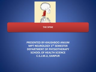
- 1. THE SPINE PRESENTED BY-KHUSHBOO ANJUM MPT NEUROLOGY 1ST SEMESTER DEPARTMENT OF PHYSIOTHERAPY SCHOOL OF HEALTH SCIENCE C.S.J.M.U, KANPUR
- 2. GENERAL STRUCTURE AND STRUCTURE • The vertebral column resembles a curved rod, composed of 33 vertebrae and 23 intervertebral discs. • The vertebral column is divided into the following five regions: cervical, thoracic, lumbar, sacral, and coccygeal.
- 5. THE CURVE OF THE VERTEBRAL COLUMN • Fetal life-convex posteriorly. (FIG-A) • Adult-PRIMARY CURVE & SECONDARY CURVE • {The two curves (thoracic and sacral) that retain the original posterior convexity throughout life are called primary curves, whereas the two curves (cervical and lumbar) that show a reversal of the original posterior convexity are called secondary curves.} • Curves that have a posterior convexity (anterior concavity) are referred to as kyphotic curves; curves that have an anterior convexity (posterior concavity) are called lordotic curves. • A curved vertebral column provides significant advantage over a straight rod in that it is able to resist much higher compressive loads.
- 6. STRUCTURE OF THE CERVICAL REGION • The cervical vertebral column consists of seven vertebrae in total. • Morphologically and functionally, the cervical column is divided into two distinct regions: the upper cervical spine, or craniovertebral region, and the lower cervical spine. • The craniovertebral region includes the occipital condyles and the first two cervical vertebrae, C1 and C2, or, respectively, the atlas and axis. • The lower cervical spine includes the vertebrae of C3 to C7. The vertebrae from C3 to C6 display similar characteristics and are therefore considered to be the typical cervical vertebrae. • The atlas, axis, and C7 exhibit unique characteristics and are considered the atypical cervical vertebrae. • All of the cervical vertebrae have the unique feature of a foramen (transverse foramen) on the transverse process, which serves as passage for the vertebral artery.
- 7. CRANIOVERTEBRAL REGION • ATLAS- The atlas is different from other vertebrae in that it has no vertebral body or spinous process and is shaped like a ring (Fig. 4-22). The atlas also possesses a facet on the internal surface of the anterior arch for articulation with the dens (odontoid process) of the axis.
- 8. AXIS- The axis is atypical in that the anterior portion of the body extends inferiorly and a vertical projection called the dens arises from the superior surface of the body (Fig. 4-23)
- 9. 7TH CERVICAL VERTEBRA / VERTEBRA PROMINENS • The 7th cervical vertebra, (C7) is the largest and most inferior. • This spinous process can be easily seen and felt at the base of the neck, making it a prominent landmark of the skeleton (giving the C7 the name vertebra prominens). • spinous process ends in a rounded tubercle and is not bifid. • C7 transverse foramina are small, and do not transmit the vertebral artery.
- 10. ARTICULATIONS • The two atlanto-occipital joints consist of the two concave superior zygapophyseal facets of the atlas articulating with the two convex occipital condyles of the skull. • These joints are true synovial joints with intra- articular fibroadipose meniscoids. • There are three synovial joints that compose the atlantoaxial joints: the median atlantoaxial joint between the dens and the atlas and two lateral joints between the superior zygapophyseal facets of the axis and the inferior zygapophyseal facets of the atlas.
- 11. CRANIOVERTEBRAL LIGAMENTS • TRANSVERSE LIGAMENT- •The transverse ligament stretches across the ring of the atlas and divides the ring into a large posterior section for the spinal cord and a small anterior space for the dens. •The transverse ligament and its longitudinal bands are sometimes referred to as the atlantal cruciform ligament.
- 12. MAJOR LIGAMENTS OF VERTEBRAL COLUMN LIGAMENTS FUNCTIONS REGION Anulus fibrosus (outer fibers) Anterior longitudinal ligament Resists distraction, translation, and rotation of vertebral bodies. Limits extension and reinforces anterolateral portion of anulus fibrosus and anterior aspect of intervertebral joints. Limits extension Cervical, thoracic, and lumbar. C2 to sacrum but well developed in cervical, lower thoracic, and lumbar regions. Anterior atlantoaxial (continuation of the anterior longitudinal, ligament) Limits extension. C2 to the occipital bone
- 13. LIGAMENTS FUNCTIONS REGION Posterior longitudinal ligament Limits forward flexion and reinforces posterior portion of the anulus fibrosus. Axis (C2) to sacrum. Broad in the cervical and thoracic regions and narrow in the lumbar region. Tectorial membrane (continuation of the posterior longitudinal ligament Limits forward flexion Axis (C2) to occipital bone Ligamentum flavum Limits forward flexion, particularly in the lumbar area, where it resists separation of the laminae Axis (C2) to sacrum. Thin, broad, and long in cervical and thoracic regions and thickest in lumbar region Supraspinous ligaments Limit forward flexion Thoracic and lumbar (C7–L3 or L4). Weak in lumbar region. Ligamentum nuchae Limits forward flexion. Cervical region (occipital protuberance to C7)
- 14. • ALAR LIGAMENTS- •The two alar ligaments are also specific to the cervical region (see Fig. 4-27). These paired ligaments arise from the axis on either side of the dens and extend laterally and superiorly to attach to roughened areas on the medial sides of the occipital condyles55 and to the lateral masses of the atlas. •These ligaments are relaxed with the head in midposition and taut in flexion. • Axial rotation of the head and neck tightens both alar ligaments. Crisco JJ, Panjabi MM, Dvorak, J: A model of the alar ligaments of the upper cervical spine in axial rotation. J Biomech 24:607, 1991.
- 16. THE LOWER CERVICAL REGION TYPICAL CERVICAL VERTEBRA • BODY- The body of the cervical vertebra is small, with a transverse diameter greater than anteroposterior diameter and height. • ARCHES- Pedicles. The pedicles project posterolaterally and are located halfway between the superior and inferior surfaces of the vertebral body. Laminae. The laminae are thin and slightly curved.They project posteromedially. Zygapophyseal Articular Processes (Superior and Inferior). The processes support paired superior facets that are flat and oval and face superoposteriorly.
- 17. • Transverse Processes. A foramen is located in the transverse processes bilaterally for the vertebral artery, vein, and venous plexus. Also, there is a groove for the spinal nerves. • Spinous Processes. The cervical spinous processes are short, slender, and extend horizontally. The tip of the spinous process is bifid. The length of the spinous processes decreases slightly from C2 to C3, remains constant from C3 to C5, and undergoes a significant increase at C7. • Vertebral Foramen. The vertebral foramen is relatively large and triangular.
- 18. FUNCTIONS OF CERVICAL REGION KINEMATICS • The atlanto -occipital joint is a plane synovial joint that permits flexion -extension, some axial rotation, and lateral flexion. • Flexion-extension takes place in the sagittal plane around a medial-lateral axis. • Axial rotation takes place in the transverse plane around a vertical axis and lateral flexion takes place in the frontal plane around an anterior-posterior axis.
- 19. ARTHOKINEMATIC • At the atlanto-occipital joint, the inferior convex condyles of the occiput articulate with the two superior concave zygapophyseal articular facets of the lateral bodies of the atlas. When the head moves on the atlas ( convex surfaces moving on the concave surface) ,the occipital condyles glide in direction opposite to the movement of the top of the head. In flexion, the condyles glide posteriorly on the atlas articular surfaces. In extension the occipital condyles glide anteriorly on the atlas , whereas the back of the head moves posteriorly.
- 20. FUNCTIONS OF CERVICAL SPINE • The cervical spine plays many roles in the head-neck area, including: • Protection of the spinal cord: It is a bundle of nerve fiber that is a continuation of the brain stem. The spinal cord transverse through a canal in the vertebral column called the spinal canal. The cervical spine protects it from external compression. • Provide support to the head: The cervical spine holds the head in position and bears its weight. • Supports blood supply to the brain: The cervical spine carries a vertebral artery in its transverse foramen and helps them to send blood to the brain. • Supports Head-Neck movements: Cervical spine, along with the neck muscle, provides a wide range of motion in this region. REHMAN S,DAS J M ,ANATOMY,HEAD AND NECK,CERVICAL SPINE,2021
- 22. A TYPICAL VERTEBRA • The structure of a typical vertebra consists of two major parts: an anterior, cylindrically shaped vertebral body and a posterior, irregularly shaped vertebral or neural arch. • The vertebral body is designed to be the weight- bearing structure of the spinal column. It is suitably designed for this task, given its block like shape with generally flat superior and inferior surfaces.
- 23. COMPONENT AND FUNCTION OF TYPICAL VERTEBRA
- 24. INTERVERTEBRAL DISC • The intervertebral disc has two principle functions: to separate two vertebral bodies, thereby increasing available motion, and to transmit load from one vertebral body to the next. • The disc thickness varies from approximately 3 mm in the cervical region, where the weight-bearing loads are the lowest, to about 9 mm in the lumbar region, where the weight-bearing loads are the greatest. • The discs are smallest in the cervical region and largest in the lumbar region.
- 25. • The intervertebral disks are composed of three parts: (1) the nucleus pulposus, (2) the anulus fibrosus , and (3) the vertebral end plate. • All three structures are composed of water, collagen, and proteoglycans (PGs); however, the relativeproportions of each vary.
- 26. ARTICULATIONS • Two main types of articulations are found in the vertebral column: cartilaginous joints of the symphysis type between the vertebral bodies, including the interposed discs, and diarthrodial, or synovial, joints between the zygapophyseal facets located on the superior articular processes of one vertebra and the zygapophyseal facets on the inferior articular processes of an adjacent vertebra above. The joints between the vertebral bodies are referred to as the interbody joints. The joints between the zygapophyseal facets are called the zygapophyseal (apophyseal or facet) joints . Synovial joints also are present where the vertebral column articulates with the ribs, with the skull, and with the pelvis at the SIJs.
- 27. LIGAMENTS AND JOINT CAPSULES • Six main ligaments are associated with the intervertebral and zygapophyseal joints. They are the • Anterior longitudinal • Posterior longitudinal ligaments and PLL; • Ligamentum flavum; • Interspinous • Supraspinous • Intertransverse ligaments .
- 28. • Anterior and posterior longitudinal ligaments-The anterior longitudinal ligament runs along the anterior and lateral surfaces of the vertebral bodies from the sacrum to the second cervical vertebra. The ligament is compressed in flexion( Fig. A) and stretched in extension (see Fig. B). • The PLL runs on the posterior aspect of the vertebral bodies from C2 to the sacrum and forms the ventral surface of the vertebral canal. It also consists of at least two layers: a superficial and a deep layer.
- 30. FUNCTION KINEMATICS • The motions available to the column as a whole are flexion and extension, lateral flexion, and rotation. • These motions are often coupled motions. • The intervertebral discs increase movement between two adjacent vertebrae. If the vertebrae lay flat against each other, the movement between them would be limited to translation alone. • This arrangement adds tremendous range of motion. Bogduk N: Clinical Anatomy of the Lumbar Spine and Sacrum, 3rd ed. New York, Churchill Livingstone, 1997.
- 31. • FLEXION In vertebral flexion, anterior tilting and gliding of the superior vertebra occur and cause widening of the intervertebral foramen and separation of the spinous processes. • EXTENSION In extension, posterior tilting and gliding of the superior vertebra occur and cause narrowing of the intervertebral foramen, and the spinous processes move closer together.
- 32. LATERAL FLEXION • In lateral flexion, the superior vertebra laterally tilts ,rotates , and translates over the adjacent vertebra below.
- 33. KINETICS • The vertebral column is subjected to axial compression , tension, bending, torsion, and shear stress not only during normal functional activities but also at rest. . Gracovetsky SA: The resting spine: A conceptual approach to the avoidance of spinal reinjury during rest. Phys Ther 67:549, 1987.
- 34. MOVEMENTS AND RANGES OF JOINTS JOINTS MOVEMENTS RANGES CERVICAL SPINE FLEXION 0-45° EXTENSION 0-45° LATERAL FLEXION 0-45° ROTATION 0-60° THORACIC AND LUMBAR SPINE FLEXION 0-80° EXTENSION 0-25° LATERAL FLEXION 0-35° ROTATION 0-45°
- 35. JOINTS MOVEMENTS RANGES SHOULDER FLEXION 0-180° (150°-180°) EXTENSION 0-45° (40°-60°) ABDUCTION 0-180° (150°-180°) ADDUCTION 0 INTERNAL ROTATION 0-90° (70°-90°) EXTERNAL ROTATION 0-90° (70°-90°) ELBOW FLEXION 0-130° (120°-150°) EXTENSION 135°-0
- 36. JOINTS MOVEMENTS RANGES FOREARM(SUPERIOR AND INFERION RADIO-ULNAR JOINT) SUPINATION 0-90° PRONATION 0-90° WRIST FLEXION 0-90° (10°-90°) EXTENSION 0-70° (50°-70°) ULNAR DEVIATION 0-40° (25°-40°) RADIAL DEVIATION 0-20° (15°-25°)
- 37. JOINTS MOVEMENTS RANGES MCP(METACARPOPHALEN GEAL) FLEXION 0-90° EXTENSION 0-20° (15°-30°) ABDUCTION 0-20° ADDUCTION 0 PIP FLEXION 0-110° (90°-120°) EXTENSION 0 DIP FLEXION 0-90° EXTENSION 0
- 38. REFRENCES • SPINE PICTURE https://gfycat.com/discover/human-spine-gifs • Levangie.P.K, Norkin.C.C : Joint structure & function,4th ed.2005,141-192. • https://commons.wikimedia.org/wiki/File:Cer vical_vertebrae_animation.gif.
- 39. khushboo anjum, MPT neurology 1st semester,C.S.J.M.U ,JAN;2022,KANPUR