Liver function tests
•Als PPTX, PDF herunterladen•
5 gefällt mir•1,124 views

LIVER FUNCTION TEST

Empfohlen
Weitere ähnliche Inhalte
Was ist angesagt?
Was ist angesagt? (20)
Ähnlich wie Liver function tests
Ähnlich wie Liver function tests (20)
Mehr von International Medicine School - Management and Science University
Mehr von International Medicine School - Management and Science University (20)
Kürzlich hochgeladen
Kürzlich hochgeladen (20)
Liver function tests
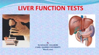
- 1. By Dr KHALED ALGARIRI CAMS- QASSIM UNIVERSITY March 2020
- 2. INTRODUCTION The liver is a reddish brown wedge shaped organ with four lobes of unequal size and shape. A human liver normally weighs 1.44–1.66 kg (3.2– 3.7 lb). Liver function tests are blood tests used to help diagnose and monitor liver disease or damage. The tests measure the levels of certain enzymes and proteins in our blood. Some of these tests measure how well the liver is performing its normal functions of producing protein and clearing bilirubin, a blood waste product.
- 3. FUNCTIONS OF THE LIVER 1-Synthetic functions : synthesis of plasma proteins, cholesterol, triacyl glycerol, lipoprotein 2-Metabolic function: protein metabolism, ketogenesis, TCA cycle, production of ATP 3-Detoxification & excretion: ammonia to urea, bilirubin, cholesterol, drug metabolites. 4- Homeostasis : blood glucose regulation 5-Storage function: Vitamin A,D,K,B12 6-Production of bile salts
- 4. Why it's done Screening: They are non invasive yet sensitive modality for liver dysfunction. Pattern of disease: They are helpful to recongnize pattern of various diseases. Like being helpful in differentiating between acute viral hapatitis and various cholestatic disorders and chronic liver disease.(CLD) Assess severity : They are heplful to assess the severity and predict the outcome of certain diseases like primary biliary cirrhosis. Follow up: They are helpful in the follow up of certain liver diseases and also helpful in evaluating response to therapy like autoimmune hepatitis
- 5. CLASSIFICATION OF LIVER FUNCTIONS TESTS
- 6. 1.Serum bilirubin 2.Urine bilirubin 3.Urine and feacal urobilinogen 4.Urine bile salts 5.Dye excretion tests TESTS BASED ON EXCRETORY FUNCTION
- 7. TESTS BASED ON DETOXIFICATION FUNCTION Hippuric acid test Determination of blood ammonia
- 8. TESTS BASED ON SYNTHETIC FUNCTION Prothrombin time Protein time
- 9. TESTS BASED ON METABOLIC FUNCTION
- 10. ENZYMES IN DIAGNOSIS OF LIVER DISEASE SERUM TRANSAMINASES AST ALT ALP GGT 5’NT
- 11. 1.Serum bilirubin 2.Urine bilirubin 3.Urine and feacal urobilinogen 4.Urine bile salts 5.Dye excretion tests TESTS BASED ON EXCRETORY FUNCTION
- 12. Serum bilirubin Bilirubin: is the end product of heme degradation derived from breakdown senescent (aging) erythrocytes by mononuclear phagocytes system specially in the spleen, liver and bone marrow. The major pigment present in bile is the orange compound bilirubin. It is highly soluble in all cell membranes (hydrophobic) and is also very toxic. Therefore, its excretion in the bile is one of the very important functions of the liver. Classification of bilirubin into direct & indirect bilirubin is based on original van den bergh method of measuring bilirubin.
- 13. Extravascular Pathway for RBC Destruction (Liver, Bone marrow, & Spleen) Hemoglobin Globin Amino acids Amino acid pool Heme Bilirubin Fe2+ Excreted Phagocytosis & Lysis Recycled The globin is recycled or converted into amino acids, which in turn are recycled or catabolized as required
- 14. BILIRUBIN METABOLISM .Bilirubin is the excretory product formed by the catabolism of heme part of haemoglobin. .Porphyrin part of heme are converted to bilirubin in reticuloendothelial cells of liver, spleen, bone marrow. Unconjugated bilirubin is bound to serum albumin &transferred to liver where it is conjugated to glucuronate by UDP GLUCURONYL TRANSFERASE. Conjugated bilirubin is excreted into bile. A fraction of bilirubin from stool is reabsorbed into blood via portal circulation (enterohepatic circulation)
- 15. Bilirubin Is the Major Component of Bile Pigments,Steps of Execretion 1. Hemoglobin is first dissociated into heme and globin. 2. In the presence of NADPH and O2, the Heme oxygenase enzyme hydroxylates Heme and converts it into Biliverdin. 3. Biliverdin is then reduced or converted into bilirubin by biliverdin reductase enzyme. Bilirubin is transported in blood bound to albumin forming a water soluble compound called hemobilirubin (unconjugated bilirubin, free bilirubin) which is rapidly transported to hepatocytes for further metabolism .
- 16. RBCs Breakdown Hemoglobin Produces & Breakdown Heme Biliverdin Bilirubin Heme Oxygenase Biliverdin Reductase
- 17. Role of Blood Proteins in the Metabolism of Bilirubin 1. Albumin Dissolved in Blood
- 18. Blood Liver Ligandin (-) charge Ligandin (-) charge Ligandin Prevents bilirubin from going back to plasma
- 19. 4-The liver removes bilirubin from the circulation rapidly, mediated by a carrier protein (receptor), and conjugates it with glucuronic acid. This reaction is catalyzed by the enzyme glucuronyl transferase in the smooth endoplasmic reticulum to have conjugated bilirubin, which is more water soluble than bilirubin. 5-The bilirubin-glucuronide (conjugated bilirubin) is secreted into the bile canaliculi. Note: the unconjugated bilirubin is normally not secreted. 6-In the small intestine, bilirubin glucuronide is poorly absorbed. In the gut, however, bacteria deconjugate it back to bilirubin, and convert it to the highly soluble colorless compound called Urobilinogen
- 20. 7- Only 20% of Urobilinogen can be absorbed by the small intestine (this represents the enterohepatic circulation of bile pigments). 70% of the Urobilinogen can be oxidized in the large intestine to Stercobilin and stercobilinogen (by bacteria). 10 % of Urobilinogen is excreted in either urine (where it is converted to yellow urobilin in the kidney) or feces (after it is converted to Stercobilin which is responsible for the brown color of feces). (entero-hepatic circulation)
- 22. Insoluble in water Present normally in plasma Tightly complex to albumin Not filtered through renal glomeruli, is not excreted in urine Toxic substance The chief form of bilirubin in the blood Unconjugated Water soluble and Present normally in bile Loosely bound to albumin Filtered through renal glomeruli and excreted in urine Non-toxic Present in low concentration in the blood Conjugated Differences between the conjugated and unconjugated bilirubin
- 23. Diagnostic Importance of Bilirubin • Disruption of bilirubin metabolism and excretion can cause hyperbilirubinaemia and subsequent jaundice • Hyperbilirubinaemia maybe unconjugated (indirect) or conjugated (direct) depending on the type of bilirubin present in plasma.
- 24. A-Unconjugated Hyperbilirubinemia • It is due to overproduction of bilirubin by reticuloendothelial system over the capacity of the liver to remove and clear from blood. It is characterized by high level of indirect or unconjugated bilirubin. This type of bilirubin can cross the blood-brain barrier into the central nervous system and cause kernicterus
- 25. Unconjugated hyperbilirubinemia occurs in the following conditions I-Neonatal or Physiologic Jaundice: This is the most common cause of jaundice in neonatal age. It results from accelerated hemolysis and immature hepatic system for uptake, conjugation and secretion of bilirubin. II-Hemolytic jaundice (Anemia)
- 26. III-Congenital Syndromes related to uptake and conjugation of bilirubin as follow : 1- Crigler-Najjar Syndrome Type 1: Due to severe decrease in the activity of UDP- glucuronyl transferase. Type II: Due to decreased activity of UDP-glucuronyl transferase that adds the second glucuronide group. 2- Gilbert Disease It is mainly due to hepatic defect in the uptake of bilirubin by liver cells. 3-Toxic Hyperbilirubinemia This is due to toxin-induced liver dysfunctions e.g. chloroform, carbon tetrachloride and mushroom poisoning.
- 27. B-Conjugated Hyperbilirubinemia • Conjugated hyperbilirubinemia is due to reflux of direct or conjugated bilirubin into blood due to biliary obstruction, conjugated bilirubin is water soluble, so it is excreted in urine and darken its color.
- 28. Conjugated hyperbilirubinemia occurs in the following conditions 1-Obstructive Jaundice (Cholestatic Jaundice) Conjugated hyperbilirubinemia results from blockage of hepatic or common bile duct (stones and tumors). 2- Micro-obstruction of intrahepatic biliary ductules by swollen damaged hepatocytes e.g. viral hepatitis and liver cirrhosis. Both cases are associated with marked increase of conjugated bilirubin and slight to moderate increase of unconjugated bilirubin (mixed hyperbilirubinemia)
- 29. 3- Rotor’s/Dubin-Johnson syndrome – defective excretion of conjugated bilirubin into the biliary cannaliculi therefore elevated conjugated bilirubin
- 30. • Jaundice becomes clinically evident when the serum bilirubin level exceeds 2.5mg/dL. • Several types of Jaundice: – Hemolytic – Hepatocellular – Obstructive • Symptoms: – Yellow discoloration of the skin, sclera and mucous membranes – Itching (pruritus) due to deposits of bile salts on the skin – Stool becomes light in color – Urine becomes deep orange and foamy
- 31. Normal range Bilirubin type Bilirubin level Total bilirubin 0.0-1.4 mg/dL or 1.7-20.5 mcmol/L Direct bilirubin 0.0-0.3 mg/dL or 1.7-5.1 mcmol/L Indirect bilirubin 0.2-1.2 mg/dL or 3.4-20.5 mcmol/L
- 34. The presence of urine bilirubin indicates hepatobiliary disease. Unconjugated bilirubin is tightly bound to albumin and not filtered by the glomerulus and thus not present in urine. Measurable amount of conjugated bilirubin in serum are found only in hepatobiliary disease. Because the renal threshold for conjugated bilirubin is low and the laboratory methods can detect low levels of bilirubin in urine so conjugated bilirubin may be found in urine when the serum bilirubin levels are normal. This is the case in early acute viral hepatitis. URINE BILIRUBIN
- 35. URINE UROBILINOGEN Increase in urine is sensitive indicator of hepatocellular disease • It is markedly increased in hemolysis • In cholestatic jaundice urobilinogen disappears from urine • Urine strips are available • Fresh urine should be used • Ehrlich’s test – gives pink-red color
- 36. URINE UROBILINOGEN An increase in the urobilinogen in urine is a sensitive indicator of hepatocellular dysfunction. Itis a good indicator of alcoholic liver damage, well compensated cirrhosis or malignant disease of liver. In viral hepatitis it appears early in urine. It is markedly increased in hemolysis. In cholestatic jaundice urobilinogen disappears from urine. It is intermittently present in case of gallstones. Urobilinogen gives a purple reaction to Ehrlich’s aldehyde reagent
- 37. BILE SALTS Products of cholesterol metabolism Facilitate absorption of fat from intestine Constitute a substantial amount of bile in bilirubin excretion and can be used in diagnosing cholestasis Primary bile salts – cholate and chenodeoxycholate are produced in liver Metabolised by bacteria in intestine Produces secondary bile salts – lithocholate, deoxycholate and ursodeoxycholate
- 38. In normal condition – renal excretion of bile salts is negligible In cholestasis – increased renal excretion of bile salts For measuremnet – chromatography (HPLC) Hay’s test – bile salts when present lower the surface tension of urine When sulphur powder is added to the urine, sulphur particles sink to the bottom of the tube In case of normal urine, it will float on the surface
- 39. ENZYMES IN DIAGNOSIS OF LIVER DISEASE
- 40. SERUM ENZYMES REFLECT THE DAMAGE OF HEPATOCYTES A large number of enzyme estimations are available which are used to ascertain liver function. They are be divided into two groups: A). Enzymes indicating hepatocellular damage. B). Enzymes indicating cholestasis (obstruction). In liver cells injury , damage to the membrane of cells & organelles allows intracellular enzymes to leak into the blood
- 41. A- Enzymes indicating hepatocellular damage AST (Aspartate transaminase)/SGOT (serum glutamate oxaloacetate transaminase) Normal range: 10-45 U/L. AST is found in both cytoplasm & mitochondria AST/GOT also reflects damage to the hepatic cells & is less specific for liver disease. > skeletal muscle> kidneys >brain • AST help diagnose various heart, muscle or brain disorders, such as a myocardial infarct (heart attack).
- 42. Alanine transaminase ALT sGPT Normal Range: 5-40 U/L. ALT(SGPT) – found primarily in liver . ALT is specific for liver disease. Its elevations favor liver cell necrosis as a cholestasis.
- 43. Serum aminotransferases (AST-ALT) are the sensitive markers of acute hepatocellular injury. Serum alanine aminotransferase or ALT (formerly called serum glutamic-pyruvic transaminase or SGPT) is a cytosolic enzyme. When necrosis or death of cells containing these enzymes occurs, aminotransferases are released into the blood and their concentration in blood increases whose level correlates with extent of tissue damage.
- 44. Most marked elevations of ALT and AST (>15 times normal) are seen in acute viral hepatitis, toxin-induced hepatocellular damage (e.g. carbon tetrachloride), and centrilobular necrosis due to ischemia (congestive cardiac failure). Moderate elevations (5-15 times) occur in chronic hepatitis, autoimmune hepatitis, alcoholic hepatitis, acute biliary tract obstruction, and drug-induced hepatitis. Mild elevations (1-3 times) are seen in cirrhosis, nonalcoholic steatosis, and cholestasis. Clinical Significant of Transaminase Enzymes
- 45. Clinical Significant of Transaminase Enzymes Increase of AST and ALT is much more in hepatocellular jaundice (>500 units/ml) than inn cholestatic jaundice (<200 units/ml). ALT and AST are elevated in acute viral hepatitis even before the appearance of jaundice. Persistence of elevated ALT and AST beyond 6 months in a case of hepatitis indicates development of chronic hepatitis Normal ALT:AST ratio is 0.7 to 1.4.
- 46. Determination of these enzymes are helpful in distinguishing hepatocellular from cholestatic jaundice Increase in AST and ALT is much more ( >500 IU/L)in hepatocellular jaundice than in cholestatic jaundice (>200 IU/L) Persistence of elevated ALT and AST beyond 6 months in a case of hepatitis indicates development of chronic hepatitis. Clinical Significant of Transaminase Enzymes
- 47. ELEVATION OF SERUM AST MAY INDICATE Acute hemolytic anemia Cirrhosis of the liver Hepatitis Acute pancreatitis or inflammation of pancreas Acute renal failure or loss of kidney function. Heart attack Primary muscle disease Recent surgery
- 48. ELEVATION OF SERUM ALT MAY INDICATE Alcoholic liver disease Cancer of liver Hepatitis or inflammation of the liver Noncancerous tumor of the liver Use of medicines or drugs toxic to the liver Cirrhosis or scarring of the liver Death of liver tissue.
- 49. AST:ALT ratio Normal ratio is 0.7 to 1.4 Useful in Wilson disease, chronic liver disease and alcoholic liver disease AST/ALT ratio > 3:1 is highly suggestive of Alcoholic liver disease AST in Alcoholic live disease is rarely >300 IU/L. ALT is usually normal in alcoholic liver disease ; can be sometimes low due to an alcohol induced deficiency of pyridoxal phosphate AST/ALT <1 is seen in viral hepatitis.
- 50. Clinical Significant of serum AST/ALT ratio
- 51. B- ENZYMES THAT DETECT CHOLESTASIS 1. ALKALINE PHOSPHATASE 2. Gamma-GLUTAMYL TRANSPEPTIDASE 3. 5-NUCLEOTIDASE
- 52. 1-ALKALINE PHOSPHOTASE ALP occurs in in all tissues, especially liver, bone, bile duct, kidney & the placenta. The ALP used to help diagnose certain liver diseases and bone disorders. Normal range: 30 - 95 IU/L (3-13 kings unit) ALP is a hydrolase enzyme responsible for removing phosphate groups from many types of molecules, including nucleotides & proteins. Levels are significantly higher in growing children. A rise in serum ALP usually associated with elevated serum bilirubin is an indicator of biliary obstruction (obstructive/post hepatic jaundice). ALP is also elevated in cirrhosis of liver & hepatic tumors.
- 53. Physiological increase in ALP is seen in – 1. >60 yrs 2. Pregnancy 3. Blood groups O and B – after fatty meal influx of intestinal ALP into blood 4. In children and adolescent during rapid bone growth If an isolated increase in ALP is seen , identification of the source of elevated isoenzyme is helpful- by fractionation of ALP by electrophoresis. Measure serum levels of GGT and 5’NT – they are elevated in only liver disease
- 54. Isoenzymes of Alkaline Phosphatase Hepatic Isoenzyme – Travels fastest towards the anode .Its level rises in extra hepatic biliary obstruction. 2. Bone Isoenzyme-Increases due to osteoblastic activity and is normally elevated in children during periods of active growth . 3. Placental Isoenzyme - Rises during last 6 weeks of pregnancy. 4. Intestinal Isoenzyme- Rise occurs after a fatty meal. May increase during various GI disorders.
- 55. Clinical Significant of ALP Normal range Adult: 42.0-128.0 IU/L. Children: 82.0-390 IU/L In acute viral hepatitis, alkaline phosphatase is usually either normal or moderately increased. Hepatitis A may present a cholestatic picture with marked and prolonged itching and elevation of alkaline phosphatase. Tumor may secrete alkaline phosphatase into plasma and there are tumor specific isoenzymes such as Regan, Nagao and Kasahara isoenzymes. Hepatic and bony metastasis can also cause elevated levels of alkaline phosphatase. Other diseases like infiltrative liver disease, abscesses, granulomatous liver disease and amyloidosis may also cause a rise in alkaline phosphatase. Mildly elevated levels of alkaline phosphatase may be seen in cirrhosis and hepatitis ofncongestive cardiac failure. Low levels of alkaline phosphatase occur in hypothyroidism, pernicious anemia, zinc deficiency and congenital hypophosphatasia.
- 56. Enzymes Prehepatic Jaundice Hepatic Jaundice Obstructive Jaundice ALS or ALT Normal Marked Elevation Elevation ALP Normal Slightly increase Marked Elevation Enzyme assays in differential diagnosis of Jaundice
- 57. Abbreviations: GGT, γ-glutamyltranspeptidase; L-AP, liver-specific alkaline phosphatase; B-AP, bone-specific alkaline phosphatase; 5′nT, 5′- nucleotidase; HFP, hepatic function panel; THP, transient hyper-alkaline phosphatemia; Ca/P, calcium/phosphorus; PTH, parathyroid hormone
- 58. Gamma Glutamyl transpeptidase(GGT) is a membrane bound glycoprotein which catalyzes the transfer of gamma glutamyl group to other peptides, amino acids and water. Large amounts are found in the kidneys, pancreas, liver, intestine and prostate. The levels of gamma glutamyl transpeptidase are high in neonates and infants up to 1 year and also increase after 60 years of life. Men have higher values. Children more than 4 yr old have serum values of normal adults. The normal range is 0-30IU/L. In acute viral hepatitis the levels of g glutamyl transpeptidase may reach its peak in the second or third week of illness and in some patients they remain elevated for 6 weeks. 2- Gamma GLUTAMYL TRANSPEPTIDASE GGT
- 59. Clinical Significant of GGT Often clinicians are faced with a dilemma when they see elevated alkaline phosphatase levels and are unable to differentiate between liver diseases and bony disorders and in such situations measurement of gamma glutamyl transferase helps as it is raised only in cholestatic disorders and not in bone diseases. Other conditions causing elevated levels of Gamma glutamyl transpeptidase include uncomplicated diabetes mellitus, acute pancreatitis and myocardial infarction. Drugs may increase the levels of Gamma glutamyl transpeptidase..
- 60. Clinical Significant of GGT The enzyme present in serum appears to originate primarily from the Hepatobiliary system. GGT activity is elevated in all forms of liver disease like 1. Obstructive jaundice 2. Cholangitis 3. Cholecystitis 4. Biliary atresia 5. Infectious hepatitis
- 61. Clinical Significant of GGT Relatively high sensitivity and specificity because of their measurement is easy and in expensive. Elevated earlier in liver diseases. Early detection of chronic alcohol misuse. Enzyme level found correlate with the duration of the drug action.
- 62. 3- 5'-Nucleotidase Normal range: 2-15 U/L The serum activity of 5'-nucleotidase is elevated in hepatobiliary disease & this parallels ALP. It is highest in post-hepatic obstructive jaundice. The 5'-nucleotidase is normal in patients with bone disease where as serum ALP increased. GGT and 5’NT is especially used to assess the nature of ALP.
- 63. TESTS BASED ON SYNTHETIC FUNCTION Prothrombin time Protein time
- 64. SYNTHETIC FUNCTION OF LIVER Liver is involved in synthesis of many plasma proteins like BLOOD CLOTTING FACTORS, LIPO PROTEINS, ETC.. • SYNTHESIS of these compounds may be affected in pathological conditions of liver.. • i..e their concentration in plasma may decrease, however of their long HALF LIIFE and REGENARATION capacity of liver decrease may be apparent only on long standing liver dieases • THESE INCLUDE 1. Serum Albumin 2. Prothrombin 3. Serum Glubins
- 65. 1-Serum Albumin In severe liver diseases, hypoalbunemia occurs. • Since half life of albumin is 20 days, decrease in albumin occurs in chronic liver diseases. • Albumin • it is most abundant protein in serum [120 mg/kg/day] • Reduction of albumin occurs in • Impaired synthesis (malnutrition, malabsorption, hepatic dysfunction,cirrhosis) 1. Loss (ascites, protein losing-nephropathy, enteropathy) 2. May result in peripheral oedema.
- 66. Due to its slow turn over – not a good indicator of acute or mild hepatic dysfunction • In hepatitis - <3g/dl of albumin – possibility of chronic liver disease •Non hepatic causes of Hypoalbuminemia - 1. Protein losing enteropathy 2. Nephrotic syndrome
- 67. 2-Serum Globulins In chronic liver dieases globulins increase due to decrease clearance by hepatocytes IgA level increased in all types of cirrhosis IgG level increases in Auto-immune hepatitis and cirrhosis IgM elevates in biliary cirrhosis
- 68. 3-Prothrombin Time PT Since prothrombin is one coagulation factor synthesized by Liver, any damage to liver can causes decrease in synthesis of Prothrombin i.e Hypoprothrombinaria indicates liver dysfunction i.e Increased in prothrombin time Since PT is also prolonged in Vitamin-K Deficiency it is carefully ruled out by estimating PT BEFORE and AFTER Vitamin-K administration. PT also be prolonged in chronic obstructive Jaundice due to resistant in Vitamin-K reabsorption (malabsorption of Vitamin-K) THUS PT IS USEFUL IN DIAGNOSIS OF JAUNDICE AND LIVER FUNCTION
- 69. Normal value : 10 to 15 sec.
- 70. TESTS BASED ON DETOXIFICATION FUNCTION Hippuric acid test Determination of blood ammonia
- 72. Hippuric acid test Hippuric acid test • 6gm of sodium benzoate dissolved in 250ml water • Collect urine for next 4 hour Hippuric acid is a normal component of urine and is typically increased with increased consumption of phenolic compounds (tea, wine, fruit juices). These phenols are converted into benzoic acid which is then converted into hippuric acid and excreted in the urine.
- 73. THANK YOU