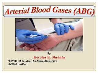
Basic ABG notes
- 1. By Kerolus E. Shehata •PGY-III IM Resident, Ain Shams University •ECFMG certified
- 2. Objectives 1)Define ABG & its indications. 2)Describe components of ABG & their normal values. 3)Acid-Base imbalance. 4)Interpret ABG changes in clinical toxicology practice.
- 3. What is the ABG? Arterial blood gas analysis is an essential part for diagnosing and managing the patient’s oxygenation status, ventilation status and acid base balance. Drawn from arteries( radial, brachial and femoral) ABG Oxygenation Ventilation Acid-Base PaO2 SaO2 PCO2 PH HCO3
- 4. ACID-BASE BALANCE The primary aim of keeping this delicate balance is to preserve the Homeostasis i.e. the highly complex interactions that maintain all body systems to functioning within a normal range. Any extreme change in this balance (PH < 6.8 or > 7.8) may result in disastrous changes e.g. denaturation of proteins & shut down of all enzymatic and metabolic processes. Such disturbed environment would be incompatible with life.
- 6. Basic Biochemistry Facts •Water (H2O) forms about 65 % of our total body weight. •When acid e.g. HCL dissolves in a solution, it ionizes into H+ (Proton) and Cl-. So, The amount of H+ (Protons) in the solution directly correlates with its acidity. •When CO2 dissolves in a water solution, it combines with H2O to form H2CO3. So, the amount of CO2 in the solution directly correlates with its acidity. •Most of our body metabolic processes make our media acidic e.g. Metabolism of Fats and CHO generates CO2 (CO2+H2O=H2CO3) Metabolism of Proteins generates many fixed acid e.g. Sulphuric, Phosphoric and Uric acids. •Bicarbonate is an amphoteric ion, meaning that it can behave as either an acid or a base, depending on the surrounding media. Since our internal body media is acidic so, It can be considered as a Base (Alkali).
- 7. TO SUM UP… •Increase in H+ or CO2 = Increase acidity •Decrease in H+ or CO2 = Decrease acidity •Increase in Bicarbonate = Increase alkalinity •Decrease in Bicarbonate = Decrease alkalinity Q: How to make the media more acidic? 1.Adding more acids e.g. H+ or CO2 2.Removing its alkaline part e.g. HCO3 Q: How to make the media more alkaline? 1.Adding more bases e.g. HCO3- 2.Removing its acidic part e.g. H+ or CO2
- 8. How can the body maintain that acid-base balance? •The 2 body systems that always try to achieve this balance are: 1)The Kidneys: through manipulating the amount of HCO3- and H+ (By secretion, excretion or reabsorption) 2)The Lungs: through manipulating the amount of CO2 (Increase or decrease the respiratory rate) •If there is a defect in one system, the other one tries to buffer its effects in order to reach the balance required for proper homeostatic functioning (the principle of Compensation). •The response of each system to make that balance varies e.g. The Lungs: Respond in minutes. The Kidneys: Respond in hours to days.
- 9. What are the components of ABG? pH Measurement of acidity or alkalinity, based on the hydrogen (H+) 7.35 – 7.45 PaO 2 The partial pressure oxygen that is dissolved in arterial plasma. 80 - 100 mm Hg PaCO2 The amount of carbon dioxide dissolved in arterial blood. 35 – 45 mmHg
- 10. What are the components of ABG? HCO 3 The calculated value of the serum concentration of bicarbonate 22 – 26 mEq/L SaO2 The arterial oxygen saturation. >95 %
- 11. pH (Power of Hydrogen) •pH is the negative logarithm of hydrogen ion concentration in a water-based solution. •Negative = Inversely related to the H+ ion concentration i.e. Increase in H+ conc. In a solution decreases the PH. •Why we use a logarithmic scale? •H+ conc. Is expressed in nanoequivalents per liter. So, we use the Log scale to shrink that large range into a simple scale (1- 14) making it easier to compare the magnitude of solution acidity or alkalinity. •For example, a pH of 3 is ten times more acidic than a pH of 4 and 100 times (10x10) more acidic than a pH value of 5.
- 12. Normal range values of the ABG strip
- 13. ACID BASE DISORDERSACID BASE DISORDERS BASIC CONCEPTS •ABG shouldn’t be used alone in the diagnosis. Correlate the clinical findings with the other lab and imaging studies to get a panoramic assessment of the patient’s condition. •ABG findings can assist not only in reaching a diagnosis, but also in determining the prognosis of a patient.
- 14. (I) Respiratory Acidosis It is defined as a pH less than 7.35 with a Paco2 greater than 45 mmHg. Acidosis is the accumulation of co2 which combines with water in the body to produce carbonic acid, thus lowering the pH of the blood.
- 15. Toxic Causes : •Any condition that results in hypoventilation can cause respiratory acidosis. (a) Central (b) Peripheral CNS depression opiates, sedatives, anesthesia, methanol, ethylene glycol...etc. 1-Respiratory muscle paralysis e.g. botulism 2- lung disease e.g. pulmonary edema 3- respiratory passage obstruction e.g. organophosphorus
- 16. Signs & symptoms of Respiratory Acidosis: •Respiratory: Respiratory distress & shallow respiration. •Nervous: (CO2 Narcosis) Headache, restlessness and confusion. If co2 is extremely high, drowsiness and unresponsiveness may be noted. •CVS: Tachycardia and Dysrhythmias due to myocardial hypoxia. Management: •Oxygen & suctioning as needed. •Pulse oximetry & ABG follow up. •Treatment of the cause e.g. pneumothorax, severe pain (Rib fracture) and CNS depressants toxicity. •If the cause can not be readily resolved, mechanical ventilation.
- 17. (II) Respiratory Alkalosis It is defined as a pH greater than 7.45 with a Paco2 lesser than 35 mmHg. Alkalosis is due to excessive wash of co2 (hyperventilation), thus increasing the pH of the blood.
- 18. Respiratory alkalosis… Cont’dCauses : Excessive wash of co2 ( hyperventilation) •Central stimulation: Psychological responses, Panic attack (Cannabis), drugs as early theophylline & salicylates toxicity…etc. •Withdrawal manifestations from depressant agents. •Increased metabolic demands e.g. fever, sepsis, pregnancy or thyrotoxicosis. (Body tries to get rid off the excess CO2 produced) •Central nervous system lesions (CO2 is a potent cerebral V.D) •MetHB, SulphHB. (compensation of Metabolic acidosis) Signs & symptoms:•CNS: Tachypnea, numbness, tingling, confusion, inability to concentrate and blurred vision (Decrease cerebral Bl. Flow). •CVS: Dysrhythmias and palpitations. •Tetanic spasms of the arms and legs (Decrease ionized calcium).
- 19. Management of Respiratory Alkalosis • Oxygen for any patient with respiratory distress of any origin. • Pulse oximetry and ABG monitoring. • Treatment of the cause. • If panic attack: calm the patient, oxygen +/- Benzodiazepines. • If carpo-pedal spasms occur, don’t give calcium because it is all a matter of distribution and not a decrease in total body calcium.
- 20. (III) Metabolic Acidosis It is defined as a pH less than 7.35 with a Hco3 less than 22 mEq/L. Toxic Causes : Any disorder that will lead to tissue hypoperfusion whatever the cause will lead eventually to increase in lactic acid production resulting in Metabolic Acidosis. 1) Late salicylate 2) Methanol 3) Ethylene glycol 4) Iron
- 21. Bicarbonate less than 22mEq/L with a pH of less than 7.35 Causes: • Renal failure (Sulphuric, phosphoric, uric acids…etc.) •Diabetic Ketoacidosis (Ketoacids) •Anaerobic metabolism (Lactic acid) •Starvation (Ketoacids) •Convulsions (Lactic acid) •Drugs: Salicylates, methanol, ethylene glycol, metformin intoxication. •Diarrhea & ATN…normal anion gap metabolic acidosis. Metabolic Acidosis… Cont’d Sign & symptoms •CNS: Headache, confusion and restlessness progressing to lethargy, then stupor or coma. •Respiratory: Acidotic (Kussmaul) breathing: Rapid and shallow •CVS: Tachycardia and Dysrhythmias
- 22. Management of Metabolic Acidosis • Treatment of the cause should be our primary aim. • Maintain adequate tissue oxygenation & Hemodynamic stability. • In severe cases, we can use Sodium Bicarbonate as a buffer to maintain a pH value that is compatible with a proper homeostatic functioning. • N.B. Correction with NaHCO3 should proceed in a cautious and non-aggressive way because pouring too much base into the circulation would cause a left shift in the O2 dissociation curve (Less release of O2 from HB into the tissues) causing more tissue hypoxia and may worsen the patient’s condition. • So, NaHCO3 correction should be guided by the Hemodynamic status of the patient and ABG monitoring to make a proper adjustment of the milliequivalents needed.
- 23. (IV) Metabolic Alkalosis It is defined as a pH greater than 7.45 with Hco3 greater than 28 mEq/L Causes It is due to excessive acid loss (repeated vomiting and nasogastric suction) OR bicarbonate retention e.g. overuse of sodium bicarbonate .
- 24. Metabolic alkalosis… Cont’dBicarbonate more than 26:28 mEq/L with a pH more than 7.45 Causes: Excess of base OR loss of acid. •Ingestion of excess antacids, excess use of bicarbonate, or use of lactate in dialysis. •Sever repeated vomiting, gastric suction, excess use of diuretics (Furosemide & HCTZ), or high levels of aldosterone. •Excess Corticosteroids use. Signs/symptoms: •CNS: Dizziness, lethargy disorientation, seizures & coma. •M/S: weakness, muscle twitching, muscle cramps and tetanic spasms. •GIT: Nausea, vomiting •Respiratory depression (Compensation). Treatment of the cause and stop the offending agent.
- 26. Step 1: Assess the pH •If below 7.35 = acidotic •If above 7.45 = alkalotic Step 2: 1- Assess the paCO2 level •If below 35 = Respiratory alkalosis element •If above 45 = Respiratory acidosis element How can I interpret an ABG Strip? 2- Assess HCO3 value •If below 22 = Metabolic acidosis element •If above 26 = Metabolic alkalosis element
- 27. Step 3: Determine if there is a compensatory mechanism working to try to correct the pH (Full or partial). Primary metabolic acidosis will have decreased pH and decreased HCO3. Compensation occurs by hyperventilation occur to decrease PaCO2 (Respiratory alkalosis). Example: Primary respiratory acidosis will have increased PaCO2 and decreased pH. Compensation occurs when the kidneys retain HCO3 (Metabolic alkalosis). N.B. Over-compensation Never happen.
- 30. Case (1) 45 years old female patient admitted with a severe attack of asthma. She has been experiencing increasing shortness of breath since admission three hours ago. Her arterial blood gas result is as follows: pH: 7.22 PaCO2: 55 mmHg HCO3: 25 mEq/L Q1: What is the primary acid-base disorder in this patient? Q2: Name 3 toxins that would give a similar ABG findings.
- 31. Comment: •PH is low = Acidosis. •PaCO2 is high = Respiratory acidosis element. •Hco3 is Normal = Normal Metabolic element. “Respiratory Acidosis, Not compensated” Some toxins that would result in respiratory acidosis: 1. Opiates & Opioids toxicity. 2. Methanol & Ethylene glycol toxicity. 3. Barbiturates & Clonidine Toxicity. 4. Botulinum toxin. 5. Paralytic snake venom.
- 32. Case ( 2) 55 years old male patient admitted with recurring bowel obstruction. He has been experiencing intractable vomiting for the last several hours. His ABG findings are: pH: 7.50 PaCO2: 42 mmHg HCO3: 33 mEq/L Q1: What is the primary acid-base disorder in this patient? Q2: What do you expect serum level of K+ and CL- to be in this patient? Q3: Name 3 toxins that would lead to intractable severe vomiting.
- 33. Comment: PH: Increases = Alkalosis PaCO2: Normal = Normal Respiratory element. HCO3: Increased = Metabolic Alkalosis element. “Metabolic alkalosis, Non Compensated” Serum K+ & Cl- would decrease in the setting of repeated vomiting. Some toxins that lead to severe intractable vomiting: 1. Theophylline intoxication. 2. Organophosphorus intoxication. 3. Acetylcholinesterase inhibitors medications (TTT of Myasthenia gravis) e.g. Neostigmine and Pyridostigmine. 4. Acute Digitalis toxicity.
- 34. Case (3) A 65 year old kidney dialysis patient who has missed his last 2 sessions at the dialysis center. The ABG findings: PH: 7.24 PaCO2: 31 mmHg HCO3: 17 mEq/L Q1: What is the primary acid-base disorder in this patient? Q2: What do you expect regarding his breathing pattern? Q3: Name 3 toxins that may lead to a similar ABG findings.
- 35. Comment: PH: Decreased = Acidosis PaCO2: Slightly decreases = Respiratory Alkalosis element HCO3: Decreased = Metabolic acidosis element “Metabolic Acidosis with mild compensatory respiratory alkalosis” Since the primary disorder is Metabolic acidosis, the respiratory system tries to compensate by increasing the R.R. to get rid off CO2 (Acid) so, respiration will be rapid and shallow acidotic (Kussmaul breathing). Some toxins that may lead to Metabolic acidosis: 1. Metformin (Lactic acid). 2. Carbon Monoxide. 3. Iron & INH. 4. Any toxin that lead to tissue hypoperfusion & tissue hypoxia (Directly or indirectly)
- 36. Case (4) 23 year old female presents with dyspnea 2 hours after ingestion of a preserved red meat. She has blue lips and nails beds. She denies any drug intake for any reason. Her ABG findings: PH: 7.31 PaCO2: 24 mmHg HCO3: 18 mEq/L Q1: What is the primary acid-base disorder in this patient? Q2: Name 2 differential diagnoses. Q3: How to differentiate between these 2 differentials? Q4: What do you expect the PaO2 and SaO2 to be if the condition was toxin-induced? Q5: What is your management plan?
- 37. Comment: PH: Decreased = Acidosis PaCO2: Decreased = Respiratory alkalosis element HCO3: Decreased = Metabolic acidosis element “Metabolic acidosis partially compensated by Respiratory alkalosis” Differential diagnosis: 1.anxiety or panic attack 2.MetHB How to Differentiate: 1.Presence or absence of metabolic acidosis. 2.MetHB level in the blood. •In MetHB, SulphHB or CarboxyHB, the PaO2 & SaO2 are NORMAL. Management: 1.Check vital signs. 2.Oxygen & Pulse oximeter. 3.Clinical, ABG and ECG monitoring 4.If no improvement, Methylene blue can be used to oxidize Fe+3 to normal ferrous HB.
Hinweis der Redaktion
- Kidney impairment must be present to maintain the metabolic alkalosis.
