Weitere ähnliche Inhalte Ähnlich wie Nutritional therapies for ocular disorders pa rça iki (20) Kürzlich hochgeladen (20) 1. Natural Therapies for Ocular Disorders
Part Two: Cataracts and Glaucoma
Kathleen Head, ND
Abstract
Pathophysiological mechanisms of cataract formation include deficient glutathione levels
contributing to a faulty antioxidant defense system within the lens of the eye. Nutrients
to increase glutathione levels and activity include lipoic acid, vitamins E and C, and
selenium. Cataract patients also tend to be deficient in vitamin A and the carotenes,
lutein and zeaxanthin. The B vitamin riboflavin appears to play an essential role as a
precursor to flavin adenine dinucleotide (FAD), a co-factor for glutathione reductase
activity. Other nutrients and botanicals, which may benefit cataract patients or help
prevent cataracts, include pantethine, folic acid, melatonin, and bilberry. Diabetic
cataracts are caused by an elevation of polyols within the lens of the eye catalyzed by
the enzyme aldose reductase. Flavonoids, particularly quercetin and its derivatives,
are potent inhibitors of aldose reductase.
Glaucoma is characterized by increased intraocular pressure (IOP) in some but not all
cases. Some patients with glaucoma have normal IOP but poor circulation, resulting in
damage to the optic nerve. Faulty glycosaminoglycan (GAG) synthesis or breakdown
in the trabecular meshwork associated with aqueous outflow has also been implicated.
Similar to patients with cataracts, those with glaucoma typically have compromised
antioxidant defense systems as well. Nutrients that can impact GAGs such as vitamin
C and glucosamine sulfate may hold promise for glaucoma treatment. Vitamin C in
high doses has been found to lower IOP via its osmotic effect. Other nutrients holding
some potential benefit for glaucoma include lipoic acid, vitamin B12, magnesium, and
melatonin. Botanicals may offer some therapeutic potential. Ginkgo biloba increases
circulation to the optic nerve; forskolin (an extract from Coleus forskohlii) has been
used successfully as a topical agent to lower IOP; and intramuscular injections of Salvia
miltiorrhiza have shown benefit in improving visual acuity and peripheral vision in people
with glaucoma.
(Altern Med Rev 2001;6(2):141-166)
Introduction
Part one of this article was published in the October 1999 issue of Alternative Medicine
Review and discussed nutritional and botanical approaches to conditions of the retina. This
second part covers alternative treatments for nonretinal disorders: senile cataracts, diabetic cata-
racts, and chronic open-angle glaucoma.
Kathleen A. Head, ND – Technical Advisor, Thorne Research, Inc.; Senior Editor, Alternative Medicine Review.
Correspondence address: PO Box 25, Dover, ID 83825. E-mail: kathi@thorne.com
Alternative Medicine Review x Volume 6, Number 2 x 2001 Page 141
Copyright©2001 Thorne Research, Inc. All Rights Reserved. No Reprint Without Written Permission
2. A large percentage of blindness in the is often a symptom, whereas posterior
world is nutritionally preventable.1 The author subcapsular cataracts tend to be most
of this comment was referring primarily to the noticeable in bright light.4 Ophthalmoscopic
use of vitamin A to prevent corneal degenera- examination is best conducted on a dilated
tion associated with a vitamin A deficiency; pupil, holding the scope approximately one
however, there is considerable evidence that foot away. Small cataracts appear as dark
many other eye conditions, which are leading defects against the red reflex, whereas a large
causes of vision impairment and blindness, cataract may completely obliterate the red
also may be preventable with nutritional reflex. Once a cataract has been established, a
supplementation, botanical medicines, diet, referral for slit-lamp examination, which
and other lifestyle changes. In addition, a num- provides more detail on location and extent of
ber of nutrients hold promise for the treatment opacity, is recommended.
of already existing cataracts and glaucoma.
Etiological/Risk Factors
Senile Cataracts Factors contributing to cataract forma-
Senile cataracts are the leading cause tion include aging, smoking,5 exposure to UV-
of impaired vision in the United States, with a B and ionizing radiation,6 oxidative stress (sec-
large percentage of the geriatric population ondary to other risk factors such as aging or
exhibiting some signs of the lesion. Over one smoking),7 dietary factors,8 increased body
million cataract surgeries are performed yearly weight (above 22-percent body fat), central
in this country alone.2 Cataracts are develop- obesity,9 and family history. Medications and
mental or degenerative opacities of the lens of environmental exposures which may contrib-
the eye, generally characterized by a gradual ute to cataract formation include steroids, gout
painless loss of vision. The extent of the vi- medications, and heavy metal exposure. Cad-
sion loss depends on the size and location of mium, copper, lead,10 iron, and nickel11 have
the cataract. Cataracts may be located in the all been found in cataractous lenses. A high
center of the lens (nuclear), in the superficial level of cadmium in the lens is associated with
cortex (cortical), or in the posterior subcapsu- smoking and can contribute to accumulation
lar area. Cataracts are also classified accord- of other heavy metals.10 Conditions which pre-
ing to their color, which is consistent with lo- dispose to cataracts include diabetes, galac-
cation and density of the cataract. Pale yellow tosemia, neurofibromatosis, hypothyroidism,
cataracts are typically slight opacities of the hyperparathyroidism, hypervitaminosis D, in-
cortex, subcapsular region, or both; yellow or fectious diseases such as toxoplasmosis, and
light brown cataracts are consistent with mod- several syndromes caused by chromosomal
erate to intense opacities of the cortex, nucleus, disorders.2
or both; and brown cataracts are associated
with dense nuclear cataracts.3 Mechanisms Involved in the
Diagnosis Pathophysiology of Cataracts
Cataracts are characterized by electro-
Symptoms include near vision image
lyte disturbances resulting in osmotic imbal-
blur, abnormal color perception, monocular
ances. Derangements in the function of the
diplopia, glare, and impaired visual acuity, and
membrane resulting in ion imbalance may be
may vary depending on location of the cataract.
due to increased membrane permeability or to
For example, if the opacity is located in the
a depression of the Na+/K+ pump because of
center of the lens (nuclear cataract), myopia
interference with the enzyme Na+/K+ ATPase.12
Page 142 Alternative Medicine Review x Volume 6, Number 2 x 2001
Copyright©2001 Thorne Research, Inc. All Rights Reserved. No Reprint Without Written Permission
3. Ocular Disorders
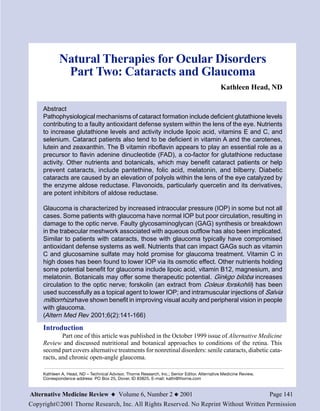
Figure 1: Synthesis of Glutathione in the Lens
creating a vicious
cycle. The
Step 1 Glutamic acid Step 2 γ-Glutamylcysteine researchers
+ + hypothesized that,
cysteine glycine “chelation therapy
+ + could be beneficial
ATP ATP in delaying
γ-Glutamylcysteine Glutathione cataractogenesis.”14
synthetase synthetase Other researchers
have confirmed
the involvement of
γ-Glutamylcysteine Glutathione transition metals,
+ + copper and iron, as
ADP ADP instigators of
+ + ascorbyl and
Pi Pi
hydroxyl radical
formation in
cataracts.15
Cataracts are also characterized by aggregates The Role of
of insoluble proteins.12 Glutathione in Lens Metabolism
Oxidative insult appears to be involved In order to fully understand the mecha-
as a precipitating factor in all cataracts. Lens nisms involved in cataract formation and the
proteins typically remain in their reduced form. link to nutritional prevention, it is important
However, in cataractous lenses, the proteins to understand the role glutathione and its en-
are found in an insoluble, oxidized form. Oxi- zyme co-factors play in metabolism within the
dation may occur as a result of many factors lens. In vitro studies of incubated lenses from
(see Etiological Factors). Higher levels of hy- animals as well as humans have helped eluci-
drogen peroxide have been found in catarac- date the mechanisms involved.
tous lenses when compared to normal con- The lens of the eye is avascular, de-
trols.13 Normally the lens contains significant pending entirely on passive diffusion, active
levels of reduced glutathione (GSH), which transport, and intra-lens synthesis for nutrients
keeps the proteins in their reduced form. How- and other substances important for metabo-
ever, there are significantly lower levels of lism. As a result, the content of the surround-
GSH in cataractous lenses. ing intraocular fluids (aqueous humor) is rel-
Advanced glycation end products evant. While levels of GSH are high in the lens,
(AGE) appear to play a role in cataract they are relatively low in the aqueous humor;
formation. Researchers have tested the thus, glutathione appears to be synthesized
hypothesis that the major AGE formed in the within the lens. Glutathione is composed of
lens has an EDTA-like structure, capable of the amino acids cysteine, glutamic acid, and
binding to copper. They found copper binding glycine, and its synthesis within the lens takes
was 20-30 percent greater in the older, place in two steps (Figure 1). Cataractous
cataractous lens protein fractions than in lenses can demonstrate dramatic decreases in
young, non-cataractous fractions. The pro- GSH, as much as 81 percent, when compared
oxidant copper precipitates further oxidation, to normal lenses.3 Researchers have examined
Alternative Medicine Review x Volume 6, Number 2 x 2001 Page 143
Copyright©2001 Thorne Research, Inc. All Rights Reserved. No Reprint Without Written Permission
4. this phenomenon in an attempt to determine have found an increase in disulfide bonds in
whether low GSH is due to decreased synthe- human cataractous lenses.17
sis or increased degradation. Decreases in the Maintaining normal cell volume and
enzymes involved in both synthesis (γ- transport of electrolytes are important factors
glutamyl transferase) and recycling (glu- in lens transparency. Glutathione may play a
tathione reductase) of GSH from oxidized glu- role in maintaining normal lens permeability
tathione (GSSG) lend credence to the theory and active cation transport by protecting sulf-
that synthesis is diminished in cataractous hydryl groups in the cell membrane from oxi-
lenses.3 These same researchers found a de- dation. Oxidation of SH groups on the surface
crease in the activity of enzymes of GSH deg- of the cell membrane results in increased per-
radation (glutathione peroxidase and glu- meability, and oxidation of important SH
tathione s-transferase) which should result in groups of Na+/K+ ATPase impedes active trans-
an increased rather than a decreased accumu- port. Reddy et al examined the effect of GSH
lation of GSH. They therefore concluded that depletion on rabbit lenses and found it directly
the loss of activity of these enzymes was not led to increased membrane permeability.18
enough to offset the losses associated with While GSH depletion did not directly impair
decreased synthesis. They also did not rule out active transport, it resulted in increased sus-
the possible loss of GSH from the lens via ceptibility of the Na+/K+ pump to oxidative
membrane leakage. damage by H2O2. Oxidation of GSH resulted
There are several ways in which glu- in a 70-percent decrease in active transport and
tathione or its depletion can affect the opacity a two-fold increase in membrane permeabil-
of the lens. A review by primary researchers ity. Other experiments have found that lens-
on glutathione metabolism and its relationship epithelial-GSH needs to be depleted by about
to cataract formation outlines three possible 60 percent for these changes to occur. The
mechanisms of cataract prevention by glu- authors point out that, “the lens has a remark-
tathione:16 (1) maintaining sulfhydryl (SH) able ability to regenerate reduced glutathione.”
groups on proteins in their reduced form pre- However, they found that, although the change
venting disulfide cross-linkage; (2) protecting in membrane permeability was reversible with
SH groups on proteins important for active the regeneration of GSH, the decrease in pump
transport and membrane permeability; and (3) activity was irreversibly affected.16
preventing oxidative damage from hydrogen H2O2 is found in the aqueous humor in
peroxide (H2O2). humans as well as other species. GSH is in-
Considering the first mechanism by volved in detoxifying this reactive oxygen spe-
which GSH can protect lenses from opacities, cies to water in a coupled reaction involving
there is an increase in high molecular weight NADPH (Figure 2). Without detoxification the
(HMW) proteins in cataractous lenses. These peroxide radicals would damage the lens mem-
protein aggregates contribute to lens opacity branes and susceptible protein groups. The
and are found particularly in dense cataracts. researchers found both normal human and rab-
Reddy and Giblin examined x-ray-induced bit lenses with high GSH content were rap-
cataracts in rabbits and found increased levels idly able to detoxify H2O2 in culture medium.16
of disulfide bonds, confirming their assertion Lenses pretreated with methyl mercury, which
that oxidation of SH groups resulted in disul- decreased the concentration of GSH by 75
fide bond formation and HMW proteins. They percent, were less able to detoxify the perox-
also found that SH groups on proteins only ide radicals.
become oxidized when levels of GSH drop
below some critical level.16 Other researchers
Page 144 Alternative Medicine Review x Volume 6, Number 2 x 2001
Copyright©2001 Thorne Research, Inc. All Rights Reserved. No Reprint Without Written Permission
5. Ocular Disorders
Figure 2: Metabolism of Hydrogen Peroxide by Glutathione
studies have
NADP GSH H2O2 quantified
(reduced significant
glutathione) levels of lutein,
zeaxanthin,
Glutathione Glutathione and alpha- and
reductase peroxidase g a m m a -
tocopherol in
the lens.21
A pro-
NADPH GSSG 2H2O spective study
(oxidized glutathione) of the effect of
carotenes and
vitamin A on
the risk of cata-
ract formation
Other researchers have postulated a was conducted as part of the Nurses’ Health
possible diffusion problem. Normally GSH is Study. A total of 77,466 female nurses, ages
synthesized and regenerated in the lens cortex 45-71 years, were included in the study, which
and then diffuses to other areas of the lens. involved food-frequency questionnaires over
Cataracts of the elderly are primarily in the a 12-year period. After other risk factors were
nucleus. Researchers examined normal human controlled for, including smoking and age,
lenses in vitro and found the older ones ap- those in the highest quintile for consumption
peared to have a barrier to diffusion of GSH of lutein and zeaxanthin had a 22-percent de-
19
from the cortex to the nucleus. creased risk of cataract extraction compared
with those in the lowest quintile.22
Specific Nutrients and Prevention of Another cohort of the Nurses’ Health
Cataracts Study followed 50,823 women, ages 45-67,
for eight years and found women in the high-
Oxidation of lens proteins is part of the
est quintile of vitamin A consumption had a
pathophysiology of cataracts. Therefore, it is
39-percent lower risk of developing cataracts
no surprise that antioxidants may help prevent
compared to women in the lowest quintile.23
the formation of cataracts.
In a similar study of male health
professionals in the United States, 36,644
Carotenes and Vitamin A: Epidemiological participants, ages 45-75 years, were followed
Evidence for eight years with periodic dietary
Levels of nutrients, including questionnaires. Men in the highest quintile for
carotenoids, have been examined in human lutein and zeaxanthin intake had a 19-percent
cataractous lenses after extraction using high decreased risk of cataract extraction when
performance liquid chromatography. Vitamins smoking, age, and other risk factors were
A and E and the carotenoids lutein and controlled for.24 Neither the women nor the
zeaxanthin were found. The newer, epithelial/ men demonstrated a decreased risk of cataract
outer cortex layer had more carotenoids, with intakes of other carotenoids (α-carotene,
tocopherol, and retinol (approximately 3-, 1.8- β-carotene, lycopene, or beta-cryptoxanthin).
, and 1.3-fold higher, respectively) than the It is hypothesized the protective effect of the
older, inner cortex/nuclear portion.20 Other carotenoids may be due to quenching reactive
Alternative Medicine Review x Volume 6, Number 2 x 2001 Page 145
Copyright©2001 Thorne Research, Inc. All Rights Reserved. No Reprint Without Written Permission
6. oxygen species generated by exposure to population of healthy volunteers, ages 55-80
ultraviolet light.25 years.31 Although results are still pending, data
The Beaver Dam Eye Study examined was collected on prior use of vitamin E and
risk for developing nuclear cataracts in 252 incidence of cataract in 1,111 participants. A
subjects who were followed over a five-year statistically significant relationship was found
period. Only a trend toward an inverse rela- between past vitamin E supplementation and
tionship between serum lutein and cryptoxan- prevention of cortical cataract but not nuclear
thin and risk of cataract development was cataract.32
noted.26 The Lens Opacities Case-Control
Study was designed to determine risk factors
Vitamin E: Animal, Epidemiological, and for cataracts in 1,380 participants, ages 40-79
Clinical Studies years. Blood chemistry and levels of vitamin
As a fat-soluble antioxidant, it is rea- E and selenium were performed on all patients.
sonable to predict a positive role for vitamin The risk of developing cataracts was reduced
E as a cataract preventive in the lens cell mem- to less than one-half (odds ratio 0.44 for
brane. Animal, epidemiological, and clinical nuclear cataracts) in subjects with higher lev-
studies help confirm this hypothesis. A pla- els of vitamin E.33 Some of these same re-
cebo-controlled animal study found 100 IU d- searchers examined the association between
alpha-tocopherol injected subcutaneously pre- antioxidants and the risk of cataract in the
vented ionizing radiation damage to the lens, Longitudinal Study of Cataract. Dietary intake,
which did occur in rats not supplemented with use of supplements, and plasma vitamin E lev-
vitamin E.27 Two other animal studies using els were assessed on 764 participants. Lens
vitamin E instilled in the eyes as drops con- opacities were examined on a yearly basis and
firmed the preventive effect of vitamin E, at the risk of development of cataract was 30-
least when used topically.28,29 percent less in regular users of a multiple vi-
Several human studies have found low tamin, 57-percent less in regular users of
levels of vitamin E intake are associated with supplemental vitamin E, and 42-percent less
increased risk for cataract development. An is those with higher plasma levels of vitamin
epidemiological investigation examined self- E.34
reported supplementary vitamin consumption In a randomized trial of 50 patients
of 175 cataract patients compared to 175 with early cataracts, subjects were assigned to
matched individuals without cataracts. The receive either 100 mg vitamin E twice daily
cataract-free group used significantly more or placebo for 30 days. There was a signifi-
vitamin E (p=0.004) and vitamin C (p=0.01) cantly smaller increase in the size of cortical
than the cataract group, resulting in at least a cataracts in the vitamin E group compared to
50-percent reduction in cataract risk in the placebo. While increases of vitamin E were
supplemented group.30 An Italian study com- found in both nuclear and cortical lens
pared 207 patients with cataracts to 706 con- homogenates after surgical removal, GSH lev-
trol subjects in a hospital setting. Vitamin E, els were increased significantly only in those
in addition to a number of other nutritional with cortical cataracts receiving vitamin E. In
factors, was associated with a decreased risk addition, the malondialdehyde (MDA) — a
for cataract.8 measure of oxidative stress — levels and glu-
The Vitamin E and Cataract Preven- tathione peroxidase levels were higher in cor-
tion Study (VECAT) is a four-year, prospec- tical cataract/vitamin E users than in the
tive, randomized, controlled trial of vitamin E nuclear cataract/vitamin E group.35 Some con-
versus placebo for cataract prevention in a clusions that can be drawn from this study are:
Page 146 Alternative Medicine Review x Volume 6, Number 2 x 2001
Copyright©2001 Thorne Research, Inc. All Rights Reserved. No Reprint Without Written Permission
7. Ocular Disorders
(1) vitamin E decreases oxidative stress in cata- It is not surprising then that a defi-
ractous lenses; (2) part of vitamin E’s protec- ciency of riboflavin has been implicated as a
tive effect is due to enhancement of GSH lev- cause of cataract formation. A study of B vita-
els; and (3) vitamin E seems to be more pro- min nutritional status of cataract patients
tective for cortical than nuclear cataracts, at (n=37) compared to age-matched controls
least in this short-term study. without cataract (n=16) found that 80 percent
of those with cataracts and only 12.5 percent
Vitamin C and Risk of Cataracts of control subjects had a riboflavin defi-
Animal experimentation has shed ciency.41 The same researcher tested for, but
some light on ascorbic acid and its role in cata- did not find, a deficiency of thiamin or pyri-
ract formation. Cataracts induced in chick doxine in cataract patients. Other researchers
embryos by the application of hydrocortisone have found a relationship between riboflavin
were prevented by the introduction of vitamin deficiency and later-stage cataracts, but not in
C to the developing embryo. In addition, vita- early cataract formation.42 The Lens Opacities
min C slowed the decline in GSH levels, which Case-Control Study found that lens opacities
occurred with the cortisone treatment.36 were associated with lower levels of ribofla-
Ascorbic acid is normally found in vin which were assessed by RBC enzyme as-
high concentrations in the aqueous humor and says and dietary intake reports.
lens in humans. A group of 44 subjects were Data collected during cancer interven-
supplemented with 2 g daily ascorbic acid. tion trials in Linxian, China, were assessed for
Significant increases in vitamin C in lens, nutrient effects on other conditions, including
aqueous humor, and plasma were noted.37 In cataracts. Two randomized, double-blind, con-
another study, lenses were exposed in vitro to trolled studies of cataract risk resulted from
light, which caused an increase in superoxide the Linxian study. In the first trial 12,141 par-
radicals and subsequent damage to the Na+/ ticipants, ages 45-74, were supplemented for
K+ pump. The damage was prevented by ad- five to six years with either a multiple vita-
dition of vitamin C in doses comparable to min-mineral or placebo. There was a statisti-
what would be found in the aqueous humor.38 cally significant 36-percent reduction in inci-
In the Nurses’ Health Study supple- dence of nuclear cataract for subjects ages 65-
mental vitamin C for a period of 10 years or 74 years given the multiple vitamin. In the
greater was associated with a 77-percent lower second trial 23,249 participants were given one
incidence of early lens opacities and an 83- of four different vitamin/mineral combina-
percent lower incidence of moderate lens tions: (1) retinol/zinc, (2) riboflavin/niacin, (3)
opacities. In this study, no significant protec- ascorbic acid/molybdenum, or (4) selenium/
tion was noted from vitamin C supplementa- alpha-tocopherol/beta carotene. Again, the
tion of less than 10 years.39 most significant effect was noted in people age
65-74, with a 44-percent decrease in nuclear
Riboflavin cataract risk in the group taking riboflavin/nia-
Riboflavin is a precursor to flavin ad- cin (3 mg riboflavin/40 mg niacin). No sig-
enine dinucleotide (FAD), which is a coen- nificant protection was noted for the other
zyme for glutathione reductase. In vitro evalu- nutrient combinations or for protection from
ations of surgically removed cataracts have cortical cataracts.43
confirmed inactivity of glutathione reductase A series of case reports from the Uni-
enzyme activity in a significant number of versity of Georgia treated 24 cataract patients
cataracts examined. Furthermore, the activity (18 with lens opacities and six with fully-de-
was restored by the addition of FAD.40 veloped cataracts) with 15 mg riboflavin daily.
Alternative Medicine Review x Volume 6, Number 2 x 2001 Page 147
Copyright©2001 Thorne Research, Inc. All Rights Reserved. No Reprint Without Written Permission
8. Dramatic improvement was reported within butter, total fat, salt, and oil (except olive oil).8
24-48 hours, and after nine months all lens The Nurses’ Health Study found regular con-
opacities disappeared.44 Larger, double-blind, sumption of spinach and kale was moderately
placebo-controlled trials are needed to confirm protective for cataracts in women.22 The Health
these seemingly dramatic improvements. Professionals Follow-up Study found spinach
and broccoli decreased risk of cataract in
Other B Vitamins men.24
Pantethine is the active coenzyme form
of pantothenic acid (vitamin B5). Several ani- Bilberry and Cataracts
mal studies have found pantethine can prevent Vaccinium myrtillus or bilberry has a
chemically-induced cataracts if given within long history of use for various eye conditions.
eight hours of exposure to lens insult.45-47 The The active components, flavonoid
proposed mechanism of action was the pre- anthocyanosides, are potent antioxidants with
vention of the formation of insoluble proteins a particular affinity for the eye and vascular
in the lens.45 tissues. The anthocyanosides are typically con-
Folic acid has been found to be low in centrated in a 25-percent standardized extract.
those prone to cataracts. An Italian epidemio- In a report of 50 patients with senile cataracts,
logical survey found those in the highest a combination of bilberry, standardized to con-
quintile for folic acid consumption were only tain 25-percent anthocyanosides, (180 mg
40 percent as likely to develop cataracts than twice daily) and vitamin E, in the form of dl-
those in the lowest quintile.8 tocopheryl acetate, (100 mg twice daily) for
four months stopped the progression of cata-
Selenium and Cataracts racts in 96 percent of the subjects treated
A decrease in glutathione peroxidase (n=25) compared to 76-percent in the control
activity has been found in the lenses of sele- group (n=25).50
nium-deficient rats. Concomitantly, an in-
crease in MDA and free radicals was also noted Melatonin
in both the selenium-deficient and vitamin-E The pineal hormone, melatonin, is a
deficient groups.48 Evaluation of selenium lev- potent antioxidant and because of its known
els in humans has found lower than normal antioxidant effects, several animal studies have
levels of selenium in sera and aqueous humor been conducted to determine its effect on pre-
in cataract patients.49 The significance of low vention of cataracts. Injections of melatonin
serum levels is unclear and the relationship have been found to inhibit both chemical- and
between selenium and cataract risk demands UVB-induced cataracts.51-53 In the UV-B and
further evaluation. melatonin study cataract formation and MDA
levels were significantly lower than the UV-B
Dietary Factors in Cataract Risk only group, leading the researchers to con-
Several epidemiological studies have clude, “These results suggest that melatonin
found dietary links to increased or decreased may protect against the UVB-induced cataract
risk of cataract. An Italian in-hospital study development by directly quenching lipid per-
examined dietary patterns and incidence of oxides and indirectly by enhancing the pro-
cataract extraction. Significant inverse rela- duction of the endogenous antioxidant GSH.”51
tionships were seen between meat, spinach, In studies examining chemically-in-
cheese, cruciferous vegetables, tomatoes, pep- duced cataracts, the animals were administered
pers, citrus fruits, and melon. An increased risk buthionine sulfoxamine (BSO), a known in-
was found in those with the highest intakes of hibitor of GSH synthesis. Half were treated
Page 148 Alternative Medicine Review x Volume 6, Number 2 x 2001
Copyright©2001 Thorne Research, Inc. All Rights Reserved. No Reprint Without Written Permission
9. Ocular Disorders
with melatonin and half were not. In one study, Flavonoids as Aldose Reductase Inhibitors
18/18 rats given BSO alone developed cata- A number of compounds, both natural
racts compared to only 1/15 in the group and synthetic, have been found to inhibit al-
treated with melatonin.52 In the other study, dose reductase. These so-called aldose reduc-
16/18 in the BSO-only group developed cata- tase inhibitors (ARIs) bind to aldose reduc-
racts, whereas only 3/18 treated with melato- tase, inhibiting polyol production.54 As a group,
nin developed cataracts.53 The researchers flavonoids are among the most potent natu-
were unsure whether the protection was due rally occurring ARIs. Several evaluations of
to a direct free-radical scavenging effect or to in vitro animal lenses incubated in high sugar
a stimulation of GSH production by melato- mediums have found flavonoids to inhibit al-
nin. dose reductase.55,56
A group of researchers examined the
Diabetic (Sugar) Cataracts effect of an orally administered ARI in inhib-
Polyol Accumulation iting polyol accumulation. They reported the
Some of the mechanisms involved in arrest of cataracts in galactosemic rats by oral
the formation of diabetic cataracts are some- feeding of a synthetic ARI.57 Building on this
what different from senile research, another group studied the effect of a
cataracts. The accumula-
tion of polyols within the
lens is a primary contrib-
uting factor. Certain tis- Figure 3: The Polyol Pathway
sues of the body, including
the lens of the eye, do not
require insulin for glucose Aldose
or other simple sugars Reductase
such as galactose to enter. D-Glucose D-Sorbitol
In diabetes sugar is in high
concentrations in the aque-
ous humor and can diffuse NADPH NADP+ NAD+
passively into the lens. The + H+
enzyme aldose reductase
within the lens converts Sorbitol
glucose to sorbitol or ga- Dehydrogenase NADPH
lactose to galactitol (Fig- + H+
ure 3). These polyols can-
not diffuse passively out of
the lens and accumulate or
D-Fructose
are converted to fructose.
The accumulation of
polyols results in an os-
motic gradient, which encourages diffusion of
fluids from the aqueous humor. The water flavonoid ARI, quercetrin (a glycoside of quer-
drags sodium with it and the swelling and elec- cetin). Rats were divided into two groups, one
trolyte imbalances result in cataract formation. receiving lab chow only, while the experimen-
tal group was fed quercetrin in rat chow plus
Alternative Medicine Review x Volume 6, Number 2 x 2001 Page 149
Copyright©2001 Thorne Research, Inc. All Rights Reserved. No Reprint Without Written Permission
10. Table 1: The Aldose Reductase Inhibitor
Activity of Some Flavonoids.60 comparable (average 380 mg/100 mL),
had not developed cataracts by the 25th
day.58 A French study examining the ef-
Molar Percent fect of oral doses of quercetin did not find
Flavonoid Concentration Inhibition
an inhibition of cataract formation in dia-
-4
Quercetrin-2-acetate 10 100 betic animals.59 In the positive study
-5
10 100
-6
100 quercetrin rather than quercetin was used,
10
10
-7
87 the former administered in a water sus-
pension which was undoubtedly more ab-
-4
Quercetrin 10 100 sorbable. The latter study was in French
-5
10 95
-6
88 with only the abstract available so a dos-
10
10
-7
55 age comparison was not possible.
Varma and associates performed
-4
Quercetin 10 100 an in vitro experiment to determine which
-5
10 83
-6
60 aspects of flavonoids conferred the most
10
10
-7
15 ARI activity, and ultimately which fla-
vonoids were the most potent in that re-
-4
Rutin 10 95 spect.60 Their earlier more limited research
-5
10 95
-6
20 had found quercetin, quercetrin, and
10
10
-7
10 myricitrin to possess the most potent ARI
activity.56
-4
Hesperidin 10 88 In the more recent experiment 44
-5
10 10
-6
0 flavonoids and their derivatives were ex-
10
10
-7
0 amined for the ability to inhibit aldose
reductase and polyol accumulation in rat
-4
Hesperidin chalcone 10 82 lenses incubated in the sugar xylose. All
-5
10 10
-6
0 flavonoids tested exhibited some inhibi-
10
10
-7
0 tory activity. The two most potent inhibi-
tors were derivatives of quercetin,
-4
Naringin 10 80 quercetrin and quercetrin-2-acetate, the
-5
10 59
-6
0 latter the most potent ARI inhibitor
10
10
-7
0 known. Table 1 demonstrates some of the
most common commercially available fla-
vonoids and their comparative inhibitions.
In decreasing order of potency they in-
clude quercetin, rutin, hesperidin and hes-
an additional 70 mg oral quercetrin daily in peridin chalcone, and naringin. Although in-
aqueous suspension. Three days after begin- hibition was also noted by isoflavones, cat-
ning flavonoid supplementation diabetes was echins, coumarins, and anthocyanins, they
chemically induced and three days later lenses were found to be much less potent than fla-
were assessed for sorbitol and fructose. The vones. When dissolved, flavones easily con-
flavonoid group demonstrated a 50-percent vert to their corresponding chalcone by the
inhibition of sorbitol and fructose accumula- opening of the center or B-ring of the flavone
tion. The control group developed cataracts by structure. Because this may occur in vivo as
the tenth day, whereas the group receiving well, the researchers tested hesperidin and
quercetrin, although their blood sugar was hesperidin chalcone and found their inhibitory
Page 150 Alternative Medicine Review x Volume 6, Number 2 x 2001
Copyright©2001 Thorne Research, Inc. All Rights Reserved. No Reprint Without Written Permission
11. Ocular Disorders
potencies were nearly identical (Table 1). The to halt progression of diabetic cataracts despite
chalcones, being more water-soluble and thus sorbitol, glucose, and fructose accumulation.64
more absorbable, may represent a more logi-
cal means of oral administration. In their re- Lipoic Acid
view Varma and associates outline a number In both in vitro lenses incubated in a
of structural factors contributing to ARI activ- glucose medium and in diabetic animal mod-
ity. In the final analysis, the pentahydroxy (five els, lipoic acid (LA) has been found to pre-
OH groups) flavones conferred the most po- vent cataract formation.65 Lipoic acid has po-
tent effect. tent antioxidant effects in both its oxidized
form (LA) and reduced form, dihydrolipoic
Vitamin C acid (DHLA). It is this property which un-
Ascorbic acid also has potential as an doubtedly is responsible for much of its pro-
aldose reductase inhibitor. An experiment was tective effect in diabetic cataracts. Packer, one
conducted in which guinea pigs were fed a of the foremost researchers on lipoic acid, hy-
normal, high vitamin C diet with 10-percent pothesizes that LA enters the lens (via a fatty
galactose or a scorbutic diet (devoid of vita- acid carrier) and is converted to DHLA. DHLA
min C) plus 10-percent galactose. The lens has the potential to regenerate ascorbic acid
epithelium of scorbutic animals had 2.5 times from ascorbyl radicals; the ascorbic acid can
as much galactitol (the polyol of galactose) on then regenerate vitamin E from tocopheryl
day 4 than those animals fed vitamin C in their radicals. Alternately, he hypothesized LA
diets.61 could directly spare vitamins C and E. The
A human study, although not on cells increases in vitamins C and E would result in
of the lens, found oral vitamin C at low doses decreased utilization of GSH and a relative
of 100-600 mg daily was able to normalize increase in its levels in the lens.63 Due to its
sorbitol levels in red blood cells within 30 days antioxidant effects and sparing of GSH and
in individuals with type 1 diabetes.62 vitamins E and C, lipoic acid has been found
to prevent chemically-induced, non-diabetic
Oxidative Stress and Diabetic cataracts in animal models as well.65
Cataracts Another mechanism by which lipoic
acid may prevent cataracts in diabetes is via
As with senile cataracts, oxidative
inhibition of aldose reductase, which was de-
stress plays an important role in the pathogen-
termined in an in vitro experiment.66 The re-
esis of diabetic cataracts. Levels of glutathione
searchers also noted the ability of aldose re-
peroxidase and vitamin C have been found to
ductase inhibitors, including lipoic acid, to
be deficient in the lenses of diabetics. Lenses
chelate transition metals such as copper. This
of humans with diabetes were found to be more
may be another way by which ARIs prevent
susceptible to oxidation of proteins, a condi-
cataracts and other complications of diabetes.
tion exacerbated by concurrent retinal dis-
ease.63
Protein Glycosylation/AGE
Glutathione in Diabetic Cataracts Generation in Diabetic Cataracts
Evidence of other mechanisms at work While glycosylated proteins (proteins
besides sorbitol accumulation is offered by the with glucose attached) and subsequent forma-
work of Ross et al. They found that GSH, ei- tion of AGE have been implicated in many
ther in vitro in lens incubation medium or in diabetic complications, there is disagreement
vivo when injected into diabetic rats, was able as to how much they contribute to diabetic or
Alternative Medicine Review x Volume 6, Number 2 x 2001 Page 151
Copyright©2001 Thorne Research, Inc. All Rights Reserved. No Reprint Without Written Permission
12. Table 2: Nutrients and Botanicals for the Prevention and
Treatment of Cataracts and ultimately
prevented cataract
Supplement Mechanism of Action formation. 67 In
vitro studies have
Glutathione Deficiency noted in cataractous lenses; important component also found lipoic
of the innate antioxidant system in the lens
acid prevents
Vitamin A Higher levels associated with a decreased risk for cataract p r o t e i n
glycosylation,
Carotenes Antioxidant; higher levels associated with decreased risk for providing yet
(Lutein and cataract
Zeaxanthin) a n o t h e r
mechanism
Vitamin E Antioxidant; increases glutathione; supplementation whereby LA may
associated with prevention p r e v e n t
Vitamin C Preserves glutathione levels; protects the Na+/K+ pump; cataracts.72
long-term supplementation (>10 years) protective
Table 2
Riboflavin Precursor to FAD, a coenzyme for glutathione reductase summarizes po-
which recycles GSH
tential nutritional
Bilberry Anthocyanoside antioxidants; study with vitamin E halted and botanical
cataract progression treatments for
cataracts.
Quercetin Aldose reductase inhibitor – diabetic cataracts
Lipoic Acid Spares vitamins C and E, increasing GSH levels; inhibits Chronic
aldose reductase and prevents protein glycosylation –
diabetic cataracts Open-Angle
Glaucoma
Currently,
there are approxi-
galactosemic cataracts. Some researchers have mately two mil-
found increased levels of glycosylated proteins lion cases of glaucoma in the United States,
and AGE associated with diabetic cataracts,67,68 but due to its insidious nature, only half may
while others have not found a strong correla- be diagnosed.4 Chronic open-angle glaucoma
tion.69,70 is the most common type, accounting for 60-
Despite the controversy, several natural 70 percent of cases. It is the leading cause of
substances that inhibit protein glycosylation blindness among African Americans2 and the
have been found to attenuate cataract second leading cause of blindness in the U.S.
formation in vitro and in animal models. population as a whole.4 Another type, acute
Acetyl-L-carnitine, which has been found to angle-closure glaucoma, is a medical emer-
be lower than normal in the lens of diabetic gency and will not be addressed here. Glau-
animals, was found in calf lenses in vitro to coma is characterized by a neuropathy of the
inhibit formation of glycosylated proteins by optic nerve, usually due to increased intraocu-
42 percent and AGE formation by 70 percent.71 lar pressure (IOP). The increased pressure is
The proteins became acetylated instead, due to an obstruction in the normal outflow of
preventing glucose from attaching. Pyruvate, fluids from the aqueous humor, with most of
a normal tissue metabolite, given orally to the outflow normally occurring in the anterior
diabetic rats decreased protein glycosylation chamber angle (Figure 4).
Page 152 Alternative Medicine Review x Volume 6, Number 2 x 2001
Copyright©2001 Thorne Research, Inc. All Rights Reserved. No Reprint Without Written Permission
13. Ocular Disorders
Figure 4: Flow of Fluids in the Eye
Aqueous humor Iris
Trabecular
Canal of Schlemm meshwork
Flow of fluid
Lens
Ciliary body
Vitreous Diffusion of fluid
humor and other constituents
Optic nerve
Patients may complain of missing words on
Diagnosis and Risk Factors reading or having trouble driving due to poor
Although IOP screening is one factor peripheral vision.
in diagnosing glaucoma, it is a fairly insensi- Risk factors for the development of
tive test. Normal IOP ranges from 7-22 mm/ glaucoma include increased intraocular pres-
Hg; however, approximately 90 percent of sure, older age, family history, being of Afri-
people with IOPs greater than 22 mm/Hg never can American descent, diabetes, hypertension,
develop glaucoma. On the other hand, some and myopia.4 Drugs associated with an in-
people with normal IOPs do develop optic creased incidence of glaucoma include corti-
nerve injury and glaucoma.4 Ophthalmoscopic costeroids and cholesterol-lowering drugs. As
examination may reveal an enlarged cup within an aside, the use of scopolamine (often used
the optic disc. If glaucoma is suspected or if by those going on a cruise to prevent seasick-
the patient is at high risk for developing glau- ness) can result in acute, angle-closure glau-
coma, referral to an optometrist or an ophthal- coma.
mologist for further evaluation is essential.
Physical symptoms are usually absent
in early stages. Once atrophy of the optic nerve
Pathophysiology
has progressed, loss of peripheral vision oc- Normally aqueous humor is produced
curs first; central vision is the last to be lost. in the ciliary processes and flows past the lens,
through the pupil, and into the anterior
Alternative Medicine Review x Volume 6, Number 2 x 2001 Page 153
Copyright©2001 Thorne Research, Inc. All Rights Reserved. No Reprint Without Written Permission
14. chamber. Outflow occurs through the angle Other researchers, examining human post-
between the cornea and the iris, through a trabeculectomy specimens (surgical procedure
meshwork of trabeculae, to the canal of for relieving increased IOP), have found evi-
Schlemm (Figure 4). The canal of Schlemm dence of a possible increase in collagen break-
is actually a very thin-walled vein that down in glaucoma.79
surrounds the entire eye. Because of its porous Reduced antioxidant defense systems
nature, large molecules are able to diffuse into are also evident in early stage glaucoma. Much
the canal and, although it is a vein, it carries of the research on the connection between an-
aqueous humor, not blood. Aqueous humor is tioxidants and glaucoma has been conducted
continually being formed and reabsorbed, and in Russia and published in Russian language
it is this balance between formation and journals, requiring dependence on abstracts.
reabsorption that regulates IOP.73 Levels of sulfhydryl groups, a reflection of
The pathological processes involved in glutathione levels, were found to be signifi-
chronic glaucoma are not completely under- cantly lower in the aqueous humor of patients
stood. It appears that morphological changes with glaucoma, particularly advanced stages
in collagen structure may precede increased of the disease, when compared to normal con-
IOP. Glycosaminoglycans (GAGs) contribute trol subjects. Red blood cell GSH levels were
to the filtration barrier. Within the trabecular also found to be lower in later stage glaucoma
meshwork is an area called the juxtacanalicular patients.80 Deficiencies of sulfhydryl groups
tissue (JCT), which is the probable site of im- have also been found in the lacrimal fluid of
pedance to aqueous outflow in glaucoma. Re- later stage glaucoma patients.81 In line with the
searchers have compared the GAG content of evidence of decreased antioxidant defense sys-
the JCT in normal and glaucomatous eyes and tems in glaucomatous eyes is the finding of
have found some significant differences. In one an increase in the lipid peroxidation product
study GAG content of five eyes from normal MDA – more than twice the normal level – in
donors was compared to five with glaucoma. the anterior chamber of patients with glau-
A significant decrease in hyaluronic acid and coma.82
increase in chondroitin sulfate was found in In some patients, particularly those
the eyes of patients with glaucoma.74 A simi- with normal IOP, an inadequate blood supply
lar study found trabecular meshwork of glau- to the optic nerve may be contributing to dam-
comatous eyes had a 77-percent lower level age and vision loss. These patients have a
of hyaluronic acid with higher levels of chon- higher than normal incidence of migraines,
droitin sulfates in trabecular meshwork, iris, suggesting vasospasm as an etiology.4
ciliary body, and sclera of eyes with glaucoma
compared to normal donors.75 The research- Conventional Treatment
ers postulated that the ability of normal aque- The goal of conventional treatment is
ous outflow is regulated by the content of to decrease intraocular pressure to avoid
GAGs and their ability to form a highly vis- damage to the optic nerve. There are a number
cous, elastic gel-like substance which acts as of topical medications used, including
a filtration system.76 Maintaining a critical cholinergic agonists, cholinesterase inhibitors,
level of hyaluronic acid appears to be essen- carbonic anhydrase inhibitors, adrenergic
tial for maintaining a well hydrated, semi-per- agonists (the newer ones are α2-selective), β-
meable meshwork.77 In vitro research on hu- blockers, and prostaglandin analogs. Osmotic
man eyes has found a decrease in GAG syn- diuretics are also used orally or IV.4 Laser
thesis, particularly hyaluronic acid, in glau- treatment may be tried instead of medications;
comatous eyes compared to normal eyes.78 and surgical intervention is usually a last resort
Page 154 Alternative Medicine Review x Volume 6, Number 2 x 2001
Copyright©2001 Thorne Research, Inc. All Rights Reserved. No Reprint Without Written Permission
15. Ocular Disorders
Table 3: Conventional Glaucoma Medications and Their Potential Side Effects4,83
Drug Category Drug Side Effect
Beta-blockers timolol, levobunolol, bronchospasm, shortness of breath
carteolol, metipranolol, fatigue, confusion, depression,
betaxolol impotence, hair loss, heart failure,
bradycardia. Timolol has also been
found to decrease HDL levels and
adversely effect the total
cholesterol:HDL ratio in women 60
years and older84
Non-selective Adrenergic epinephrine, dipivefrin high incidence of allergic or toxic
Agonists reactions
α2-Selective Adrenergic apraclonidide high rate of allergic reactions,
Agonists tachyphylaxis
brimonidine dry mouth but less likely to cause
allergic reactions
Cholinergic Agonists carbachol detached retina, GI disturbances,
headache, frequent urination
pilocarpine hypertension, tachycardia, bronchial
spasm
Cholinesterase Inhibitors physostigmine, The latter three in the list cause
neostigmine, irreversible rather than reversible
demecarium, miosis; they may be cataractogenic;
echothiophate iodide, increase risk of retinal detachment.
isoflurophate
Carbonic Anhydrase acetazolamide, fatigue, anorexia, depression,
Inhibitors methazolamide, paresthesias, electrolyte abnormalities,
(oral, IV, topical) dichlorphenamide, kidney stones, blood dyscrasias
ethoxzolamine
dorzolamide topical so does not have the side
effects of the others
Prostaglandin Analog latanoprost increased pigmentation of the iris;
worsening of uveitis
strategy but may be used initially in patients common side effects of glaucoma medications
who do not tolerate medications. Some are listed in Table 3.4,83
Alternative Medicine Review x Volume 6, Number 2 x 2001 Page 155
Copyright©2001 Thorne Research, Inc. All Rights Reserved. No Reprint Without Written Permission
16. Potential Nutrient Deficiencies in Nutrients that Effect GAGs: Vitamin C and
Glaucoma Glucosamine Sulfate
Deficiencies of specific nutrients have Due to the morphological changes seen
been found in patients with glaucoma, and in collagen structures associated with glau-
supplementation may play a role in treatment. coma it seems logical to explore the effect of
nutrients known to exert specific influences
Thiamin on GAGs. Vitamin C has probably been re-
Thiamin (vitamin B1) has been found searched more than any one single nutrient in
to be deficient in some patients with glaucoma. the treatment of glaucoma. Researchers seem
Blood levels and dietary intakes of both vita- to be in disagreement as to whether people with
mins C and B1 were examined in 38 patients glaucoma tend to have an ascorbate defi-
with glaucoma and compared to 12 controls. ciency.85,88 However, there is convincing re-
The glaucoma patients demonstrated a signifi- search on its effectiveness in treating glau-
cantly lower level of thiamin (but not vitamin coma.
C), which was not a reflection of decreased Researchers first examined the effect
intake causing the researchers to postulate an of high dose IV vitamin C on animals and then
impaired absorption of thiamin in these pa- humans, and found it successfully decreased
tients.85 As thiamin deficiency has been asso- IOP. IV doses used were in the range of 1 g/kg
ciated with degeneration of ganglionic cells body weight; oral doses used were half that
of the brain and spinal cord, the researchers (500 mg/kg body weight).89 A total of 49 eyes
postulated a possible degeneration of the op- were treated, 25 with chronic open-angle glau-
tic nerve as well. coma. The others included acute glaucoma,
hemorrhagic glaucoma, and glaucoma second-
ary to another disease process. Those with
Chromium
chronic open-angle glaucoma responded the
A deficiency of chromium has been
most dramatically. Table 4 shows the average
implicated in increased IOP in humans and
drops in IOP two hours and 4-5 hours after a
those afflicted with primary open-angle glau-
single dose of ascorbate. The higher the ini-
coma have demonstrated decreased erythro-
tial IOP, the greater the drop after ascorbate.
cyte chromium levels.86
In eyes with normal IOP, the average drop in
pressure was 3.5 mmHg. Decreases in pres-
Intervention Trials of Specific sure were maintained for as much as eight
Nutrients in Glaucoma hours. Due to the almost universal side effect
Vitamin B12 of gastric upset and diarrhea from such high
While a deficiency of vitamin B12 has doses of vitamin C, the researchers studied the
not been implicated in glaucoma, such a defi- effect of divided daily doses (0.1-0.15 g/kg 3-
ciency may cause optic atrophy and visual field 5 times daily). All but one patient experienced
defects which mimic glaucoma. An open trial decreased IOP during a two-week trial of di-
of 5 mg B12 daily in glaucoma patients found vided doses and most experienced mild gas-
no change in IOP with supplementation. How- tric upset and diarrhea that disappeared after
ever, there was no progression of visual field 3-4 days. Use of IV vitamin C would have al-
loss during five years of follow-up.87 leviated the gastric upset and diarrhea. Some
patients who had previously been resistant to
conventional drug therapy were able to main-
tain normal IOPs with the vitamin C regimen.
Page 156 Alternative Medicine Review x Volume 6, Number 2 x 2001
Copyright©2001 Thorne Research, Inc. All Rights Reserved. No Reprint Without Written Permission
17. Ocular Disorders
Table 4: Decreases in IOP after Oral Ascorbic Acid at 0.5 mg/kg
body weight89
Average Pressure Average Pressure
Initial IOP Decrease at 2 Hours Decrease at 4-5 Hours
50-69 mmHg 16 mmHg 25 mmHg
32-49 mmHg 14 mmHg 19 mmHg
20-31 mmHg 6.5 mmHg 6.5 mmHg
A previous study of oral ascorbate did seems warranted, not only to prevent poten-
not find it effective at lowering IOP in glau- tial harm but to monitor potential benefits of
coma. The researchers, however, used only 500 this supplement. Double-blind studies to con-
mg twice daily. These same researchers suc- firm the potential benefit of GS in glaucoma
cessfully lowered IOP with topical use of a are needed.
10-percent aqueous solution of vitamin C.90,91
There are several postulated mecha- Magnesium in the Treatment of Glaucoma
nisms for ascorbate’s ability to lower IOP. In Caused by Vasospasm
high doses it acts as a potent osmotic agent.88 A subcategory of glaucoma patients
Vitamin C’s ability to halt lipid peroxidation with normal IOP – those for which optic nerve
has also been hypothesized to play a role.88 damage is caused by vasospasm leading to
Vitamin C has also been found, in vitro, to decreased blood supply to the optic nerve –
stimulate synthesis of hyaluronic acid in tra- may benefit from supplemental magnesium.
becular meshwork from glaucomatous eyes.78 This patient group is often treated with cal-
Ascorbate has also been found to reduce the cium channel blocker drugs. These drugs,
viscosity of hyaluronic acid and increase out- however, are not without side effects, includ-
flow through the trabeculum.92 ing leg and periorbital edema. Ten patients,
Based on the observation that open- six with open-angle glaucoma and four with
angle glaucoma may be due in part to a hyalu- normal-tension glaucoma, were supplemented
ronic acid deficiency, a researcher has postu- with magnesium, a natural calcium channel
lated a beneficial effect of glucosamine sul- blocker.94 All patients in this preliminary open
fate (GS) for the treatment of glaucoma.93 He trial suffered from cold-induced vasospasms
cites two case reports in which glaucoma pa- of the extremities and all had marked visual
tients given 3 g/day GS for osteoarthritis re- field deficits, despite normal or drug-normal-
ported substantial drops in IOP. Because glu- ized IOP. Each subject received 121.5 mg
cosamine sulfate is also a substrate for chon- magnesium twice daily for one month. At the
droitin sulfate, which has been found to be el- end of four weeks there was a non-statistically
evated above normal in the trabecular mesh- significant trend toward improvement in vi-
work in glaucoma patients, it would seem to sual field tests (eight improved and two dete-
have the possible potential of further aggra- riorated, one because of a cataract). While it
vation of the condition. While this author has is tempting to assume magnesium can improve
not heard reports of exacerbation with glu- blood supply to the optic nerve by dilating the
cosamine sulfate, close monitoring of IOP in optic blood vessels, a larger, placebo-con-
patients with glaucoma supplemented with GS trolled trial of this subpopulation suffering
Alternative Medicine Review x Volume 6, Number 2 x 2001 Page 157
Copyright©2001 Thorne Research, Inc. All Rights Reserved. No Reprint Without Written Permission
18. from glaucoma and vasospasm seems war- melatonin secretion, resulted in a blunting of
ranted. the usual early-morning fall in IOP.
Administration of 200 mcg melatonin to half
Antioxidants in Glaucoma of the bright-light group resulted in a
Low glutathione levels may contrib- significant drop in IOP within an hour; the
ute to some of the pathological processes seen effect lasted up to four hours. This experiment
in glaucoma. Supplementation of lipoic acid may have important implications, not only for
can increase glutathione in red blood cells80 possible treatment of glaucoma with
and lacrimal fluid81 of patients with glaucoma. melatonin, but also in regard to timing of
In a Russian controlled trial of lipoic acid, 45 medications. It should also be noted that beta-
patients with stage I and II glaucoma were blockers, sometimes used for treatment of
supplemented with either 75 mg (n=26) or 150 glaucoma, decrease melatonin levels. One
mg (n=19) for two months. A control group of researcher found topical use of the beta-
glaucoma patients (n=31) were given only lo- blocker, timolol, did not work as well in the
cal hypotensive medication. Improvement in evening.100
visual function was seen in approximately 45
percent of subjects supplemented with lipoic Coenzyme Q10 May Prevent
acid.95 As mentioned previously, vitamins C Cardiac Side Effects of Timolol
and E are also glutathione sparing. The issue The beta-blocker medication timolol
of oral supplementation of glutathione is a may have significant cardiac side effects, in-
controversial one with some researchers con- cluding bradycardia and heart failure.4 Sixteen
tending oral doses of GSH are not effective at glaucoma patients on topical timolol were
raising plasma and tissue levels of GSH. How- given 90 mg CoQ10 for six weeks. CoQ10
ever, both animal96,97 and human98 studies have delayed the appearance of negative inotropic
found oral doses of glutathione lead to in- effects, including bradycardia, associated with
creased plasma and tissue levels. timolol, preventing the negative cardiac effects
of the drug without interfering with its effect
Melatonin and Diurnal Rhythms in on IOP.101
Intraocular Pressure
Intraocular pressure normally varies Omega-3 Fatty Acids
throughout the day, with the lowest pressure Epidemiological and animal studies
occurring in the very early morning hours. IOP point to a possible protective effect of omega-
also parallels fluctuations in cortisol levels, 3 fatty acids in glaucoma. Both topical admin-
high cortisol conferring higher IOPs. Diurnal istration of prostaglandin E3 and D3 – end
variations in IOP are more pronounced in products of omega-3 fatty acid metabolism –
people with glaucoma (>10 mmHg) compared and IM injections of cod liver oil – high in
to 3-7 mmHg variations in non-glaucomatous omega-3 fatty acids – led to decreased IOP in
eyes. Because melatonin levels peak around 2 rabbits.102,103 Epidemiological evidence has
a.m., a time when IOP is on a downward trend, found a low prevalence of chronic open-angle
researchers studied its effect on IOP.99 A series glaucoma among Eskimos on a native diet high
of experiments on subjects with normal IOPs in omega-3 fatty acids.104 These studies have
and no diagnosis of glaucoma were conducted. led researchers to consider the potential for
Baseline IOPs showed maximum pressures omega-3 fatty acids in the prevention and treat-
from 4-6 p.m. and minimum pressures from ment of glaucoma. Further investigation is
2-5 a.m. In the first experiment, exposure to warranted.
bright light, which suppressed normal
Page 158 Alternative Medicine Review x Volume 6, Number 2 x 2001
Copyright©2001 Thorne Research, Inc. All Rights Reserved. No Reprint Without Written Permission
19. Ocular Disorders
Botanical Treatment of Glaucoma supplementation. Subjects crossed over to the
There are several botanicals that may other treatment after a two-week washout pe-
hold promise for the treatment of glaucoma. riod. Ginkgo significantly increased blood
Most studies are merely preliminary, and flow to the ophthalmic artery with no change
larger, controlled studies are needed. seen in the placebo group.107 Although Ginkgo
did not alter IOP, it may provide potential ben-
Vaccinium Myrtillus (bilberry) efit in glaucoma patients who suffer from de-
Vaccinium myrtillus holds promise in creased ocular blood flow.
the treatment of glaucoma, although there has
been just one very limited study. Eight patients Coleus Forskohlii
with glaucoma were given a single dose of 200 The triterpene forskolin from the plant
mg anthocyanosides from bilberry (most ex- Coleus forskohlii stimulates the enzyme ade-
tracts are 25-percent anthocyanosides so as- nylate cyclase.108 Adenylate cyclase then
suming this standardization was used, the pa- stimulates the ciliary epithelium to produce
tients were given 800 mg bilberry standard- cyclic adenosine monophosphate (cAMP),
ized extract). Electroretinography improve- which in turn decreases IOP by decreasing
ments were noted.105 Due to the fact that the aqueous humor inflow.109
article is in Italian, further details are unavail- Results of studies using topical
able. While this evidence is very preliminary, forskolin applications to decrease IOP have
it is not unreasonable to imagine the been mixed. A study of 2-, 1-, and 0.5-percent
anthocyanosides’ collagen-stabilizing effect forskolin solutions applied to the eyes of nor-
exerting a positive effect on the trabecular mal rabbits found significant, dose-dependent
meshwork, facilitating aqueous outflow. decreases in IOP within a half hour, peaking
Anthocyanosides also exert potent antioxidant in 2-3 hours, and lasting up to 10 hours.110 On
effects. the other hand, a 1-percent forskolin solution
failed to significantly decrease IOP in glauco-
Ginkgo Biloba matous monkeys after two days of treatment.111
Forty-six patients, some with severe To date, human studies on forskolin’s
visual field disturbances, and some with seri- effect on IOP have been limited to healthy
ous retinal vascular degeneration, were given volunteers. Several studies have found it ef-
160 mg/day Ginkgo extract for four weeks, fective at lowering IOP and decreasing aque-
then 120 mg/day. Progress was assessed ous outflow in this population. Meyer et al
monthly by measuring visual acuity, visual compared the effect of 1-percent forskolin
fields, fundoscopic exam, IOP, blood pressure, versus placebo in 10 healthy volunteers in a
and pulse rate. Although only mild improve- randomized, crossover trial. In the first study,
ments were noted, these were deemed relevant both the placebo group and the forskolin group
due to the severity of the ocular damage at the experienced a decrease in IOP, which was at-
beginning of the study.106 tributed to the local anesthetic oxybuprocaine.
A potential therapeutic effect of a In the second trial, proxymetacaine was used
Ginkgo extract for the treatment of glaucoma as the topical anesthetic and forskolin was
was evaluated in a phase I, placebo-controlled, found to significantly decrease IOP compared
crossover trial in 11 healthy volunteers. Sub- to placebo.112
jects were treated with either 40 mg Ginkgo In 20 healthy volunteers one dose of a
extract three times daily or placebo for two 1-percent forskolin solution had no effect,
days. Ocular blood flow via Color Doppler whereas two instillations five minutes apart led
Imaging was measured before and after to significant decreases in IOP and aqueous
Alternative Medicine Review x Volume 6, Number 2 x 2001 Page 159
Copyright©2001 Thorne Research, Inc. All Rights Reserved. No Reprint Without Written Permission
20. Table 5: Potential Nutrient and Botanical Approaches to Glaucoma
Supplement Route of Administration Mechanism
Vitamin C IV or oral Osmotic agent; enhance hyaluronic acid
synthesis and reduce its viscosity
Vitamin B12 Oral or IM Correct a deficiency which may cause
optic nerve atrophy
Lipoic acid Oral Antioxidant; increase glutathione levels
Magnesium Oral May decrease vasospasm and increase
blood supply to the optic nerve
Melatonin Oral Normal diurnal rhythms of IOP fluctuation
reflect melatonin rhythms; antioxidant
CoQ10 Oral Prevent cardiac side effects of timolol
Bilberry Oral Anthocyanosides exert antioxidant and
collagen stabilizing effect
Ginkgo Oral Increase blood flow to the ophthalmic
artery and ultimately to the optic nerve
forskolin Topical eye drops Decrease IOP by stimulation of cAMP
Salvia IM or IV Increase microcirculation to the retinal
miltiorrhiza ganglions, improving visual acuity and
visual fields
flow rate.113 In eight healthy subjects one drop While topical use of forskolin in
of forskolin significantly decreased IOP and animals and healthy humans appears
flow rate was diminished an average of 34 promising, clinical studies on its use in
percent.114 Another study, however, did not find glaucoma patients are lacking. Furthermore,
forskolin to have a significant effect at decreas- while oral standardized extracts of Coleus
ing flow rate in a group of 15 healthy volun- forskohlii are known to raise cAMP as its
teers given one dose of 1-percent forskolin in mechanism of action in various disease
each of three situations: during the day, at night conditions, it is not clear whether oral dosages
while sleeping, and following pretreatment have any effect on cAMP levels in the eye.
with timolol.115 More research on this important topic is
Page 160 Alternative Medicine Review x Volume 6, Number 2 x 2001
Copyright©2001 Thorne Research, Inc. All Rights Reserved. No Reprint Without Written Permission
21. Ocular Disorders
needed. Forskolin eye drops are available already existing cataracts. Maintaining normal
through compounding pharmacies. glutathione levels seems of utmost importance
in prevention of cataracts. Nutrients such as
Salvia Miltiorrhiza lipoic acid, vitamins C and E, and selenium
Salvia miltiorrhiza is a commonly used increase glutathione levels or act as co-factors
botanical in Chinese medicine. Injected IV this for glutathione-dependent enzymes. Since
botanical appears to improve microcirculation prevention of lens opacities is easier than
of the retinal ganglion cells. Two experiments treating already existing cataracts, use of these
on rabbits found it protected the optic nerve and other antioxidants to prevent cataracts
from the damaging effects of increased IOP, should be researched more thoroughly,
with better results when used in conjunction particularly in view of the high number of
with a medication to lower IOP.116,117 cataract surgeries performed each year and the
In a human study, 121 patients with fact that cataracts are the leading cause of
mid- or late-stage glaucoma with medication- visual impairment in the United States.
controlled IOP received daily IM injections of Even with the use of conventional
a 2 g/mL solution of Salvia miltiorrhiza alone medications, maintaining normal IOP and pre-
or in combination with other Chinese herbs venting damage to the optic nerve in glaucoma
(four different groups). After 30 days visual patients can be challenging. There are several
acuity had improved in 43.8 percent of the eyes nutrients and botanicals that hold promise in
and visual field improvement was noted in improving circulation to the optic nerve and
49.7 percent of eyes. There were no signifi- even lowering IOP including vitamins B12 and
cant differences among the four herbal prepa- C, melatonin, lipoic acid, Ginkgo biloba,
rations; but the effect was a statistically sig- Vaccinium myrtillus, topical forskolin, and IM
nificant (p<0.01) improvement compared to Salvia miltiorrhiza. Many of these studies have
23 untreated eyes. Follow-up on 19 eyes oc- been performed on normal, healthy eyes. Pro-
curred 7-30 months after the 30-day treatment spective trials on patients with glaucoma are
and 14/19 eyes maintained or demonstrated needed.
improvement in visual fields, suggesting a pos-
sible long-term benefit from this herbal treat- References
ment. Double-blind evaluations of oral admin- 1. Somers A. Avoidable blindness. Aust N Z J
istration of Salvia seem warranted.118 Ophthalmol 1988;16:31-35.
2. Horton J. Disorders of the Eye. In: Fauci AS,
Table 5 summarizes potential nutri- Braunwald, E, Isselbacher KJ, et al, eds.
tional and botanical treatments for glaucoma. Harrison’s Principles of Internal Medicine.
14th ed. New York, NY: McGraw-Hill;
1998:168.
Conclusions 3. Rao GN, Sadasivudu B, Cotlier E. Studies of
Considerable epidemiological, in vitro, glutathione s-transferase, glutathione peroxi-
and animal data point to the benefit of the dase and glutathione reductase in human
normal and cataractous lenses. Ophthalmic Res
possible prevention and treatment of cataracts 1983;15:173-179.
with herbal and particularly nutritional 4. Beers MH, Berkow R, eds. Merck Manual,
supplementation. Much of the research has Centennial Edition. Whitehouse Station, NJ:
been in vitro due to the ease of cataractous lens Merck Research Laboratories; 1999.
evaluation in this experimental model. 5. Christen WG, Manson JE, Seddon JM, et al. A
However, there is a definite paucity of good, prospective study of cigarette smoking and risk
prospective clinical studies on the use of of cataract in men. JAMA 1992;268:989-993.
nutrients and botanicals for the treatment of
Alternative Medicine Review x Volume 6, Number 2 x 2001 Page 161
Copyright©2001 Thorne Research, Inc. All Rights Reserved. No Reprint Without Written Permission
22. 6. Taylor HR. Epidemiology of age-related 19. Sweeney MJ, Truscott RJW. An impediment to
cataract. Eye 1999;13:445-448. glutathione diffusion in older normal human
7. Bhuyan KC, Bhuyan DK. Molecular mecha- lenses: a possible precondition for nuclear
nism of cataractogenesis: III. Toxic metabo- cataract. Exp Eye Res 1998;67:587-595.
lites of oxygen as initiators of lipid 20. Yeum KJ, Shang FM, Schalch WM, et al. Fat-
peroxidation and cataract. Curr Eye Res soluble nutrient concentrations in different
1984;3:67-81. layers of human cataractous lens. Curr Eye
8. Tavani A, Negri E, La Vecchia C. Food and Res 1999;19:502-505.
nutrient intake and risk of cataract. Ann 21. Bates CJ, Chen S, Macdonald A, Holden R.
Epidemiol 1966;6:41-46. Quantitation of vitamin E and a carotenoid
9. Schaumberg DA, Glynn RJ, Christen WG, et pigment in cataractous human lenses, and the
al. Relations of body fat distribution and effect of a dietary supplement. Internat J Vit
height with cataract in men. Am J Clin Nutr Nutr Res 1996;66:316-321.
2000;72:1495-1502. 22. Chasen-Taber L, Willet WC, Seddon JM, et al.
10. Cekic O. Effect of cigarette smoking on A prospective study of carotenoid and vitamin
copper, lead, and cadmium accumulation in A intakes and risk of cataract extraction in US
human lens. Br J Ophthalmol 1998;82:186- women. Am J Clin Nutr 1999;70:509-516.
188. 23. Hankinson SE, Stampfer JM, Colditz GA, et
11. Cekic O, Bardak Y, Totan Y, et al. Nickel, al. Nutrient intake and cataract extraction in
chromium, manganese, iron and aluminum women: a prospective study. BMJ
levels in human cataractous and normal lenses. 1992;305:335-339.
Ophthalmol Res 1999;31:332-336. 24. Brown L, Rimm EB, Seddon JM, et al. A
12. Auricchio G, Libondi T. The physiologic and prospective study of carotenoid intake and risk
pharmacologic factors protecting the lens of cataract extraction in US men. Am J Clin
transparency and the update approach to the Nutr 1999;70:517-524.
prevention of experimental cataracts: a review. 25. Moeller SM, Jacques PF, Blumberg JB. The
Metab Pediatr Syst Ophthalmol 1983;7:115- potential role of dietary xanthophylls in
124. cataract and age-related macular degeneration.
13. Spector A, Garner WH. Hydrogen peroxide J Am Coll Nutr 2000;19:522S-527S.
and human cataract. Exp Eye Res 1981;33:673- 26. Lyle BJ, Mares-Perlman JA, Klein B, et al.
681. Serum carotenoids and tocopherols and
14. Saxena P, Saxena AK, Cui XL, et al. Transition incidence of age-related nuclear cataract. Am J
metal-catalyzed oxidation of ascorbate in Clin Nutr 1999;69:272-277.
human cataract extracts: possible role of 27. Ross WM, Creighton MO, Trevithick JR.
advance glycation end products. Invest Radiation cataractogenesis induced by neutron
Ophthalmol Vis Sci 2000;41:1473-1481. or gamma irradiation in the rat lens is reduced
15. Garner B, Davies MJ, Truscott RJ. Formation by vitamin E. Scannin Microsc 1990;4:641-
of hydroxyl radicals in the human lens is 649.
related to the severity of nuclear cataract. Exp 28. Nagata M, Kojima M, Sasaki K. Effect of
Eye Res 2000;70:81-88. vitamin E eye drops on naphthalene-induced
16. Reddy VN, Giblin FJ. Metabolism and cataract in rats. J Ocul Pharmacol Ther
function of glutathione in the lens. Human 1999;15:345-350.
Cataract Formation. Pitman, London (Ciba 29. Ohta Y, Yamasaki T, Niwa T, et al. Preventive
Foundation Symposium) 1984;106:65-87. effect of topical vitamin E-containing lipo-
17. Spector A, Roy D. Disulfide-linked high some instillation on the progression of
molecular weight protein associated with galactose cataract. Comparison between 5-
human cataract. Proc Natl Acad Sci USA week- and 12-week-old rats fed a 25%
1978;75:3244-3248. galactose diet. Exp Eye Res 1999;68:747-755.
18. Reddy VN, Garadi R, Chakrapani B, Giblin 30. Robertson JM, Donner AP, Trevithick JR.
FJ. Effect of glutathione depletion on cation Vitamin E intake and risk of cataracts in
transport and metabolism in the rabbit lens. humans. Ann N Y Acad Sci 1989;570:372-382.
Ophthalmic Res 1988;20:191-199.
Page 162 Alternative Medicine Review x Volume 6, Number 2 x 2001
Copyright©2001 Thorne Research, Inc. All Rights Reserved. No Reprint Without Written Permission