
Empfohlen
Weitere ähnliche Inhalte
Was ist angesagt?
Was ist angesagt? (20)
Ähnlich wie 2. thrombosis, embolism, infarction dr. sinhasan- mdzah
Ähnlich wie 2. thrombosis, embolism, infarction dr. sinhasan- mdzah (20)
Mehr von kciapm
Mehr von kciapm (20)
Kürzlich hochgeladen
Kürzlich hochgeladen (20)
2. thrombosis, embolism, infarction dr. sinhasan- mdzah
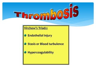
- 1. Virchow’s Triad::: Endothelial Injury Stasis or Blood turbulence Hypercoagulability
- 2. Loss of endothelium exposes Subendothelial collagen Etiology: Hypertension Endotoxins Scarred valves Hyperhomocystinemia Hypercholesterolemia Smoking Radiation 1. ENDOTHELIAL INJURY
- 3. Turbulence Arterial thrombus Stasis Venous thrombus Alteration in Flow causes…. Disrupt the laminar blood flow Cause concentration of Clotting factors Permit build of thrombi Promote endothelial cell activation
- 4. PRIMARY/ GENETIC: Factor V Leiden mutation Prothrombin mutation Antithrombin III deficiency Protein C & Protein S deficiency ACQUIRED CAUSES: Prolonged bed rest- Immobilization Myocardial Infarction Surgery, Fracture, Burns Cancer, Cardiac valves, DIC, SLE Hyperestrogen states, Smoking, Sickle cell anemia, Nephrotic syndrome.
- 5. Previous extensive transmural myocardial infarction. The stasis of blood in the aneurysm predisposes to mural thrombosis.
- 6. Deep thrombi occur in large veins of leg DVT are asymptomatic in 50 % of patients Trousseau Syndrome: Tumor associated procoagulant release Increased risk of Thromboembolic phenomenon in disseminated cancers. Also known as migratory thrombophlebitis Ca Pancreas, Prostate, Stomach, Breast, Lung, Osteosarcoma, AML- M3.
- 10. “Detached Intravascular Solid, Liquid or gas mass carried by blood to a distant site.” 99% are Thrombo-embolus Consequences of embolus is ischemic necrosis of affected tissue.
- 11. Venous Embolism/ Pulmonary (DVT) Arterial Embolism (Post MI) Paradoxical Embolism (Venous will become arterial: due to ASD/ VSD) Fat embolism Amniotic fluid embolism Air embolism Septic embolism Foreign body embolism. Types of Embolism:
- 12. Most commonly from venous emboli from leg veins (DVT) “Saddle embolus” obstructs main Pulmonary artery Once a Pulmonary embolus occurs, patient will be prone for recurrent emboli episodes. Multiple emboli or shower of small emboli in small pulmonary arteries.
- 13. Most arise from Intra cardiac mural thrombi Left ventricular wall infarction and Mitral stenosis predisposes to thrombi and embolus Arterial emboli travel to wide variety of sites Lower limbs, Brain, intestines, kidney, spleen…….any organ.
- 14. Microscopic fat globules enter circulation following fracture of long bones Fat embolism syndrome:: Symptoms appear 1- 3 days after injury Pulmonary insufficiency: Tachypnea, Dyspnea, Tachycardia Neurologic symptoms: Irritability, Restlessness, Delirium, Coma Low platelets: Petechial skin rash Fatal in 10% of individuals
- 15. Fracture long bones: Imp. Soft tissue trauma Burns Parenteral lipid infusion Sickle cell crisis Acute pancreatitis Liposuction Decompression sickness
- 16. Gas bubbles in circulation 100 ml of air is needed to produce clinical effect Chest wall injury, Neck injury, Therapeutic, Intra-operative Decompression sickness seen in Deep sea divers
- 17. Amniotic fluid into ruptured uterine veins Grave, but uncommon complication Important obstetric complication Sudden onset of severe dyspnea, Cyanosis, Hypotension, Shock, Seizures, Coma. If survives… Pulmonary edema, DIC AMNIOTIC FLUID EMBOLISM
- 19. An infarct is an area of ischemic necrosis caused by occlusion of arterial supply or venous drainage Can be due to Thrombus, embolus, vasospasm, atheroma, compression of vessels, etc., Venous blockade Congestion
- 20. Classified based on the color 1. Red (Hemorrhagic) infarct 2. White (Anemic) infarct Red infarcts are seen in:: - Dual blood supply.. Lung, Small intestine - Loose tissues.. Lung - With venous occlusions (ovarian torsion) - Previously congested tissue
- 21. White Infarcts:: - Arterial occlusions - Organs with end arterial blood supply - Solid organs.. Heart, Spleen, Kidneys, Brain Microscopy:: - Coagulative necrosis - Liquefactive necrosis