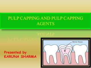
Pulp capping and pulp capping agents
•Als PPTX, PDF herunterladen•
12 gefällt mir•545 views

SEMINAR

Empfohlen
Weitere ähnliche Inhalte
Was ist angesagt?
Was ist angesagt? (20)
Ähnlich wie Pulp capping and pulp capping agents
Ähnlich wie Pulp capping and pulp capping agents (20)
Mehr von DR KARUNA SHARMA
Mehr von DR KARUNA SHARMA (20)
Kürzlich hochgeladen
Kürzlich hochgeladen (20)
Pulp capping and pulp capping agents
- 1. PULP CAPPING AND PULP CAPPING AGENTS Presented by KARUNA SHARMA
- 2. Pulp therapies in primary molars
- 3. Objectives Esthetics Masticaton Speech Psychology Preservation of the arch space Growth of Skeletal & Dental complex(Bar nett,1985) Effects on succedenous tooth (Whine, 1981)
- 4. Title Indirect Pulp Capping Direct Pulp Capping Pulpotomy Pulpectomy Classification of pulp therapy procedures in primary teeth Conservative procedures Radical procedures Weine.Endodontic therapy 6th Edition.Mosby Publication.
- 5. Title Protective liner Indirect Pulp Capping Direct Pulp Capping Pulpotomy Pulpotomy pulpectomy Pulpectomy Classification of pulp therapy procedures in primary teeth Vital pulp therapy for primary teeth diagnosed with a normal pulp or reversible pulpitis Nonvital pulp treatment for primary teeth diagnosed with irreversible pulpitis or necrotic pulp Clinical guidelines on pulp therapy for primary and young permanent molars AAPD 2009
- 7. Protective liner Itota T, Nakabo S, Torii Y, Narukami T, Doi J, Yoshiyama M. Effect of fluoride-releasing liner on demineralized dentin. Quintessence Int 2006;37(4):297-303. Weiner RS, Weiner LK, Kugel G. Teaching the use of bases and liners: A survey of North American dental schools. J Am Dent Assoc 1996;127(11):1640-5. A protective liner is a thinly-applied liquid placed on the pulpal surface of a deep cavity preparation, covering exposed dentin tubules, to act as a protective barrier between the restorative material or cement and the pulp. Placement of a thin protective liner such as calcium hydroxide, dentin bonding agent, or glass ionomer cement is at the discretion of the clinician.
- 8. Protective liner Indications In a tooth with a normal pulp, when all caries is removed for a restoration, a protective liner may be placed in the deep areas of the preparation to minimize injury to the pulp, promote pulp tissue healing, and/or minimize postoperative sensitivity.
- 9. Objectives • The placement of a liner in a deep area of the preparation is utilized to preserve the tooth’s vitality, promote pulp tissue healing and tertiary dentin formation, and minimize bacterial microleakage. • Adverse post-treatment clinical signs or symptoms such as sensitivity, pain, or swelling should not occur.
- 10. Pulp Capping
- 12. Indirect Pulp Capping Definition Objectives Mechanism Indications Contraindications Materials Techniques Success of Indirect pulp capping
- 13. Definition
- 14. “It is defined as procedure wherein small amount of carious dentin is retained in deep areas of the cavity to avoid pulp exposure, followed by placement of a suitable medicament and restorative material that seals off the carious dentin and encourages pulp recovery” -Ingle Definition John I Ingle Leif K Bakland Endodontics, Fifth Edition
- 15. “It is defined as application of a suitable medicament over a thin layer of remaining carious dentin(affected dentin), after deep excavation of infected dentin without exposure to the pulp” -Mathewson,1995
- 16. Objectives (AAPD) Tooth vitality should be preserved Restorative material should seal the involved dentin completely No post-treatment senstivity, pain & swelling No harm to the succedenous tooth
- 17. Title Removal of the carious dentin Remaining carious dentin is covered with biocompatible material Physiologic remineralisation can occur only if the affected dentin contains sound collagen fibers and living odontoblastic processes Sound collagen fibres – base to appatite crystals Living odontoblastic processes supply calcium phosphate from vital pulp for remineralisation Mechanism
- 18. Reparative dentin Highest amount of dentin formed during first month (Traubman 1967). New dentin formation faster – Primary teeth ↑ than permanent teeth Males > females The rate of reparative dentin formation is 1.4 micron per day.
- 19. A- Soft necrotic B- Firm, semi soft C-Hard, discolored Kopel, 1976
- 21. INFECTED LAYER AFFECTED LAYER Highly demineralized Superficial layer Lacking sensation Stained by 0.5% fuschin in propylene glycol Intertubular dentin greatly demineralized Deteriorated collagen fibers Unmineralizable Intermediately demineralised Deeper layer Sensitive Does not stain Partially demineralized Sound collagen cross linkage Remineralizable 21
- 23. Indications (Kopel HM, 1985) History • Tolerable dull pain with mild discomfort from chemical and thermal stimuli • No history of spontaneous pain Clinical examination • Large carious lesion without any frank pulpal exposure • Positive response to electric pulp sensitivity, thermal stimulation and test cavity • Normal appearance of adjacent gingiva • Normal to percussion Radiographic examination • Large carious lesion in close proximity to the pulp • Normal lamina dura • Normal periodontal ligament space • No periapical radiolucency John I Ingle Leif K Bakland Endodontics, Fifth Edition
- 24. Contraindications History • Sharp, penetrating pain • Prolonged spontaneous pain, particularly at night Clinical examination • Excessive tooth mobility • Parulis in the gingiva • Discoloration. Radiographic examination • Large carious lesion with apparent pulp exposure • Interrupted lamina dura • Widened periodontal ligament space • Radiolucency at the furcation areas. John I Ingle Leif K Bakland Endodontics, Fifth Edition
- 25. Techniques
- 26. Title Techniques Two appointment technique One appointment technique John I Ingle Leif K Bakland Endodontics, Fifth Edition
- 27. Title Two appointment technique (First sitting) Carious peripheral dentin should be removed by sharp spoon excavator Stop the excavation as soon as firm resistance of sound dentin is felt Administer local anesthesia and isolate with a rubber dam. Establish cavity outline with a high speed hand piece. Remove the majority of soft, necrotic, infected dentin with a large round bur in a slow speed hand piece without exposing the pulp. . Cover the remaining affected dentin with a hard setting calcium hydroxide dressing. Fill or base the remainder of the cavity with a reinforced ZOE cement and Do not disturb this sealed cavity for 6 to 8 weeks.
- 29. 1st Appointment
- 30. Title Two appointment technique (Second sitting) Between the appointments history must be negative and temporary restoration should be intact. Bitewing radiographs of the treated tooth should be assessed for the presence of reparative dentin The remaining affected carious dentin should appear dehydrated and "flaky“,The area around the potential exposure should appear whitish and may be soft, this is "predentin". The cavity preparation should be irrigated and gently dried. Carefully remove all temporary filling material, especially the calcium hydroxide dressing over deep portions of the cavity floor. . Cover the entire floor with a hard setting calcium hydroxide dressing. A base should be placed with a reinforced ZOE or glass ionomer cement, and tooth should receive a final restoration.
- 31. 2nd Appointment
- 35. Title Indications Poor patient cooperation Inability to remove carious dentin in the first appointment Disadvantages According to Leng et al (1980) The re-entry and re-excavation may potentially increase the chances of pulp exposure. Two appointment technique
- 36. TitleTwo appointment technique Current literature indicates that there is inconclusive evidence that it is necessary to reenter the tooth to remove the residual caries.
- 37. These investigators suggested that reentry to remove the residual minimal carious dentin after capping with calcium hydroxide may not be necessary if the final restoration maintains a seal and the tooth is asymptomatic. One appointment technique
- 38. TitleOne appointment technique Carious peripheral dentin should be removed by sharp spoon excavator Stop the excavation as soon as firm resistance of sound dentin is felt Administer local anesthesia and isolate with a rubber dam. Establish cavity outline with a high speed hand piece. Remove the majority of soft, necrotic, infected dentin with a large round bur in a slow speed hand piece without exposing the pulp. . Cover the remaining affected dentin with a hard setting calcium hydroxide dressing. A base should be placed with a reinforced ZOE or glass ionomer cement, and tooth should receive a final restoration.
- 41. Title Calcium Hydroxide Zinc oxide Eugenol Materials
- 42. Title Dentin bonding agents (Falster CA et al 2002) Resin modified glass ionomer (Farooq 2000) Light cured composites (Lado & Stanley 1987) Materials
- 43. Title Materials Light cured composites
- 44. Title Materials Dentin bonding agents J.J. Marchi .Analysis of Primary Tooth Dentin After Indirect Pulp Capping. Journal of Dentistry for Children-75:3, 2008
- 45. Title Materials Resin modified glass ionomer J.J. Marchi .Analysis of Primary Tooth Dentin After Indirect Pulp Capping. Journal of Dentistry for Children-75:3, 2008
- 50. In histological sections 4 histological layers: Carious decalcified dentin Rhythmic layers of irregular reparative dentin Regular tubular dentin Normal pulp with a slight increase in fibrous element.
- 51. Success of I.P.C
- 52. Intact restoration Negative history of pain. No radiographic evidence of abnormal root resorption. No radiological evidence of radicular disease. Success of Indirect pulp capping
- 53. Indirect pulp capping Indirect pulp capping has been shown to have a higher success rate than pulpotomy in long term studies. It also allows for a normal exfoliation time. Therefore, indirect pulp treatment is preferable to a pulpotomy when the pulp is normal or has a diagnosis of reversible pulpitis.
- 55. Direct Pulp Capping Definition Studies Objectives Indications Contraindications Materials Techniques Success of direct pulp capping
- 56. Title “Direct pulp capping is the placement of a biocompatible agent on the healthy pulp tissue that has been inadvertently exposed from caries excavation or traumatic injury.” -Fuks (1988) Definition John I Ingle Leif K Bakland Endodontics, Fifth Edition
- 57. TitleGuidelines (AAPD) Guidelines developed by the American Academy of Pediatric Dentistry (AAPD) recommend that direct pulp capping should be reserved for small mechanical or traumatic exposures in primary teeth.
- 58. Title Maintain the vitality of underlying pulp tissue regions. Encourage the pulp to wall off the exposure site by initiating a dentin bridge. To seal the pulp against bacterial leakage Objectives John I Ingle Leif K Bakland Endodontics, Fifth Edition
- 59. Indications (Kopel HM, 1985) History • Mild discomfort from chemical and thermal stimuli • Absence of spontaneous pain Clinical examination • “Pinpoint”mechanical exposures • The exposed pulp tissue should be bright red in color Radiographic examination • Large carious lesion in close proximity to the pulp • Normal lamina dura • Normal periodontal ligament space • No periapical radiolucency John I Ingle Leif K Bakland Endodontics, Fifth Edition
- 60. Contraindications History • Sharp, penetrating pain that persists after withdrawing stimulus • Prolonged spontaneous pain, particularly at night Clinical examination • Large pulp exposure • Excessive tooth mobility • Purulis in the gingiva • Non responsiveness to pulp testing techniques. Radiographic examination • Large carious pulp exposure • Interrupted or broken lamina dura • Widened periodontal ligament space • Radiolucency at the root apices or furcation areas.
- 61. Title Technique It is prudent to remove peripheral masses of carious dentin before beginning the excavation where an exposure may occur. The area should be appropriately irrigated with non irritating solutions such as normal saline to keep the pulp moist. Kalins, Frisbee, Delgado et al DEBRIDEMENT Kopel HM. Cosiderations of Direct pulp capping agents in Primary teeth: A Review. J Dent Children.
- 62. Title Technique BLEEDING & CLOTTING Hemorrhage at the exposure site can be controlled with cotton pellet pressure. A biocompatible material is placed directly in contact with pulp tissue
- 63. Title Technique Exposure enlargement Frigoletto noted that small exposures & good blood supply provide the best healing potential. There have been recommendations that the exposure site must be enlarged. (Cvek & zilberman, 1980) Kopel HM. Considerations of Direct pulp capping agents in Primary teeth: A Review. J Dent Children.
- 64. fast setting ZOE cement to achieve a hermetic seal and lastly placing final restoration (SS crown) Placing a hard set CaOH material over the exposure Hemostasis with NaOCl Flushing out dentinal debris with mild solutions Excavate the caries Administer local anesthesia and isolate with a rubber dam. Technique
- 65. Title
- 67. • Beveridge and Brown demonstrated that cold stimuli could decrease intrapulpal pressure by 28 mm Hg. • This was considered to be a direct result of the vasoconstriction of the pulpal blood vessels in response to the cold stimulus.
- 68. • In the hydrodynamic theory, Brannstrom postulated that the capillary action of fluid in the dentinal tubules resulted in the transmission of pain between the tubules and vital pulp tissue in the more apical portions of the pulp chamber and root canal system,even though there was necrotic tissue or hemorrhage in the intervening area.
- 69. • This would seem to account for the sensitivity to tactile stimuli and changes in temperature, even though a large portion of the coronal pulp might be necrotic. • The coefficient of expansion of dentin fluid is estimated to be approximately ten times greater than that of the tubule wall. • Cooling of dentin would result in a contraction of the tubules’ contents with a resultant flow of the fluid away from the pulpal tissues.
- 70. • During acute inflammation, there is an accumulation of polymorphonuclear neutrophils and dilatation of capillaries which allows plasma proteins to escape into connective tissue spaces. • The resultant edema could cause pressure on the pulpal and/or periapical nerve fibers and elicit a painful response. • Van Hassell found that in human teeth the intrapulpal pressure could increase an average of 15 mm Hg in an area of local inflammation.
- 71. • The application of a cold stimulus could, in turn, result in contraction of the dentinal fluid and decrease the pressure within the pulp, yielding a rapid transient reduction of pain.
- 73. INTRODUCTION Historically, the first pulp capping procedure was performed in 1756, by the Phillip pfaff, who packed a small piece of gold over an exposed vital pulp to promote healing. However, the success of the pulp capping procedure greatly depends upon the circumstances under which it is performed and the prognosis depends upon the age, type, site and size of pulp exposure.
- 74. PROPERTIES Stimulate reparative dentin formation Maintain pulpal vitality Release fluoride to prevent secondary caries Bactericidal or bacteriostatic Adhere to dentin Adhere to restorative material Resist forces during restoration placement and during the life of restoration. Sterile Radiopaque Provide bacterial seal
- 75. PULP CAPPING AGENTS 1. Ca (OH)2(1960’s) 2. Zinc oxide eugenol cement (1960-70’s) 3. Corticosteroids and antibiotics (1970’s) 4. Polycarboxylate cement (1970’s) 5. Inert materials (1970’s)(Isobutyl cyanoacrylate and Tri calcium phosphate ceramic) 6. Collagen(1980)
- 76. PULP CAPPING AGENTS 7.Bonding agents(1995) 4-META-MMA-TBB adhesives and hybridizing dentin bonding agents 8.Lasers (1995-2010) CO2 Nd: YAG 9.Mineral trioxide aggregate (1996-2008) 10.Growthfactors(1900-2007)Bone Morphogenic Protein (BMP 2,4,7) 11.Emdogain(2001-2011)
- 77. Calcium Hyroxide • Calcium hydroxide (Ca (OH) 2) was introduced to the dental profession in 1921 by Hermann and has been considered the “gold standard” of direct pulp capping materials for several decades, against which new materials should be, tested
- 79. Calcium hydroxide has the unique potential to induce mineralization even in tissues that have not been programmed to mineralize.
- 80. Sciaky and Pisanti in 1960 observed that calcium hydroxide do not become incorporated in the mineralized repaired tissue, which derives its mineral content solely from the dental pulp,through blood supply.
- 81. CaOH Ca ions Reduced capillary permeability Reduced serum flow Reduced levels of inhibitory pyrophosphate mineralization Hydroxyl ions Neutralizes acid produced by osteoclasts Optimum ph for pyrophosphate activity Increased levels of calcium ion dependent pyrophosphatase
- 82. Zinc Oxide Eugenol (ZOE) Cement • Tronstad and Mjör stated that ZOE cement is more beneficial for inflamed and exposed pulp. • However in the literature Glass and Zander, Hembree and Andrews, Watts, Holland et al., found that ZOE, in direct contact with the pulp tissue, produced chronic inflammation, lack of calcific barrier, and end result is necrosis.
- 83. Title These agents include Neomycin and Hydrocortisone, Cleocin, Cortisone, Ledermix, Penicillin and Keflin. Corticosteroids & Antibiotics
- 84. Title Reduces pulp inflammation Vanocmycin + Ca(OH)2 stimulated a more regular reparative dentin bridge. Corticosteroids & Antibiotics
- 85. TitlePolycarboxylate cements Kopel HM. Cosiderations of Direct pulp capping agents in Primary teeth: A Review. J Dent Children. bonds to the tooth structure McWalter, G et al., found that it lacks an antibacterial effect and calcific bridge formation .
- 86. TitleHybridizing bonding agents J.J. Marchi .Analysis of Primary Tooth Dentin After Indirect Pulp Capping. Journal of Dentistry for Children-75:3, 2008
- 87. Title Miyakoshi et al have shown the effectiveness of 4- META-MMA-TBB in obtaining an effective biologic seal. Pulp showed reparative dentin deposition without pulp pathosis. Heitman and Unterbrink studied a GLUTARALDEHYDE containing Dentin bonding agent. All teeth were vital after 6 month post operative period. Hybridizing bonding agents J.J. Marchi .Analysis of Primary Tooth Dentin After Indirect Pulp Capping. Journal of Dentistry for Children-75:3, 2008
- 88. Title Have cytotoxic effect Absence of calcific bridge formation In vivo studies have demonstrated that the application of an adhesive resin directly onto a site of pulp exposure, or to a thin layer of dentin (less than 0.5 mm), causes dilatation and congestion of blood vessels as well as chronic inflammatory pulpal response Hybridizing bonding agents J.J. Marchi .Analysis of Primary Tooth Dentin After Indirect Pulp Capping. Journal of Dentistry for Children-75:3, 2008
- 89. TitleCollagen Fibers Carmichael DJ reported that collagen fibers are less irritating than Ca (OH)2 and promotes mineralisation but does not help in thick dentin bridge formation Fuks et al found dentin bridge after 2 months in 73% cases.
- 90. Title Materials like Iso-butyl Cyanoacrylate and Tri- Calcium Phosphate ceramic have been used as pulp capping agents. Reduces pulp inflammation Stimulate dentin bridge formation Inert Materials
- 91. Title None of these materials have been promoted to the dental profession as a viable technique Inert Materials
- 92. Title M.T.A Bone Morphogenic Proteins (B.M.P) Cell Inductive Agents
- 93. MTA (Mineral Trioxide Aggregate) Torabinejad centered his research in the development of MTA at the loma linda university, California in 1995.
- 94. Composition Tricalcium silicate Dicalcium silicate Tricalcium aluminate Tetracalcium aluminoferrite Bismuth oxide Traces of silica
- 95. • exists in both white and gray form • difference between the two forms is thelack of iron in the tetracalcium aluminoferrite in the white version. • Can be used for root end fillings perforation repair,pulpotomy and apexification treatment.
- 96. Title MTA (Mineral Trioxide Aggregate) Pitts et al (1996) & Sluyk et al (1998) It allows some micro leakage but superior sealing ability to amalgam, ZnOE / IRM - superior to CH in animal models (Torabinejab M et al, 1999 &Junn D.J et al, 1998) Cell Inductive Agents
- 97. TitleTitle Major Advantages: Excellent sealing ability Good Compressive Strength Good Biocompatibility Pitt Ford et al documented Superior bridge formation and preservation of pulp vitality with MTA when compared with Ca(OH)2 Cell Inductive Agents
- 98. TitleTitle Major Advantages: has significant antimicrobial property Hydrophilic Is alkaline(12.5) and may induce dentinogenesis. The presence of blood has little impact on the degree of leakage of MTA. Thus it can be used as an alternative to calcium hydroxide in both direct pulp capping and pulpotomy preocedures. Cell Inductive Agents
- 99. TitleTitle Bone morphogenic protein (BMP) BMP belongs to super family transforming growth factor beta (TGF-b). - TGF b is a potent modulator of tissue repair in different situations. BMP-2, 4, and 7 plays a role in the differentiation of adult pulp cells into odontoblasts during pulpal healing. Cell Inductive Agents Kopel HM. Cosiderations of Direct pulp capping agents in Primary teeth: A Review. J Dent Children.
- 100. TitleTitle - Urist (1965), observed demineralized bone matrix could stimulate new bone formation when implanted to ectopic sites (muscles) demineralized dentin – from both bone & dentin - Soren J et al (1997), dentin formation by recombinant human osteogenic protien-1. Cell Inductive Agents Kopel HM. Cosiderations of Direct pulp capping agents in Primary teeth: A Review. J Dent Children.
- 101. Recombinant Insulin Like Growth Factor-I Lovschall H, et al., evaluated recombinant insulin like growth factor-I (rhIGF-I) in rat molars and concluded that dentin bridge formation was equal to dycal after 28 days. TitleTitleCell Inductive Agents
- 102. TitleTitle Emdogain
- 103. • EMD is enamel matrix derivative secreted from Hertwig’s epithelial root sheath during porcine tooth development. • It is an important regulator of enamel mineralization and plays an important role during periodontal tissue formation.
- 104. TitleTitleNew Materials.. Emdogain “Amelogenin” & “Amelin” Stimulates dental stem cells for regeneration of periodontium Rangel AG et al. Direct Pulp capping in Primary Molars with Enamel Matrix derivative. JCPD 2009.34(1).9-12.
- 105. • It stimulates the regeneration of acellular cementum, periodontal ligaments, and alveolar bone. • Nakamura Y et al., concluded that amount of hard tissue formed in EMD treated teeth was more than twice that of the calcium hydroxide treated control teeth
- 106. • Al-Hezaimi K evaluated Calcium hydroxide, ProRoot White MTA and white Portland cement after EMD application on the exposed pulp. MTA produced a better quality reparative hard tissue response with the adjunctive use of EMD compared with calcium hydroxide
- 107. • Melcer et al., suggested between the years 1985 and 1987 that the carbon dioxide (CO2) (1W) laser used for direct pulp capping . • Yasuda Y, et al., did a study to examine the effect of CO2 laser irradiation on mineralization in dental pulp cells in rats and the results suggested that CO2 laser irradiation stimulated mineralization in dental pulp cells . Laser
- 108. • Neodymium-doped yttrium-aluminium- garnet laser emits an infrared beam at a wavelength of 1064nm can be of therapeutic benefit for direct pulp capping and pulpotomy in clinical practice
- 109. TitleTitle Orban’s Oral Histology & Embryology 11th Edition. Grossman’s Endodontic Practice twelfth edition Text book of pediatric dentistry Nikhil Marwah 2nd edition Guidelines on pulp therapy for primary and immature permanent teeth AAPD Antonio Nancy.Tencate’s Oral Histology.6th Edition. Kopel HM. Cosiderations of Direct pulp capping agents in Primary teeth: A Review. J Dent Children. Rangel AG et al. Direct Pulp capping in Primary Molars with Enamel Matrix derivative. JCPD 2009.34(1).9-12. John I Ingle Leif K Bakland Endodontics, Fifth Edition References
- 110. Thank You