HUMAN TISSUE ANATOMY & PHYSIOLOGY
•
11 gefällt mir•3,423 views

HUMAN TISSUE ANATOMY & PHYSIOLOGY, EPITHELIAL TISSUE, MUSCLE TISSUE, CONNECTIVE TISSUE & NERVOUS TISSUE FOR PHARMACY STUDENTS #B.PHARM, #PHARM.D

Empfohlen
Empfohlen
Weitere ähnliche Inhalte
Was ist angesagt?
Was ist angesagt? (20)
Ähnlich wie HUMAN TISSUE ANATOMY & PHYSIOLOGY
Ähnlich wie HUMAN TISSUE ANATOMY & PHYSIOLOGY (20)
Mehr von Kameshwaran Sugavanam
Mehr von Kameshwaran Sugavanam (20)
Kürzlich hochgeladen
Kürzlich hochgeladen (20)
HUMAN TISSUE ANATOMY & PHYSIOLOGY
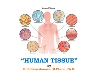
- 2. © 2015 Pearson Education, Inc. BODY TISSUES “Groups of cells with similar structure and function” Study of tissue is known as histology The tissues combined together to form various organs Eg: Kidney, Stmach Four primary types: 1. Epithelial tissue (epithelium) 2. Connective tissue 3. Muscle tissue 4. Nervous tissue
- 4. EPITHELIAL TISSUE: • The epithelium covers the external body surface and lines the internal organs, tubules, vessels & body cavities. • Cells are closely packed and are arranged in one or more layers • Epithelial layers contain no blood vessels, • They must receive nourishment from the underlying connective tissue, through the basement membrane
- 5. • The basement membrane separates the epithelial tissue from the underlying connective tissue • The lower surface of the epithelium rests on a basement membrane
- 6. © 2015 Pearson Education, Inc. CLASSIFICATION OF EPITHELIUM Number of cell layers – Simple—one layer – Stratified—more than one layer Shape of cells –Squamous :Flattened –Cuboidal : Cube-shaped –Columnar : Column-like
- 8. SIMPLE EPITHELIUM Cells arranged in single layer SIMPLE SQUAMOUS EPITHELIUM: Structure: Single layer of flat cells Location usually forms membranes • Alveoli of the lungs • Linings of blood vessel and lymphatic vessels • line and cover organs in ventral cavity Functions: Diffusion, Filtration, or Secretion in membranes
- 9. SIMPLE CUBOIDAL EPITHELIUM: Structure: Single layer of cube-like cells Location: Common in glands and their ducts • Forms walls of kidney tubules • Covers the surface of ovaries Functions: secretion Absorption ciliated types propel mucus reproductive cells
- 10. SIMPLE COLUMNAR EPITHELIUM: Structure: Single layer of tall cells, • Contains Goblet cells - secrete mucus Location: • Lines the mucus membrane of stomach, intestine, uterus Functions: Secretion absorption ciliated types propel mucus reproductive cells
- 11. PSEUDOSTRATIFIED COLUMNAR EPITHELIUM Structure: – All cells rest on a basement membrane – Single layer, but some cells are shorter than others giving a false (pseudo) impression of stratification Location: • Respiratory tract, where it is ciliated and known as pseudostratified ciliated columnar epithelium Functions: Absorption secretion
- 12. STRATIFIED EPITHELIUM “arrangement of cells over one another” STRATIFIED SQUAMOUS EPITHELIUM: Structure: It comprises of multiple layers of flattened squamous cells 2types Keratinized stratified squamous epithelium Non – Keratinized stratified squamous epithelium
- 13. • Two types: Keratinized stratified squamous epithelium: • Contains tough keratin fibres • Which gives protective qualities to the skin • Eg: skin Non – Keratinized stratified squamous epithelium • These cell does not contain keratin • The cell surface remains moist • Eg epithelium lines vagina, mouth, esophagus
- 14. STRATIFIED CUBOIDAL EPITHELIUM: Structure: Two or more layers of cuboidal cells Eg: it is found in the pharynx Duct of sweat gland Function: • Protection
- 15. STRATIFIED COLUMNAR EPITHELIUM: Structure: Comprises of multiple layer of columnar cells surface cells are columnar, cells underneath vary in size and shape Eg: Mucus layer of anus Few parts of male urethra Functions protection
- 16. TRANSITIONAL EPITHELIUM: Transitional – variable appearance It present in the area which are subject to changes in stress and tension STRUCTURE: They are multiple layer of cells & elastic in nature Its ideal for lining urinary bladder LOCATION: Urinary bladder FUNCTION: Allow bladder to stretch while accumulation of urine
- 17. GLANDULAR EPITHELIUM – It is specialized for performing secretary activity – One or more glandular cells responsible for secretion Two major gland types – Endocrine gland • Ductless; secretions diffuse into blood vessels • eg: thyroid, adrenals, and pituitary – Exocrine gland • Secretions empty through ducts to the epithelial surface • Eg: sweat and oil glands, liver, and pancreas
- 18. CONNECTIVE TISSUE • Found everywhere in the body • Includes the most abundant and widely distributed • Found in every organ of the body
- 19. © 2015 Pearson Education, Inc. Connective Tissue Characteristics • Variations in blood supply – Some tissue types are well vascularized – Some have a poor blood supply or are avascular • Extracellular matrix – Nonliving material that surrounds living cells
- 20. © 2015 Pearson Education, Inc. Extracellular Matrix Two main elements 1. Ground substance—mostly water along with adhesion proteins and polysaccharide molecules 2. Fibers • Produced by the cells • Three types: 1. Collagen (white) fibers 2. Elastic (yellow) fibers 3. Reticular fibers (a type of collagen)
- 21. © 2015 Pearson Education, Inc. Connective Tissue Types • From most rigid to softest, or most fluid: – BONE – CARTILAGE – DENSE CONNECTIVE TISSUE – LOOSE CONNECTIVE TISSUE – BLOOD
- 22. • Bone (osseous tissue) – Composed of: • Osteocytes (bone cells) sitting in lacunae (cavities) • Hard matrix of calcium salts • Large numbers of collagen fibers – Functions to protect and support the body
- 23. CARTILAGE: – Less hard and more flexible than bone – Found in only a few places in the body – Chondrocyte (cartilage cell) is the major cell type Types • HYALINE CARTILAGE • ELASTIC CARTILAGE • FIBROCARTILAGE
- 24. HYALINE CARTILAGE The word hyaline derived from greek Hyaline – glass hyaline cartilage is the most widespread type of cartilage It forms covering of ends of bone at joints It forms the support ring of respiratory tubes LOCATIONS: Larynx Entire fetal skeleton prior to birth Epiphyseal plates (end portion of long bone) FUNCTIONS – more flexible skeletal element than bone
- 25. Chondrocyte (cartilage cell) Lacunae (b) Diagram: Hyaline cartilage
- 26. Fibrocartilage The strongest & most durable cartilage of the body Matrix of the fibrocartilage tissue is densely packed with white collagen fibres Location: • Forms cushion like intervertebral discs between vertebrae of the spinal column
- 27. ELASTIC CARTILAGE – Highly compressible – Few collagen fibres present – Contains large number of very fine elastic fibres – provides flexibility Location: External ear larynx
- 29. Dense connective tissue (dense fibrous tissue) – The fibres are closely packed in the matrix – Fibroblasts (synthesis ECM & Collagen)are less in number Types: – Dense irregular connective tissue – Dense regular connective tissue – Elastic dense regular fibrous tissue Locations: • Tendons—attach skeletal muscle to bone • Ligaments—attach bone to bone at joints and are more elastic than tendons • Dermis—lower layers of the skin
- 31. LOOSE CONNECTIVE TISSUE TYPES Two types: Areolar connective tissue Adipose tissue Reticular connective tissue
- 32. LOOSE FIBROUS CONNECTIVE TISSUE: – Also known as - Areolar tissue • Most widely distributed connective tissue • The tissues are stretchable – loose connective tissue • Functions as a universal packing tissue and “glue” to hold organs in place • Layer of areolar tissue called lamina propria underlies all membranes • All fiber types form a loose network • Can soak up excess fluid (causes edema)
- 33. Figure 3.19e Connective tissues and their common body locations. Mucosa epithelium Lamina propria Fibers of matrix Nuclei of fibroblasts Elastic fibers Collagen fibers Fibroblast nuclei (e) Diagram: Areolar Photomicrograph: Areolar connective tissue, a soft packaging tissue of the body (270×)
- 34. ADIPOSE TISSUE • Adipose tissue primarily consist of fat cells – adipocytes • Adipocyte – contains large vesicle filled with triglycerides • Accumulation of more fat may increase the cell size • White fat present in high amount – stores energy for body • It provides support and protection to body organs FUNCTIONS – Insulates the body – Protects some organs – Serves as a site of fuel storage
- 35. Figure 3.19f Connective tissues and their common body locations. Nuclei of fat cells Vacuole containing fat droplet Vacuole containing fat droplet Nuclei of fat cells (f) Diagram: Adipose Photomicrograph: Adipose tissue from the subcutaneous layer beneath the skin (570×)
- 36. RETICULAR CONNECTIVE TISSUE Reticular – like a net It forms a mesh like network by thin branching reticular fibre Locations: – Spleen – Lymph – Bone marrow
- 37. BLOOD (VASCULAR TISSUE) – Blood is a liquid connective tissue – It does not contain any fibre or ground substances – It mainly comprises of – Plasma – liquid portion forms around 55% – Blood cells – solid portion – forms 45% – It includes WBCs, RBCs. Platelets Functions – Transport of gas (Oxygen & CO2), nutrients, waste – Regulate body temperature & pH – WBC gives immunity to body
- 38. MUSCLE TISSUE • Muscular tissue present in all parts of body • This system assist the skeletal system in movement of the body • Contraction& relaxation are the character of this tissues • Pumping of blood by heart, movement in the GIT are done by these muscles
- 39. THREE TYPES OF TISSUES: 1. Skeletal muscle 2. Cardiac muscle 3. Smooth muscle
- 40. Skeletal muscle – Voluntarily (consciously) controlled – The muscle fibres are long and cylindrical shaped – They are striated (Stripes) – They are multinucleated cell – They attached to the skeleton and pull on bones or skin – Produces gross body movements or facial expressions
- 41. CARDIAC MUSCLE: – These are cross striated muscle – Uninucleate in nature, branching – Involuntarily controlled – Found only in the heart – These muscles helps in generation of contraction – Pumps blood through blood vessels
- 42. Smooth (visceral) muscle – These cells thin and spindle shaped – Non striated muscle, Involuntarily controlled – Uninucleate in nature – They contain actin (thin) & myosin (thick) filaments – helps in contraction – Found in walls of hollow organs such as stomach, uterus, and blood vessels – Peristalsis, a wavelike activity, is a typical activity
- 43. NERVOUS TISSUE • These tissues are responsible for rapid communication & coordination between various parts of body • Neurons are located within the organs of central nervous system • Eg: brain and spinal cord • A typical neuron contains following structure – Cell body or soma – Myelin sheath – Node of ranvier
- 44. CELL BODY OR SOMA: • A plasma membrane encloses the cell body • It has centrally located nucleus • Cytoplasm of the cell body consist of the granules – Nissl bodies • 2 Cytoplasmic extensions emerge from cell body Axon: • It terminates nerve impulse away from the cell body or soma Dendrites: • They are either one or more in number and carry nerve signals towards the body
- 45. MYELIN SHEATH: It covers the axon forming a whitish fatty, non cellular layer around the axon NODE OF RANVIER: it is the gap between the two adjacent schwann cells
- 46. Functions: involved in the transportation of nerve impulse Between neuron to neuron & Between neuron to effector organ
- 47. THANK YOU