

Empfohlen
Weitere ähnliche Inhalte
Was ist angesagt?
Was ist angesagt? (20)
Andere mochten auch
Andere mochten auch (20)
Ähnlich wie Anthoceros
Ähnlich wie Anthoceros (20)
Mehr von Jayakara Bhandary
Mehr von Jayakara Bhandary (20)
Kürzlich hochgeladen
Kürzlich hochgeladen (20)
Anthoceros
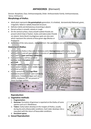
- 1. ANTHOCEROS (Hornwort) Division: Bryophyta, Class: Anthoceratopsida Order: Anthocerotales Family: Anthocerotaceae Anthoceratopsida, Anthocerotaceae, Genus: Anthoceros Morphology of thallus: • Adult plant represents the gametophytic generation. It is thalloid, dorsiventrally flattened, green, irregularly lobed or radially dissected structure structure. • Definite midrib and dichotomous branching is absent. • Dorsal surface is smooth, velvety or rough. • On the ventral surface, many s smooth walled rhizoids are present which help in fixation. Scales and tuberculate rhizoids are absent. Some bluish mucilaginous spots are also seen which represent the colonies of blue blue-green alga (Nostoc or Anabaena). • At the end of the rainy season, elonga elongated horn- like sporophytes are seen on the gametophyte. Anatomy of thallus: • Internally, the thallus is few cells in thickness and is homogeneous without any tissue differentiation and cell specialisation. It consists of parenchymatous tissue covered by single layer each of upper and lower epidermis. • Each parenchyma cell has a distinct nucleus, large chloroplast and a single pyrenoid. • Smooth-walled rhizoids arise from the lower epidermal cells, in the middle region of ventral surface. • On the ventral side, mucilage filled cavities are present which open outside by h slime pores. Endophytic algae such as Nostoc, Anabaena, etc. are present in re these mucilage cavities. Reproduction: a) Vegetative methods: 1. Fragmentation: 2. Gemmae: Formation of gemmae is reported on the thallus of some : species such as A. Glandulosus Glandulosus. 3. Tubers: Corky tubers develop on the margins of thallus, usually : during dry and unfavourable conditions. Each tuber develops into a new plant with the return of favour favourable conditions. 4. Persistent apices: b) Sexual Reproduction:
- 2. Sexual reproduction is oogamous type. Many species are monoecious while some are dioecious. In monoecious species, antheridia develop much earlier to archegonia (Protandrous condition). Sex organs develop inside the gametophytic thallus. Structure of Antheridia: • Develop singly or in groups, in the upper region of the thallus, inside closed cavities called antheridial chambers. • Each antheridium has an ovoid body and a multicellular, slender stalk. The body is covered by a single layered antheridial wall. • Inside the body, a mass of androcytes or spermatocytes are present. Each spermatocyte is a bi-flagellate ‘coma’ shaped structure with a single haploid nucleus. Structure of Archegonia: • They are present sunken in the thallus on the upper side, close to the apex, in acropetal order. • Each archegonium is flask-like in appearance with a basal swollen venter region and a narrow neck. • The venter has a basal egg cell and an upper venter canal cell. 4-6 neck canal cells are arranged in a row at the neck part. • There is no jacket cells covering the archegonium. The vegetative cells of the thallus provide protection to the archegonium. Cover cells or lid cells are found at the tip of the archegonium. Fertilisation: Rain splash mechanism, as in Riccia/Marchantia. Structure of Sporophyte: The diploid zygote develops into a sporophyte on the upper surface of the gametophyte. Mature sporophytes are elongated (2-3 cm long), horn-like structures with a bulbous base. It is covered at the base by a tubular involucre which is developed from the gametophytic tissue. The sporophyte is differentiated into 3 distinct regions: 1. Foot: It is the basal, bulbous structure which is found deeply embedded in the gametophytic tissue. It helps in attaching the sporophyte to gametophyte and in absorption of water and nutrients from it. Foot is made up of parenchyma cells. 2. Intermediate or intercalary zone: It is a narrow zone of meristematic cells located between the basal foot and the upper capsule. These cells help in the continuous growth of the sporophyte. 3. Capsule: It is the fertile, major and conspicuous part of the sporophyte which is long and cylindrical. It is green when young, but turns grey or brown on maturity. The capsule is composed of the following structures: a) Columella: It is the central solid core of sterile cells, consisting of 16 vertical rows of cells. It extends from the base to almost to the tip of the capsule.
- 3. b)Sporogenous tissue: It is the mass of fertile spore- forming cells surrounding the columella, like a dome. At the base of the capsule, it is single layered and called archesporium. It becomes 2- 4 layered and develops into diploid spore mother cells upwards. Towards the tip of the capsule, the spore mother cells divide by meiosis and produce haploid spores. Along with spores, chains of sterile cells called pseudo-elaters are also present. They are without spiral thickenings and are nutritive in function. c) Capsule wall: It is the outer wall of the capsule which is 4-6 layers in thickness. The outermost layer is called epidermis which is interrupted by stomata. The inner layers consist of chlorenchymatous cells and are photosynthetic. Therefore, the sporophyte of anthoceros is partially autotrophic or semi-independent. Dehiscence of Sporophyte and germination of spores: The spores ripen and mature basipetally, from top downwards. The mature part looses water, shrinks and ruptures or splits longitudinally, exposing the spores. The splits extend downwards. The exposed spores are blown away by the wind. The dispersed spores germinate during the next moist season. During germination, they absorb water and swell. The exospore ruptures and the endospore comes out in the form of a germinal tube. The tip of the germinal tube divides repeatedly forming a young thallus. ©Dr. M. Jayakara Bhandary Associate Professor of Botany Government College, Karwar