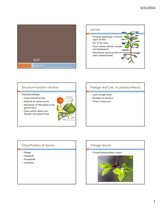
Leaf
•
0 gefällt mir•3,478 views

This document discusses leaf structure and function. It begins by defining leaves and their basic anatomy. It then covers leaf classification, morphology, histology, and development. The key structures discussed include the epidermis, mesophyll, vascular bundles, petiole, and abscission zone. Gymnosperm and angiosperm leaves are compared in terms of their tissues and support structures. Leaf development starts from the shoot apical meristem and progresses through initiation, outgrowth, and maturation of tissues.

Empfohlen
Weitere ähnliche Inhalte
Was ist angesagt?
Was ist angesagt? (20)
Andere mochten auch
Ähnlich wie Leaf
Ähnlich wie Leaf (20)
Mehr von Jasper Obico
Mehr von Jasper Obico (20)
Kürzlich hochgeladen
Kürzlich hochgeladen (20)
Leaf
- 1. 3/21/2010 Leaves Principal appendage or lateral organ of stem Part of the shoot Tissue systems: dermal vascular dermal, and fundamental Determinate apical growth (vs. stem—indeterminate) LEAF Biology 101 Structure-function relation Foliage leaf (rel. to photosynthesis) PHOTOSYNTHESIS Lacks storage tissues Large external surface Develops no periderm Extensive air space system Primary tissues only Abundance of chloroplasts i th Ab d f hl l t in the ground tissue Close spatial relation bet. Vascular and ground tissue Classification of leaves Foliage leaves Foliage Principal photosynthetic organs Cataphylls Hypsophylls cotyledons t l d 1
- 2. 3/21/2010 Cataphylls Hypsophylls Cata= down; phyllon= leaf Hypso= high Leaves inserted at low levels of shoot Leaves inserted at high levels of the plant Scales on bud and underground stem) Floral bracts (protection) Protection or storage Prophylls Pro= before First cataphylls on lateral branch Monocots– 1 prophyll Eudicots E di t – 2 prophyllh ll Cotyledon Phyllomes First leaf of the plant General terms Include foliage leaves, scales, bracts, floral appendages 2
- 3. 3/21/2010 FOLIAGE LEAF morphology Blade/lamina– flattened structure Petiole Leaf sheath Simple and compound leaf (leaflets) Cladodes Stipules phyllode Histology of MATURE leaf Epidermis ANGIOSPERM LEAF Epidermal cells Guard cells with subsidiary cells Trichomes Ti h Silica and cork cells (Gramineae) Bulliform cells Fiber like cells 3
- 4. 3/21/2010 Epidermis Wall structure of epidermis Terrestrial Presence of cutin in the outer walls Living tissue a. Thin – mesophytes and water plants No well differentiated chloroplasts b. thick, lignified – xerophytes c. Sili ifi d – grasses and allies Silicified d lli Aquatic May show more abundant chloroplasts Mesophyll Mesos= in the middle Living, lacunose parenchyma with chloroplasts Mesophyte Dicots– palisade and spongy Palisade-- development is affected by light P li d d l t i ff t d b li ht -- more chloroplasts (sun vs. shade plants) Vascular system Vascular bundles or group of it = veins Single vein– conifers, Equisetum Dicot– largest vein occur in median position (midvein) with (midvein)—with rib 4
- 5. 3/21/2010 Histologic composition Monocots– usually equal in size or may vary (larger Collateral bundles– x is adaxial; p is abaxial veins alternalte with smaller ones); median bundle Bicollateral – adaxial phloem occurs only in large may be larger than others veins Largest veins (distribution of bundles) - circular - irregular - crescent shape (if single) Veins (dicots) Larger veins Smaller veins May have primary entirelyprimary and secondary tissues Tracheary elements Vessels and sievetubes are tracheids; phloem part may parenchyma only at ultmate endings 5
- 6. 3/21/2010 Bundle sheaths Part of ground tissue Also called border parenchyma (dicots) May contain chloroplasts In monocots (Gramineae), two types exists: 1. Parenchymatous– with chloroplasts 2. thick-walled sheath/ mestom sheath—inner; surrounded by parenchymatous sheath also --- procambial origin Supporting structures PETIOLE Not so developed as in the stem Comparable to stem Flat blades– vascular system Ground tissue =~ cortex of stem Dicots -- less chloroplasts – the bundle sheaths and extensions -- supporting structures: collen or scleren ti t t ll l -- collenchyma (large veins) Vascular bundles -- sclereids -- collateral (Syringa) Monocots– large amounts of sclerenchyma -- fibers (assoc. with vascular bundles) -- bicollateral -- concentric (most dicots) Distribution of vascular tissues Petiole Continuous or multi-stranded arc (open toward 1 Collateral bundle– x is adaxial; phloem is adaxial) abaxial Form a circle (with addtl. Bundles within circle) Bicollateral bundle—on both sides of the xylem Numberous and arranged in several superposed arc If in arcs or circles– phloem oriented periphery circles scattered *rachis and pedicels of leaflets—similar to petiole but with less tissue 6
- 7. 3/21/2010 Pinus leaf Xeromorphic GYMNOSPERM LEAF Low ratio of surface to volume Epidermis heavily cuticularized/ thick-walled Pinus leaf Presence of hypodermis—thick wall; compact yp ; p (except with stoma) Guard cells sunken (overtopped by subsidiary cells) Vascular bundles surrounded by transfusion and endodermis respectively mesophyll not differentiated Other features Resin ducts Vascular bundles– x adaxial side; p abaxial side Xylem is endarch Transfusion tissue 2 kinds of cells: a] living parenchyma cells with non- lignified walls and b] thin-walled but lignified tracheids Development of the leaf with bordered pits Parenchyma cells- deeply staining Tracheids (near xylem) Albuminous cells (near phloem) – dense cytoplasm and prominent nuclei Universally present in gymnos Function: water storage or auxiliary conducting system 7
- 8. 3/21/2010 Origin from SAM Periclinal division in the flank meristem Lateral protrusion—occurs NEAR the surface Leaf buttress formed Leaf develops Tunica and corpus Early growth and histogenesis Participates in the formation of leaf primordium After initiation cell division, enlargement and If single layer tunica– corpus maturation If three-layered tunica-- tunica Stages of leaf development a. a Formation of foliar buttress b. Formation of leaf axis c. Formation of lamina Adaxial meristem Marginal meristem a. marginal initials b. b b submarginal i l initials As the leaf axis is elevated above the buttress---procambium is differentiated 8
- 9. 3/21/2010 Vascularization Leaf abscission Procambium of midvein differentiates first in the Separation of leaf from the stem without injury to leaf axis the living tissues As the lamina is formed, the procambium While giving protection to newly exposed surface differentiates in the middle layers y from dessication and infection The development progresses BASIPETALLY Occurs in abscission zone Abscission zone Histologic structure Occurs within the Contains minimum of strengthening tissues petiole or at its base Parenchymatous except in vascular tissues Facilitating separation Vascular elements (tracheids) are short a. Histologic structure of g Weak W k portion ti petiole b. Presence of separation layer Separation layer Protection of the surface exposed Cell walls are chemically modified a. Formation of scar or cicatrice Cell walls increase in volume, swell, assume - deposition of suberin, lignin, or wound gum gelatinous appearance b. Periderm formation beneath the scar cells separate from each other or are easily broken Calcium pectate water soluble pectin 9