Week 2. Diffusion magnetic resonance imaging, tractography, mapping the brain's connectome.
1. 2012.10.30.
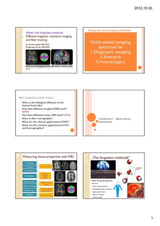
Diagnostic neuroimaging modalities
Water:
Water : the forgotten molecule
Diffusion magnetic resonance imaging CT – Computed Tomography
Brain anatomy
Structural MRI
Fine brain anatomy
and fiber tracking Stereotactic reference frame Vascular structure
Dr. András Jakab, MD, PhD
Multi-modal imaging
Intra-operative imaging Diffusion, perfusion MRI
Dr.Berényi Ervin, MD, PhD
field
spectrum for
modalities, open MRI, low- Fine pathological
information
1.Diagnostic imaging
Positron Emission
MR Spectroscopy
2.Research
Tomography PET
Brain metabolism
Brain function
Brain metabolism
Biochemical mapping
3.Neurosurgery
Electro encephalography,
Functional MR imaging fMRI
Multimodal Imaging in Neurosciences credit course, LORETTA,
Brain function
Magnetoencephalography
2012
KEY LEARNING POINTS TODAY
1. What is the biological diffusion in the
human brain like?
2. How does diffusion weighted MRI work?
(DWI)
3. How does diffusion tensor MRI work? (DTI)
4. What is fiber tractography? CHAPTER I. – BIOLOGICAL
5. What are the clinical applications of DWI? DIFFUSION
6. What are the research applications of DTI
and tractographies?
Measuring tissue properties with MRI „The forgotten molecule”
molecule”
T1 relaxation
T2 relaxation Structural MRI
Proton density
Diffusion-
Tissue diff i
Ti diffusion weighted
imaging
Water has special properties:
Diffusion direction
Diffusion tensor
•H-bonds
imaging
Diffusion anisotropy • Favours polar molecules
Diffusion spectral • Topologically binds to proteins?
Diffusion maps imaging, HARDI
• High surface friction
Metabolites MR spectroscopy • High heat capacity
• Soluble oxygen
1
2. 2012.10.30.
„The forgotten molecule”
molecule”
Water in biological tissues
Kémiai Nobel –díj 2003: Peter Agre
(aquaporins), Roderick MacKinnon
(K-csat.)
Diffusion:
Diffusion: the biological basics Diffusion:
Diffusion: basic physics
1827, Robert Brown 1905: „the miraculous year”
Vis vitalis? Or someting “On a Heuristic Point of View on the Creation
and Conversion of Light”(17 March
else? 1905)(Photo-Electric Effect) Nobel prize in
physics, 1921
Random motion
“On the Electrodynamics of Moving Bodies” (30
„Thermal motion” F.M.Exner, 1900 June 1905)
“Does the inertia of a body depend on its Albert Einstein, 1905
energy content?”(27 September 1905)(Theory of
Special Relativity) E = mc²
“Investigation on the Theory of the Brownian
Movement: On the motion of small particles
suspended in liquids at rest …”(11 May 1905)
Einstein, 1905
The molecular-kinetic theory of
temperature : Brownian motion =
molecular diffusion
0
?
RANDOM WALK
Source Karla Miller, FMRIB, University of Oxford
2
4. 2012.10.30.
CHAPTER II. – DIFFUSION MRI
End of part I. - any questions?
1. Patient (water + fat = lot of spins)
Diffusion-
Diffusion-weighted MR imaging (DWI)
THE MRI RECIPE 2. Excite (Shout at the patient with a
Repeat this! This is called SEQUENCE radiofrequency coil)
3. Wait until the excited spins „relax” Le Bihan & Breton, CRASS, 1985
4. During relaxation, the spins (water +
fat =patient) shout back at you, they
Intravoxel incoherent motion
send an ECHO Movement during imaging =
5. You listen to the echo and record it
(this is the k-space acquisition) the MRI signal is affected
,
Human, made of
ECH
excitable spins
6. Decode the i l t image!
6 D d th signal, get i ! Spatially
S ti ll variant magnetic gradient
i t ti di t
(HOproton spins) encodes the location of spins
Stationary vs. Moving molecules are
coded
Forrás: Karla Miller, FMRIB, University of Oxford Forrás: Karla Miller, FMRIB, University of Oxford
4
5. 2012.10.30.
The image contrast formation during Stejskal-
Stejskal-Tanner sequence
DWI Diffusion-weighting in the MRI sequence
◦ Apply a gradient for delta time (-> DTI!)
If particles move during the imaging time,
◦ Apply in many directions (at least 3)
the signal is attenuated
If diffusion is slower, the signal is higher
(
(more particles „stay in place”)
p „ y p )
◦ So what does DWI tells us?
◦ = the diffusion magnitude = diffusion coefficient
So what does diffusion MRI
measure?
measure?
1. How fast is diffusion in tissues?
2. Is there a pathology, where diffusion is
restricted (cells are dead)?
Image source: Peter Barsi
…. WHAT ELSE??
Why tissue diffusion is special?
special?
It has directional preference = anisotropic!!
What types of diffusion are there?
there?
Free diffusion (isotropic)
Restricted diffusion
Hindered diffusion (barriers)
Forrás: Karla Miller, FMRIB, University of Oxford
5
6. 2012.10.30.
Displaying the diffusion’s direction
Diffusion Imaging Biological diffusion shows various magnitudes in different directions.
In an isotropic medium (inside a glass of water for example) water
molecules naturally move according to Brownian motion. Herpes encephalitis
forrás: emedicine.medscape.com
In biological tissues however the diffusion is very often anisotropic.
For example a molecule inside the axon of a neuron has a low
probability to cross a myelin membrane.
Apply gradient in a given direction dark regions correspond to where
non Brownian motion has occured.
S. Maier and M. Kubicki
ACA stroke
forrás: RADsounds Wiki
Healthy brain
Diffusion Tensors & Anisotropy
Diffusion tensors (?)
Examining diffusion from many directions = direction encoding
DTI allows researchers to quantify the diffusion of water in brain
tissue
Diffusion for each image voxel is
described by 3 perpendicular vectors
λ1
λ1
λ2
λ2 Peter Basser (NIH)
λ3
λ3
TENSORS: the key
to display direction
Anisotropic diffusion dependency
Isotropic diffusion occurs when
occurs when water
there is no restriction to water
movement is restricted to
movement (e.g., ventricles, CSF)
one primary direction
(e.g., myelinated fibers)
Mapping diffusion
Diffusion weighted image Spatial representation of Calculating tensors for each
acquisition diffusion magnitudes image point
Displaying tensorial information on scalar
images
6
7. 2012.10.30.
Image source: Brian Wandell, Stanford
So what is needed for diffusion
tensor imaging? What can we measure with DTI?
Diffusion MAGNITUDE: APPARENT DIFFUSION COEFFICIENT
- Many diffusion weighting
directions during a scan The orderedness of diffusion: DIFFUSION ANISOTROPY
Diffusion direction (color coded maps)
-At least 6 directions Diffusion magnitudes parallel and perpendicular to fibers
- Suggested: 24-64 directions
- Processing of images
- Special display modes
CHAPTER III. – TRACTOGRAPHY
End of part II. - any questions?
7
8. 2012.10.30.
Diffusion tractography
Tract = brain pathway, axonal pathways
Tractography = displaying white matter pathways through post-
procesisng of diffusion tensor images
Brain connections?
Computerized data and image processing
Diffusion tractography: examples Diffusion tractography: examples
Displaying the cortico-spinal tract
Displaying language pathways
Right handed Left handed
NeuroImage 35 (2007) 1064–1076
8
9. 2012.10.30.
Konnektivity (c) Jakab A.
Beyond tensors…
tensors…
Making the resolution of axons better
Q-ball imaging, High angular diffusion
imaging (HARDI)
End of part III. - any questions?
9
10. 2012.10.30.
Summary so far:
T1, T2-weighted MRI: relaxation time of tissue water
protons (i.e. in „watery” of „fatty” environment)
Diffusion weighted MRI (DWI) : the magnitude of
diffusion, restricted diffusion
Diffusion tensor MRI (DTI): the orientation of diffusion
Tractography: displaying white matter pathways
CHAPTER IV. – PRACTICAL USE OF
Diffusion spectrum imaging (DSI): the precise spatial
DIFFUSION MRI (DWI, DTI) characteristics of diffusion with high angular resolution
DWI, DTI and tractography can be combined
with oter imaging modalities (CT, MRI, PET…)
9.1a 9.1b 9.1c 9.1d
1. DWI in ischaemic stroke
Stroke: ischaemia
Cells are dying, Na/K pump disfunction, cells swell,
extracellular spaces are reduced (cytotoxic edema!)
Diffusion becomes restricted in 15-30 minutes
Ischaemic stroke is the single best clinical application of DWI
Diffusion weighted image FLAIR Image T1-wtd image Post-contrast Axial T1-wtd so far!
(DWI) image
Diffusion weighted image (DWI)
9.1e 9.1f
47 year-old left
handed gentleman
with one day history
Image source: Peter Barsi
of somnolence, left
facial droop and
slurred speech.
Q9.1. Diagnosis Please MRA Circle of Willis Post-contrast coronal T1
wtd image
2. DWI differentiates abscesses and 3. DWI differentiates epidermoid cyst
tumors from arachnoid cysts
Necrotic high-grade
Abscess tumor
Image source: SS. Jhawar et al.
Image source: Peter Barsi
Content of an abscess = „soup”, dense pus Content of an epidermoid tumor = dense cells
Content of a necrotic tumor center = liquid Content of a arachnoid cyst = liquid
10
11. 2012.10.30.
White matter characterization with DTI?
Anizotrópia - Anizotrópia - - Anizotrópia - -
Anizotrópia ++ Anizotrópia + Diffúzió - Diffúzió ? / n Diffúzió - -
DTI applications 1. DTI in neuro-oncology
neuro-
Structure, anatomy of white matter • DTI can characterize brain tumors
preoperatively
◦ Development, anatomy of white matter
– FA is reduced: vasogenic edema or infiltration?
◦ Disrupted anatomy of white matter (i.e. for
– Tumor FA: cellularity, proliferation activity
neurosurgeons)
– „current literature”:
current literature :
• Frakcionális anizotrópia: Cellularitás és proliferációs aktivitás (MIB-2)
• MRS és DTI: Proliferációs aktivitás (Ki-67)
Tissue integrity •
•
FA: Low grade vs. Anaplasticus glioma
ADC hisztogramok: 1p/19q l.o.h. oligodendrogliomákban
◦ Demyelinisation (Multiple sclerosis) •
•
„Oligo-like” és „astro-like” komponens megjelenítése
ADC hisztogram: Terápiás válasz (Bevacizumab), progressziómentes időtartam becslése
◦ Axonal damage, Wallerian degeneration – Displaying white matter tract’s relation to tumors
◦ Edemea
◦ Characterizing pathological tissues
mean FA: 0.1887
mean ADC(x1k): 1.4791
11
12. 2012.10.30.
DTI in neuro-oncology
neuro- DTI: clinical examples
A. Jakab, M. Emri, P. Molnár, E. Berényi. Glioma
grade assessment by using histogram
analysis of diffusion tensor imaging-
derived maps. Neuroradiology. 2010 Sep 21.
What is the tumor’s
grade?
Low? (WHO I-II)
High? (WHO III-IV)
• Forrás: Nucifora et al. (2007) Diffusion-Tensor MR Imaging and Tractography: Exploring Brain
Microstructure and Connectivity. Radiology. 245, 367-384.
12
13. 2012.10.30.
Theodor Meynert (1833-1892)
• Role of the frontal lobe
• White matter connections
underlie intelligence?
• Disconnection in syndromes
• Forrás: Nucifora et al. (2007) Diffusion-Tensor MR Imaging and Tractography: Exploring Brain
Microstructure and Connectivity. Radiology. 245, 367-384.
• Forrás: Nucifora et al. (2007) Diffusion-Tensor MR Imaging and Tractography: Exploring Brain
Microstructure and Connectivity. Radiology. 245, 367-384.
Cerebral palsy in a 20-month-old girl with
spastic hemiplegia.
• Forrás: Nucifora et al. (2007) Diffusion-Tensor MR Imaging and Tractography: Exploring Brain • Forrás: Lee et al. Diffusion-Tensor MR Imaging and Fiber Tractography: A New Method of
Microstructure and Connectivity. Radiology. 245, 367-384. Describing Aberrant Fiber Connections in Developmental CNS Anomalies
13
14. 2012.10.30.
Ohdo syndroma
Small girl, epileptic seizures • Source: Berényi E, Jakab A
DTI and neuroscience research
Understanding brain development in
utero
14
15. 2012.10.30.
Fiber tracking of a living fetus in
utero
Language pathways
Left side
Right side
Displaying the arcuate fasciculus
Normal anatomy vs. Aphasia! Right dominance!
Jakab A, Béres M, Spisák T, Kis SA, Emri M, Berényi E, Handedness and interhemispheric differences in the
Image credits: Gregor Kasprian, University of
anatomical connectivity of perisylvian language areas: a network-based approach, Magn. Reson. Mat. Phys.
Vienna
Biol. Med. 24 (S1):276, 2011.
Malformations
• The anatomy of the arcuate in severe
brain developmental diseases in
children
• Schizencephaly, polymicrogyria,
lissencephaly, …
• Jakab A, M Paldino – Harvard Medical
A M.Paldino
School, Children’s Hospital
1 Parcellating grey matter from connectivity Segmenting the thalamus
• Visualizing the connections from subcortex to cortex
• Thalamus
• Specific cortical connections
Behrens et al. Non-invasive mapping of connections between human thalamus and cortex
New type of image contrast
using diffusion imaging Nature Neuroscience 6, 750 - 757 (2003)
15
16. 2012.10.30.
Biological correspondence: regions Biological correspondence: regions
defined by Atlas vs. connectivity defined by Atlas vs. connectivity
Biological correspondence: regions Cortico-thalamic connections
depicted using probabilistic
defined by Atlas vs. connectivity tractography. Points represent the
center-of-gravities of each
connectivity map.
Axial (top) view, isometric 3d
display.
Connections of the precentral and Applications: neurosurgical planning
postcentral gyrus and the spatial
relationship between connectivity- (thalamotomies)
based landmarks and the MNI152-
transformed Morel Atlas data (VLpv, Kincses ZT et al. (2012)
VPLa, VPLp nuclei). Points represent Target Identification for
the center-of-gravities of each Stereotactic Thalamotomy
connectivity map. Using Diffusion
Superior view, isometric 3d display. Tractography- PloS One.
7(1): e29969. doi:10.1371/
journal.pone.0029969
Jakab A et al. (2012)
Generation of Individualized
Thalamus Target Maps by
Using Statistical Shape Models
and Thalamocortical
Tractography.
AJNR American Journal of
Neuroradiology.
doi: 10.3174/ajnr.A3140
16