Ulcerative Colitis
•Als PPTX, PDF herunterladen•
28 gefällt mir•22,297 views

Image result for ulcerative colitis Ulcerative colitis (UL-sur-uh-tiv koe-LIE-tis) is an inflammatory bowel disease (IBD) that causes inflammation and ulcers (sores) in your digestive tract. Ulcerative colitis affects the innermost lining of your large intestine (colon) and rectum. Symptoms usually develop over time, rather than suddenly.

Empfohlen
Weitere ähnliche Inhalte
Was ist angesagt?
Was ist angesagt? (20)
Ähnlich wie Ulcerative Colitis
Ähnlich wie Ulcerative Colitis (20)
Mehr von MR. JAGDISH SAMBAD
Mehr von MR. JAGDISH SAMBAD (20)
Kürzlich hochgeladen
Kürzlich hochgeladen (20)
Ulcerative Colitis
- 2. Ulcerative colitis is a recurrent ulcerative & inflammatory disease of the mucosal & submucosal layers of the colon & rectum. The peak incidence is between 30 & 50 years of age. 10% to 15% of the patients develop carcinoma of the colon.
- 3. ETIOLOGY Genetic predisposition. Environmental factors may trigger disease (viral or bacterial pathogens, dietary). Immunologic imbalance or disturbances. Defect in intestinal barrier causing hypersensitive mucosa & increased permeability. Defect in repair of mucosal injury, which may develop into a chronic condition.
- 4. Etiological factors superficial mucosa of colon diffuse inflammations, or shedding of the colonic epithelium. Bleeding occurs ulcerations. (The mucosa becomes edematous & inflamed. ) The disease process usually begains in the rectum & spreads proximally to involve the entire colon.
- 5. Diarrhea painful straining Increased bowel sounds There often is weight loss, fever, dehydration, hypokalemia, anorexia, nausea & vomiting, iron- deficiency anemia Crampy abdominal pain. Anal area may be irritated & reddened; left lower abdomen may be tender on palpation. There is tendency for the patient experience remissions & exacerbations. Increased risk of developing colorectal cancer. May inhibit extracolonic manifestations of eye (irritis), joint (polyarthritis), & skin complaints (erythema nodosum, pyoderma gangrenosum).
- 6. DIAGNOSTIC EVALUATION:- Diagnosis is based on a combination of laboratory, radiologic, endoscopic, & histologic findings. Laboratory Tests:- Stool examination to rule out enteral pathogens; fecal analysis positive for blood during active disease. Complete blood count- hemoglobin & hematocrit may be low due to bleeding; WBC may be increased. Elevated erythrocyte sedimentation rate (ESR). Decreased serum levels of potassium, magnesium, & albumin
- 7. Other Diagnostic Tests:- Barium enema to assess extent of disease & detect Pseudopolyps, carcinoma, & strictures.
- 8. Flexible proctosigmoidoscopy/colonoscopy findings reveal mucosal erythema & edema, ulcers, inflammation that begins distally in the rectum & spreads proximally for variable distances. CT scan can identify complications such as toxic megacolon. Rectal biopsy – differentiates from other inflammatory diseases or cancer.
- 9. General Measures:- Bed rest, I.V. fluid replacement, clear liquid diet. For patients with severe dehydration & excessive diarrhea, fluid may be recommended to rest the intestinal tract & restore nitrogen balance. Treatment of anemia- iron supplements for chronic bleeding, blood replacement for massive bleeding.
- 10. Drug Therapy Sulfasalazine (Azulfidine)- mainstay drug for acute & maintenance therapy. Given orally & is systemically absorbed. Oral salicylates, such as mesalamine (Pentasa), olsalazine (Dipentum Mesalamineenema available for protosigmoiditis; suppository for proctitis. Corticosteroids- treated with 5-aminosalicylic acid preparations to benefit from their potential steroid- sparing effects. Immunosuppressive drugs- purine analogues, 6- mercaptopurine, azathioprine may be indicated when patient is refractory or dependent on corticosteroids. Antidiarrheal medications may be prescribed to control diarrhea, rectal urgency & cramping, abdominal pain;
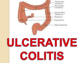
- 11. I. Noncurative approaches (possible curative, reconstructive procedure at later date): a)Temporary loop colostomy for decompression if toxic megacolon present without perforation. b)Subtotal colectomy, ileostomy, & Hartmann’s pouch. c)Colectomy with ileorectal anastomosis.
- 12. Loop colostomy Colostomy Hartmann’s pouch
- 13. . Reconstructive procedures – curative: II. Reconstructive procedures – curative: a)Total proctocolectomy with permanent end- ileostomy. b)Total proctocolectomy with continent ileostomy c) Total colectomy with ileal reservoir- anal (or ileal reservoir-distal rectal) anastomosis – procedure of choice. Multiple reservoir shapes can be surgically created; however, the J-shaped pouch (reservoir) is the easiest to construct. d)The ultimate surgical goal is to remove the entire colon & rectum to cure patient of
- 14. COMPLICATIONS Perforation, hemorrhage Toxic megacolon- fever, tachycardia, abdominal distention, peritonitis, leukocytosis, dilated colon on abdominal X-ray – life-threatening Abscess formation, stricture, anal fistula Malnutrition, anemia, electrolyte imbalance Skin lesions (erythema nodosum, pyoderma gangrenosum) Arthritis, ankylosing spondylitis Colon malignancy Liver disease (sclerosing colagitis) Eye lesions (conjunctivitis) Growth retardation in prepubertal children Possible infertility in females.
- 16. Assessment Review nursing history for patterns of fatigue & over-work, tension, family problems that may exacerbate symptoms. Assess food habits & use of any dietary or herbal supplements used as alternative therapies that may have a bearing on triggering symptoms (milk intake may be a problem). Many patient use vitamins, herbs & homeopathic remedies without realizing the effect on bowel function. Determine number & consistency of bowel movements, any rectal bleeding present. Listen for hyperactive bowel sounds; assess weight.
- 17. Nursing Diagnoses Chronic pain r/t disease process Imbalanced Nutrition: less than body requirement r/t diarrhea, nausea & vomiting Deficient fluid volume r/t diarrhea & loss of fluid & electrolytes Risk for infection r/t disease process, surgical procedures Ineffective coping r/t fatigue, felling of helplessness, & lack of support system.
- 18. Nursing Intervention Promoting Comfort:- Follow prescribe treatment of reducing or eliminating food & fluid & instituting parenteral feeding or low reside diets to the intestinal tract. Give sedatives & tranquilizers, as prescribed, not only to provide general rest , but also to slow peristalsis. Be aware of skin breakdown around anus. Cleanse the skin gently after each bowel movement. Apply a protective emollient such as petroleum jelly etc. Relieve painful rectal spasms Report any evidence of sudden abdominal distention Reduce physical activity Provide commode or bathroom next to bed because urgency of movement may be problem.
- 19. Achieving Nutritional Requirements:- Maintain acutely ill patient on parenteral replacement of vitamins, fluids, & electrolytes. When resuming oral fluids & food, select those that are nonirritating to the mucosa. Avoid dairy products if patient is lactose intolerant. Provide a well-balanced, low-residue, high protein diet to correct malnutrition. Determine which foods the patient can tolerate, & modify diet plan accordingly. Possible avoids cold fluids, which may increase intestinal motility. Administer prescribed medications for symptomatic relief of diarrhea.
- 20. Maintain fluid Balance:- Maintain accurate intake & output records Check weight daily Monitor serum electrolytes, & report abnormalities. Observer for decrease skin turgor, dry skin, oliguria, decreased temperature, weakness, increase hemoglobin, hematocrit, BUN, & specific gravity, which all are signs of fluid loss leading to dehydration.
- 21. Minimizing Infection & Complications:- Give antibacterial drugs as prescribed. Administer corticosteroids as prescribed. Provide conscientious skin care after severe diarrhea. Administer prescribed therapy to correct existing anemia. Observe for signs of colonic perforation & hemorrhage – abdominal rigidity, distention, hypotension, tachycardia.
- 22. Providing Supportive Care:- Recognise psychological needs of the patient. - Fear, anxiety, & discouragement. - Hypersensitivity may be evident. Acknowledge patient’s complaints. Encourage the patient to talk; listen & offer psychological support. Answer questions about the permanent or temporary ostomy, if appropriate. Initiate patient education about living with chronic disease. Include the patient as a part of the health care team to provide continuity of care. Offer educational & emotional support to family members Refer for psychological counseling, as needed.
- 23. Home Care Considerations:- Pouchitis:- Patient undergoing one of the continent restorative procedure (Kock, or ileal reservoir & anal anastomosis) must be alert for a common late postopaerative complication called pouchitis. The symptoms include increased stool output, cramps & malaise. It is thought to be related to stasis within the pouch/ reservoir & usually responds to metronidazole . Assess for these symptoms & notify health care
- 24. Food Blockage:- Patient with a temporary or permanent ileostomy must be alert for signs & symptoms of a food blockage. - This is a mechanical blockage of undigested foodstuffs at the level of the fascia. - It is most likely to occur in the first 6 weeks postoperatively when the bowel is edematous. Symptoms may include spurty, watery stoolwith strong odor, decreased or no stool output, abdominal discomfort, cramping or bloating, & stomal swelling. Nausea & vomiting are late symptoms & requires immediate attention. Treatment includes: - Avoiding solid foods & drinking clear liquids when symptoms occur. Patient with ileostomies must never take laxatives. - Applying a pouching system with a larger opening to
- 25. - Gently massaging the abdomen around the stoma & pulling the knees to chest & rocking the body back & forth. - A warm shower or bath may help with relaxation. - If the blockage lasts for more than 2 to 3 hours or if nausea/vomiting occurs, seek medical attention immediately It is best to instruct the patient how to prevent a food blockage by limiting certain foods the first few months after surgery – Chinese vegetables, skins & seeds, fatty meats, been hulls, popcorn & other foods that do not digest well. Instruct the patient to avoid problem foods, chew food well, drink plenty of fluids while eating, eat possible problem foods in small amounts, & reintroduce problem foods slowly into the diet.
- 27. Patient Education & Health Maintenance:- Teach patient about chronic aspect of ulcerative colitis & each component of care prescribed. Encourage self-care in monitoring symptoms, seeking annual checkup, & maintaining health. Alert patient to possible postoperative problems with skin care, aesthetic difficulties, & surgical revisions. Encourage patient to share experiences with others undergoing similar procedures.
- 28. ASSIGNMENT Q. Write the Nursing Care Plan of Patient with ulcerative colitis?
- 29. BIBLIOGRAPHY 1. Richard Hatchett & David thompson.med-surg. nursing.first edition(2002); publish by churchil Livingstone sydney P.N 552-560. 2. Shaffer’s.Medical-Surgical.seven editions. BI publications New Delhi (2002). P.N. 439-444. 3. Lippincott. Manual of nursing practice. Eight edition (2006). P.N. 673-677. 4. Brunner & Suddarth’s.Medical-Surgical nursing.10th edition.Lippincott williams & wilkins publication (2004). P.N. 1042-1054. 5. www.google.com. 6. www.pubmed.com.