SlideShare ist ein Scribd-Unternehmen logo
1 von 5
Publication for the
 Philips MRI Community
 Issue 46 – 2012/2


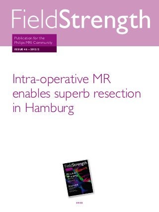
Intra-operative MR
enables superb resection
in Hamburg
Asklepios clinic takes full advantage of dual-use Achieva MR-OR scanner


                                                   the
                                          tion for        ty
                                 Pub lica          mm uni
                                          MR I Co
                                 Phi lips
                                                     /2
                                           46 – 2012
                                  Issu e


                                          an
                                   More th g
                                   M R imagin         e MR ena
                                                                bles
                                               perativ            burg
                                      Intra-o            n in Ham
                                                resectio
                                       superb
                                                                    y team
                                                          n oncolog
                                                 radiatio              g
                                        Herlev                can brin
                                                       t MRI
                                         exp lains wha


                                                       R
                                               Neuro M  ic neuro
                                                                  imaging
                                                                          gets boo
                                                                            enix
                                                                                   st

                                                Pediatr              at Pho
                                                            nia 3.0T
                                                from Inge         pital
                                                          n’s Hos
                                                 Childre
                                                                          into
                                                                  rs look          I
                                                           earche          g 7T MR
                                                   Osu Res     of Ms usin
                                                         nisms
                                                   mecha


                           This article is part of FieldStrength issue 46
                                               2012/2
6
            User experiences


        Paul Kremer, MD, PhD, head
        of the neurosurgical department at
        Asklepios Clinic


        Intra-operative MR enables
        superb resection in Hamburg
        Asklepios clinic takes full advantage of dual-use Achieva MR-OR scanner


        When constructing a new neuro-OR building at Asklepios Clinic (Hamburg, Germany), the decision
        was made to include an intra-operative MR suite. To offset the potentially high costs of an MR system
        that might only be used a few times per week, a dual-use suite was designed for use both intra-
        operatively and by outpatients. So far, more than 1,000 patients have been scanned, about 30 of
        whom were examined intra-operatively.


        Paul Kremer, MD, PhD, is head of the              Dr. Kremer says the intra-operative MRI is           “When using an MRI scanner only for intra-
        neurosurgical department at Asklepios Clinic,     usually used for patients who are having surgery     operative use, just one or two times a week,
        one of the first non-academic neurosurgery        for gliomas, both malignant and benign. “It’s very   it’s too expensive,” says Dr. Kremer. “But the
        departments with intra-operative MRI. He          important to check the resection because it’s        door between the OR room and the MR room
        helped to implement the intra-operative MRI       difficult during the microsurgery to determine       allows us to directly go from the OR into the
        suite in Heidelberg in 1995, then brought the     the tumor margins. It’s very difficult. That is      MR room for intra-operative MRI. And when
        concept to Asklepios, where the intra-operative   why we perform intra-operative MRI, including        the door is closed, the scanner can be used
        MR suite was installed in July 2011.              different image types – T1- or T2-weighted           independently for regular MRI examinations.
                                                          imaging – to check the resection. In Heidelberg      This is a very good approach.”
        “The difference between our center and            several studies were performed on the benefits
        others in the region is that we have a head       for the patient, and these indeed showed a large     Special solutions were designed for intra-
        and neck center, which includes ENT, facial       benefit for the patient.”                            operative MR. “We use a trolley to transport
        maxillofacial surgery, neurosurgery,                                                                   the tabletop with the patient from the OR to
        neurology, neuroradiology, neuropediatrics        Special solutions for intra-operative MRI            the MRI system, and it works very smoothly,”
        and neuropathology,” says Dr. Kremer.             There are several good reasons to have a dual-       Dr. Kremer explains. “Also very important is
        “We do intra-operative MRI on Tuesdays            use MR scanner, from both a scheduling aspect        head fixation for neuro procedures. Fixation
        and Fridays so we can share the imaging           and an economic one, but several challenges          devices are usually made of metal, but we have
        time with the other departments.”                 had to be overcome first.                            found an MR-compatible solution.”


    6    FieldStrength - Issue 46 - 2012/2
MR-OR

“The door between the OR and MR room allows direct transfer.
 When the door is closed, the scanner can be used independently
 for regular MRI examinations. This is a very good approach.”


Pre-op coronal T2W                                                            Pre-op axial T2W	


Intra-op T1W                                                                  Intra-op FLAIR	


Resection of anaplastic astrocytoma
A 31-year-old female with an incompletely resected tumor in another neurosurgical unit some months before. Pre-operative
T2-weighted MRI demonstrates a huge residual tumor mass right temporal. The tumor was resected under neuro-navigational
guidance. Intraoperative T1-weighted and FLAIR images show a complete tumor resection, however, some fluid contents within
the resection area make interpretation of the intra-operative images more difficult. The patient could be discharged from the
hospital 10 days after surgery with the recommendation of following chemotherapy.


                                                                                                                                        CONTINUE
                                                                                                                                        FieldStrength   7
User experiences


    Dr. Kremer explains that at Asklepios, the        all working at the same time,” says Dr. Kremer.   “We bring the patient into the magnet, which
    MR room and the OR room share an air-             “But the results are very impressive.”            is done quickly, then perform the imaging, and
    conditioning system, so the air in the magnet                                                       then the patient comes back into the OR and
    room is filtered by the same system as the OR.    Resolution to see small remnants                  we can start the surgery again. If there are
    “First, the MR room is cleaned the day before     “We have found the image quality is very good,”   some tumor remnants we resect again; if not,
    a procedure,” says Dr. Kremer, “Then we           says Dr. Kremer. “In Heidelberg we used a 0.2T    we close the wound. We go back and forth as
    clean the magnet again one hour before the        magnet, and the imaging quality was quite nice.   much as we need to.”
    procedure. The room is closed for about half      Now we have the Philips Achieva 1.5T magnet
    an hour, and all the air is treated again. The    and the image quality is outstanding. It helps    Generally, the same sequences are used
    patient’s head and surgical wound still is open   that the head of the patient is fixed into the    before and during the surgery. “If the tumor
    but covered, and the patient is transported       head fixation system so the patient is lying      has shown contrast enhancement before, we
    into the magnet room.”                            motionless. We have the resolution to see small   also scan sequences with gadolinium during
                                                      tumor remnants – it’s really impressive.”         surgery; if there’s no enhancement we use
    “It sounds like a difficult process, with the                                                       T2-weighted or FLAIR sequences. We can also
    sterilization system, the trolley system, the     “The time it takes for intra-operative imaging    do fiber tracking if we want.” In the near future,
    head fixation system and the navigation system    is about half an hour,” explains Dr. Kremer.      the clinic plans to begin using fMRI as well.


                                   Pre-op                                                  Intra-op


                                     Post-op                                               Glioma resection
                                                                                           A 44-year-old male demonstrating a multifocal recurrence of
                                                                                           a malignant glioma underwent surgery. Intraoperative images
                                                                                           show a small residual enhancing mass at the level of the corpus
                                                                                           callosum, which was resected directly after intraoperative MRI.
                                                                                           Postoperative contrast-enhanced T1-weighted MRI did not
                                                                                           show any enhancing lesion residues.


8    FieldStrength - Issue 46 - 2012/2
MR-OR


“After intra-operative imaging the patient comes back
 into the OR. If there are some tumor remnants we resect
 again. We go back and forth as much as we need to.”


              Philips intra-operative MRI takes
              next step with Ingenia MR-OR*
              Philips has been a leader in interventional MR since 1995, and has been offering both 1.5T and 3.0T
              MR-guided neurosurgery. An MR-OR suite for intra-operative MRI adds value to neurosurgical
              facilities, supporting resection procedures that can save precious time for both surgeon and patient:
              when intra-operative MR reveals incomplete resection, the resection can be completed in the same
              procedure and reduce the need for subsequent surgery. When Philips introduced Ingenia, the first
              digital broadband MR system, the next generation MR-OR was conceived as well.


              Wide-bore Ingenia 1.5T and 3.0T use dStream architecture,        MR-OR provides MRI during neurosurgery, enabling
              so that the signal is digitized in the coil at the patient and   surgeons to see the results of the surgery before finishing
              transported via fiber-optic cables, increasing SNR by up         it. Smooth in-line transfer of the patient between MR and
              to 40% compared to its predecessor. MR-OR using Ingenia          OR keeps transfer times down to just a few minutes.
              1.5T or 3.0T provides faster, easier and comfortable intra-
              operative MRI. The dual-room concept is designed for             With front and rear docking capabilities to increase
              high-end intra-operative MR with smooth workflow. It is          flexibility and throughput, as well as a 70 cm bore, the
              developed to offer MR and OR, which can be used together         Ingenia MR-OR system is developed for both brain
              or alone to promote high usage and cost effectiveness.           and spine neurosurgery, with a focus on fast, smooth
              For intra-operative use, the Ingenia is combined with a          workflow. It’s the most versatile and fastest intra-
              Maquet OR table, a choice of two types of head frames,           operative MRI ever developed by Philips.
              and coils. It can be combined with neuro navigation systems
              such as those from BrainLAB or Medtronic.


              * Earliest availability of Ingenia MR-OR is expected end 2012.


                                                                                                                                FieldStrength   9

Weitere ähnliche Inhalte

Andere mochten auch

Uso de las radiaciones ionizantes en medicina
Uso de las radiaciones ionizantes en medicinaUso de las radiaciones ionizantes en medicina
Uso de las radiaciones ionizantes en medicinaMiguel Martín Landrove
 
[PHPConferenceBR] Automação de testes com Codeception
[PHPConferenceBR] Automação de testes com Codeception[PHPConferenceBR] Automação de testes com Codeception
[PHPConferenceBR] Automação de testes com CodeceptionJúlio de Lima
 
TDC2015: Testes em APIs REST com Rest-Assured
TDC2015: Testes em APIs REST com Rest-AssuredTDC2015: Testes em APIs REST com Rest-Assured
TDC2015: Testes em APIs REST com Rest-AssuredJúlio de Lima
 
Com fer uns bunyols de quaresma
Com fer uns bunyols de quaresmaCom fer uns bunyols de quaresma
Com fer uns bunyols de quaresmajvidalialcover
 
Tipos de contenedores
Tipos de contenedoresTipos de contenedores
Tipos de contenedoresastrid2014
 
Hypothesis ORCID presentation
Hypothesis ORCID presentationHypothesis ORCID presentation
Hypothesis ORCID presentationORCID, Inc
 
Mi materia favorita♥
Mi   materia   favorita♥Mi   materia   favorita♥
Mi materia favorita♥Kerly lisseth
 
DevQA - Da zona de conforto ao comprometimento com a Qualidade
DevQA - Da zona de conforto ao comprometimento com a QualidadeDevQA - Da zona de conforto ao comprometimento com a Qualidade
DevQA - Da zona de conforto ao comprometimento com a QualidadeKamilla Queiroz Xavier
 
Lightning Talk Session - arXiv (S. Warner)
Lightning Talk Session - arXiv (S. Warner)Lightning Talk Session - arXiv (S. Warner)
Lightning Talk Session - arXiv (S. Warner)ORCID, Inc
 
5 years. voc. weather clothes
5 years. voc. weather clothes5 years. voc. weather clothes
5 years. voc. weather clothesjvidalialcover
 
l'entusiasme dels cinc anys
l'entusiasme dels cinc anysl'entusiasme dels cinc anys
l'entusiasme dels cinc anysjvidalialcover
 

Andere mochten auch (15)

Uso de las radiaciones ionizantes en medicina
Uso de las radiaciones ionizantes en medicinaUso de las radiaciones ionizantes en medicina
Uso de las radiaciones ionizantes en medicina
 
Hoja de vida
Hoja de vidaHoja de vida
Hoja de vida
 
[PHPConferenceBR] Automação de testes com Codeception
[PHPConferenceBR] Automação de testes com Codeception[PHPConferenceBR] Automação de testes com Codeception
[PHPConferenceBR] Automação de testes com Codeception
 
TDC2015: Testes em APIs REST com Rest-Assured
TDC2015: Testes em APIs REST com Rest-AssuredTDC2015: Testes em APIs REST com Rest-Assured
TDC2015: Testes em APIs REST com Rest-Assured
 
Com fer uns bunyols de quaresma
Com fer uns bunyols de quaresmaCom fer uns bunyols de quaresma
Com fer uns bunyols de quaresma
 
Tipos de contenedores
Tipos de contenedoresTipos de contenedores
Tipos de contenedores
 
Guia#1 Mantenimiento
Guia#1 MantenimientoGuia#1 Mantenimiento
Guia#1 Mantenimiento
 
Hypothesis ORCID presentation
Hypothesis ORCID presentationHypothesis ORCID presentation
Hypothesis ORCID presentation
 
Mi materia favorita♥
Mi   materia   favorita♥Mi   materia   favorita♥
Mi materia favorita♥
 
DevQA - Da zona de conforto ao comprometimento com a Qualidade
DevQA - Da zona de conforto ao comprometimento com a QualidadeDevQA - Da zona de conforto ao comprometimento com a Qualidade
DevQA - Da zona de conforto ao comprometimento com a Qualidade
 
Lightning Talk Session - arXiv (S. Warner)
Lightning Talk Session - arXiv (S. Warner)Lightning Talk Session - arXiv (S. Warner)
Lightning Talk Session - arXiv (S. Warner)
 
5 years. voc. weather clothes
5 years. voc. weather clothes5 years. voc. weather clothes
5 years. voc. weather clothes
 
l'entusiasme dels cinc anys
l'entusiasme dels cinc anysl'entusiasme dels cinc anys
l'entusiasme dels cinc anys
 
la festa de carnaval
la festa de carnavalla festa de carnaval
la festa de carnaval
 
Irm
IrmIrm
Irm
 

Mehr von Jhon Arriaga Cordova (20)

Resonancia no ecoplanar en colesteatoma.pptx
Resonancia no ecoplanar en colesteatoma.pptxResonancia no ecoplanar en colesteatoma.pptx
Resonancia no ecoplanar en colesteatoma.pptx
 
Oído.pptx
Oído.pptxOído.pptx
Oído.pptx
 
Toshiba Visions 23
Toshiba Visions 23Toshiba Visions 23
Toshiba Visions 23
 
FieldStrength 49
FieldStrength 49FieldStrength 49
FieldStrength 49
 
FieldStrength 50 advanced neuro
FieldStrength 50 advanced neuroFieldStrength 50 advanced neuro
FieldStrength 50 advanced neuro
 
Ct somatom sessions-33
Ct somatom sessions-33Ct somatom sessions-33
Ct somatom sessions-33
 
Somatom sessions 32
Somatom sessions 32Somatom sessions 32
Somatom sessions 32
 
Somatom sessions 31
Somatom sessions 31Somatom sessions 31
Somatom sessions 31
 
Somatom sessions 30
Somatom sessions 30Somatom sessions 30
Somatom sessions 30
 
Somatom sessions 26
Somatom sessions 26Somatom sessions 26
Somatom sessions 26
 
Somatom sessions 25
Somatom sessions 25Somatom sessions 25
Somatom sessions 25
 
Somatom sessions 24
Somatom sessions 24Somatom sessions 24
Somatom sessions 24
 
Somatom session 29
Somatom session 29Somatom session 29
Somatom session 29
 
Somatom session 29
Somatom session 29Somatom session 29
Somatom session 29
 
Somatom session 28
Somatom session 28Somatom session 28
Somatom session 28
 
Somatom session 27
Somatom session 27Somatom session 27
Somatom session 27
 
Somatom session 27
Somatom session 27Somatom session 27
Somatom session 27
 
Neurography Supplement to Issue 50
Neurography Supplement to Issue 50Neurography Supplement to Issue 50
Neurography Supplement to Issue 50
 
ISMRM Edition - Issue 49
ISMRM Edition - Issue 49ISMRM Edition - Issue 49
ISMRM Edition - Issue 49
 
Pediatric Imaging - Issue 47
Pediatric Imaging - Issue 47Pediatric Imaging - Issue 47
Pediatric Imaging - Issue 47
 

Fs 46 03_asklepios_intra-operative_mr

  • 1. Publication for the Philips MRI Community Issue 46 – 2012/2 Intra-operative MR enables superb resection in Hamburg Asklepios clinic takes full advantage of dual-use Achieva MR-OR scanner the tion for ty Pub lica mm uni MR I Co Phi lips /2 46 – 2012 Issu e an More th g M R imagin e MR ena bles perativ burg Intra-o n in Ham resectio superb y team n oncolog radiatio g Herlev can brin t MRI exp lains wha R Neuro M ic neuro imaging gets boo enix st Pediatr at Pho nia 3.0T from Inge pital n’s Hos Childre into rs look I earche g 7T MR Osu Res of Ms usin nisms mecha This article is part of FieldStrength issue 46 2012/2
  • 2. 6 User experiences Paul Kremer, MD, PhD, head of the neurosurgical department at Asklepios Clinic Intra-operative MR enables superb resection in Hamburg Asklepios clinic takes full advantage of dual-use Achieva MR-OR scanner When constructing a new neuro-OR building at Asklepios Clinic (Hamburg, Germany), the decision was made to include an intra-operative MR suite. To offset the potentially high costs of an MR system that might only be used a few times per week, a dual-use suite was designed for use both intra- operatively and by outpatients. So far, more than 1,000 patients have been scanned, about 30 of whom were examined intra-operatively. Paul Kremer, MD, PhD, is head of the Dr. Kremer says the intra-operative MRI is “When using an MRI scanner only for intra- neurosurgical department at Asklepios Clinic, usually used for patients who are having surgery operative use, just one or two times a week, one of the first non-academic neurosurgery for gliomas, both malignant and benign. “It’s very it’s too expensive,” says Dr. Kremer. “But the departments with intra-operative MRI. He important to check the resection because it’s door between the OR room and the MR room helped to implement the intra-operative MRI difficult during the microsurgery to determine allows us to directly go from the OR into the suite in Heidelberg in 1995, then brought the the tumor margins. It’s very difficult. That is MR room for intra-operative MRI. And when concept to Asklepios, where the intra-operative why we perform intra-operative MRI, including the door is closed, the scanner can be used MR suite was installed in July 2011. different image types – T1- or T2-weighted independently for regular MRI examinations. imaging – to check the resection. In Heidelberg This is a very good approach.” “The difference between our center and several studies were performed on the benefits others in the region is that we have a head for the patient, and these indeed showed a large Special solutions were designed for intra- and neck center, which includes ENT, facial benefit for the patient.” operative MR. “We use a trolley to transport maxillofacial surgery, neurosurgery, the tabletop with the patient from the OR to neurology, neuroradiology, neuropediatrics Special solutions for intra-operative MRI the MRI system, and it works very smoothly,” and neuropathology,” says Dr. Kremer. There are several good reasons to have a dual- Dr. Kremer explains. “Also very important is “We do intra-operative MRI on Tuesdays use MR scanner, from both a scheduling aspect head fixation for neuro procedures. Fixation and Fridays so we can share the imaging and an economic one, but several challenges devices are usually made of metal, but we have time with the other departments.” had to be overcome first. found an MR-compatible solution.” 6 FieldStrength - Issue 46 - 2012/2
  • 3. MR-OR “The door between the OR and MR room allows direct transfer. When the door is closed, the scanner can be used independently for regular MRI examinations. This is a very good approach.” Pre-op coronal T2W Pre-op axial T2W Intra-op T1W Intra-op FLAIR Resection of anaplastic astrocytoma A 31-year-old female with an incompletely resected tumor in another neurosurgical unit some months before. Pre-operative T2-weighted MRI demonstrates a huge residual tumor mass right temporal. The tumor was resected under neuro-navigational guidance. Intraoperative T1-weighted and FLAIR images show a complete tumor resection, however, some fluid contents within the resection area make interpretation of the intra-operative images more difficult. The patient could be discharged from the hospital 10 days after surgery with the recommendation of following chemotherapy. CONTINUE FieldStrength 7
  • 4. User experiences Dr. Kremer explains that at Asklepios, the all working at the same time,” says Dr. Kremer. “We bring the patient into the magnet, which MR room and the OR room share an air- “But the results are very impressive.” is done quickly, then perform the imaging, and conditioning system, so the air in the magnet then the patient comes back into the OR and room is filtered by the same system as the OR. Resolution to see small remnants we can start the surgery again. If there are “First, the MR room is cleaned the day before “We have found the image quality is very good,” some tumor remnants we resect again; if not, a procedure,” says Dr. Kremer, “Then we says Dr. Kremer. “In Heidelberg we used a 0.2T we close the wound. We go back and forth as clean the magnet again one hour before the magnet, and the imaging quality was quite nice. much as we need to.” procedure. The room is closed for about half Now we have the Philips Achieva 1.5T magnet an hour, and all the air is treated again. The and the image quality is outstanding. It helps Generally, the same sequences are used patient’s head and surgical wound still is open that the head of the patient is fixed into the before and during the surgery. “If the tumor but covered, and the patient is transported head fixation system so the patient is lying has shown contrast enhancement before, we into the magnet room.” motionless. We have the resolution to see small also scan sequences with gadolinium during tumor remnants – it’s really impressive.” surgery; if there’s no enhancement we use “It sounds like a difficult process, with the T2-weighted or FLAIR sequences. We can also sterilization system, the trolley system, the “The time it takes for intra-operative imaging do fiber tracking if we want.” In the near future, head fixation system and the navigation system is about half an hour,” explains Dr. Kremer. the clinic plans to begin using fMRI as well. Pre-op Intra-op Post-op Glioma resection A 44-year-old male demonstrating a multifocal recurrence of a malignant glioma underwent surgery. Intraoperative images show a small residual enhancing mass at the level of the corpus callosum, which was resected directly after intraoperative MRI. Postoperative contrast-enhanced T1-weighted MRI did not show any enhancing lesion residues. 8 FieldStrength - Issue 46 - 2012/2
  • 5. MR-OR “After intra-operative imaging the patient comes back into the OR. If there are some tumor remnants we resect again. We go back and forth as much as we need to.” Philips intra-operative MRI takes next step with Ingenia MR-OR* Philips has been a leader in interventional MR since 1995, and has been offering both 1.5T and 3.0T MR-guided neurosurgery. An MR-OR suite for intra-operative MRI adds value to neurosurgical facilities, supporting resection procedures that can save precious time for both surgeon and patient: when intra-operative MR reveals incomplete resection, the resection can be completed in the same procedure and reduce the need for subsequent surgery. When Philips introduced Ingenia, the first digital broadband MR system, the next generation MR-OR was conceived as well. Wide-bore Ingenia 1.5T and 3.0T use dStream architecture, MR-OR provides MRI during neurosurgery, enabling so that the signal is digitized in the coil at the patient and surgeons to see the results of the surgery before finishing transported via fiber-optic cables, increasing SNR by up it. Smooth in-line transfer of the patient between MR and to 40% compared to its predecessor. MR-OR using Ingenia OR keeps transfer times down to just a few minutes. 1.5T or 3.0T provides faster, easier and comfortable intra- operative MRI. The dual-room concept is designed for With front and rear docking capabilities to increase high-end intra-operative MR with smooth workflow. It is flexibility and throughput, as well as a 70 cm bore, the developed to offer MR and OR, which can be used together Ingenia MR-OR system is developed for both brain or alone to promote high usage and cost effectiveness. and spine neurosurgery, with a focus on fast, smooth For intra-operative use, the Ingenia is combined with a workflow. It’s the most versatile and fastest intra- Maquet OR table, a choice of two types of head frames, operative MRI ever developed by Philips. and coils. It can be combined with neuro navigation systems such as those from BrainLAB or Medtronic. * Earliest availability of Ingenia MR-OR is expected end 2012. FieldStrength 9