Romanowsky Stain /certified fixed orthodontic courses by Indian dental academy
•
14 gefällt mir•3,024 views

The Indian Dental Academy is the Leader in continuing dental education , training dentists in all aspects of dentistry and offering a wide range of dental certified courses in different formats.for more details please visit www.indiandentalacademy.com

Empfohlen
Weitere ähnliche Inhalte
Was ist angesagt?
Was ist angesagt? (20)
Andere mochten auch
Andere mochten auch (20)
Ähnlich wie Romanowsky Stain /certified fixed orthodontic courses by Indian dental academy
Ähnlich wie Romanowsky Stain /certified fixed orthodontic courses by Indian dental academy (20)
Mehr von Indian dental academy
Mehr von Indian dental academy (20)
Kürzlich hochgeladen
Kürzlich hochgeladen (20)
Romanowsky Stain /certified fixed orthodontic courses by Indian dental academy
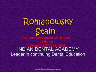
- 1. RomanowskyRomanowsky StainStain Colour responses of blood cells to Romanowsky staining INDIAN DENTAL ACADEMYINDIAN DENTAL ACADEMY Leader in continuing Dental EducationLeader in continuing Dental Education www.indiandentalacademy.comwww.indiandentalacademy.com
- 2. REFERENCESREFERENCES Shrish M. Kawathalkar; Essentials of haematology - 1Shrish M. Kawathalkar; Essentials of haematology - 1stst editionedition Jaypee brother medical publishers.Jaypee brother medical publishers. de Gruchi’s Clinical Haematology in Medical Practice- 5de Gruchi’s Clinical Haematology in Medical Practice- 5thth editionedition Blackwell Science Ltd.Blackwell Science Ltd. P. N. Marshall, S. A. Bentley, and S. M. Lewis ; StainingP. N. Marshall, S. A. Bentley, and S. M. Lewis ; Staining properties and stability of a standardised Romanowsky stainproperties and stability of a standardised Romanowsky stain Journal of Clinical Pathology, 1978, 31, 280-282Journal of Clinical Pathology, 1978, 31, 280-282 www.indiandentalacademy.comwww.indiandentalacademy.com
- 3. P. N. Marshall, S. A. Bentley, and S. M. Lewis ; An evaluation ofP. N. Marshall, S. A. Bentley, and S. M. Lewis ; An evaluation of some commercial Romanowsky Stains J. clin. Path., 1975, 28,some commercial Romanowsky Stains J. clin. Path., 1975, 28, 680-685.680-685. Text book Of Practical Haematology ; ninth edition, Dacie &Text book Of Practical Haematology ; ninth edition, Dacie & Lewis.Lewis. www.indiandentalacademy.comwww.indiandentalacademy.com
- 4. Colour responses of blood cells toColour responses of blood cells to Romanowsky stainingRomanowsky staining Cellular componentCellular component NucleiNuclei ChromatinChromatin NucleoliNucleoli CytoplasmCytoplasm ErythroblastErythroblast ReticulocyteReticulocyte ErythrocyteErythrocyte ColourColour PurplePurple BlueBlue Dark blueDark blue BlueBlue Dark pinkDark pink www.indiandentalacademy.comwww.indiandentalacademy.com
- 6. PromyelocytePromyelocyte MetamyelocyteMetamyelocyte MyelocyteMyelocyte MonocyteMonocyte NeutrophilNeutrophil BasophilBasophil LymphocyteLymphocyte GranulesGranules BasophilBasophil EosinophilEosinophil NeutrophilNeutrophil Toxic granulesToxic granules PlateletPlatelet BlueBlue PinkPink PinkPink Grey-blueGrey-blue Pink/purplePink/purple BlueBlue Blue/clearBlue/clear Purple / bluePurple / blue Red-orangeRed-orange PurplePurple Dark BlueDark Blue PurplePurple www.indiandentalacademy.comwww.indiandentalacademy.com
- 7. Neutrophil Basophil Eosinophil Monocyte Lymphocyte www.indiandentalacademy.comwww.indiandentalacademy.com
- 8. ToxicToxic GranulationGranulation SepsisSepsis InflammationInflammation Changes in granulocytesChanges in granulocytes seen in blood film ofseen in blood film of patients with inflammatorypatients with inflammatory conditions.conditions. These are dark blue, coarseThese are dark blue, coarse granules in neutrophils.granules in neutrophils. www.indiandentalacademy.comwww.indiandentalacademy.com
- 9. Other inclusionsOther inclusions Howell-Jolly bodyHowell-Jolly body Dohle bodyDohle body PurplePurple Light blueLight blue www.indiandentalacademy.comwww.indiandentalacademy.com
- 10. Howell- jolly bodies are nuclearHowell- jolly bodies are nuclear remnants in erythrocytes.remnants in erythrocytes. The nuclear remnants in normal redThe nuclear remnants in normal red cells is removed by spleen.cells is removed by spleen. These bodies are seen inThese bodies are seen in splenectomy, asplenia, hereditarysplenectomy, asplenia, hereditary spherocytosis, megaloblasticspherocytosis, megaloblastic anemia,abnormal erythropoisis.anemia,abnormal erythropoisis. www.indiandentalacademy.comwww.indiandentalacademy.com
- 11. Howell jolly bodies are round, smooth,Howell jolly bodies are round, smooth, pyknotic purple bodies.pyknotic purple bodies. Size: 0.5-1 micron.Size: 0.5-1 micron. They are nuclear remnants or aggregatesThey are nuclear remnants or aggregates of chromosomes that have been separatedof chromosomes that have been separated from mitotic spindle and remain in RBC.from mitotic spindle and remain in RBC. www.indiandentalacademy.comwww.indiandentalacademy.com
- 12. Dohle BodiesDohle Bodies 11. Burn. Burn 2. Infection2. Infection 3. Physical trauma3. Physical trauma 4. Neoplastic diseases4. Neoplastic diseases 5. Chediak-higashi5. Chediak-higashi Syndrome.Syndrome. Döhle bodies are light blue-gray, oval, basophilic, leukocyte inclusions located in the cytoplasm of neutrophils. They measure 1-3 µm in diameter. They are thought to be remnants of the rough endoplasmic reticulum. They composed of agglutinated ribosomes. www.indiandentalacademy.comwww.indiandentalacademy.com
- 13. MICROCYTEMICROCYTE Iron deficiency anemia Thalassemia Lead poisoning www.indiandentalacademy.comwww.indiandentalacademy.com
- 15. BURR CELLBURR CELL 1. Chronic renal disease1. Chronic renal disease 2. Hyperlipidemia2. Hyperlipidemia www.indiandentalacademy.comwww.indiandentalacademy.com
- 16. BURR CELLBURR CELL A/c echinocytes , red cells with abnormalA/c echinocytes , red cells with abnormal cell membrane characterized by thornycell membrane characterized by thorny projection.projection. It is reversible condition.It is reversible condition. Loss of intracorpuscular water due toLoss of intracorpuscular water due to osmotic imbalance.osmotic imbalance. Red cell receptors bind to HDL whichRed cell receptors bind to HDL which induces this shape in hyperlipidemia.induces this shape in hyperlipidemia. www.indiandentalacademy.comwww.indiandentalacademy.com
- 17. TARGET CELLTARGET CELL 1. Thalassemia1. Thalassemia 2. Iron deficiency2. Iron deficiency anaemiaanaemia 3. Liver disease3. Liver disease 4. Asplenia4. Asplenia www.indiandentalacademy.comwww.indiandentalacademy.com
- 18. TARGET CELLTARGET CELL A/C codocyte.A/C codocyte. The appearance is due to decrease in HbThe appearance is due to decrease in Hb content relative to surface area of redcontent relative to surface area of red cell.cell. Decrease in cell volume without alteringDecrease in cell volume without altering membrane area.membrane area. In blood vessel codocyte is bell shapeIn blood vessel codocyte is bell shape and in smear appears target shaped.and in smear appears target shaped. www.indiandentalacademy.comwww.indiandentalacademy.com
- 19. Sickle cell AnemiaSickle cell Anemia 1. Sickle Cell Anaemia1. Sickle Cell Anaemia www.indiandentalacademy.comwww.indiandentalacademy.com
- 20. Sickle cell AnemiaSickle cell Anemia Red cells containing HbSRed cells containing HbS when subjected towhen subjected to deoxygenated state leads todeoxygenated state leads to polymerization (viscous gelpolymerization (viscous gel state) of HbS molecule.state) of HbS molecule. Upon oxygenationUpon oxygenation depolymerization (fluiddepolymerization (fluid state) of Hbs occurs leadingstate) of Hbs occurs leading to normal shape of RBCs.to normal shape of RBCs. www.indiandentalacademy.comwww.indiandentalacademy.com
- 21. Bite CellsBite Cells HaemolyticHaemolytic disorder withdisorder with GG6DP deficiency6DP deficiency www.indiandentalacademy.comwww.indiandentalacademy.com
- 22. Bite CellsBite Cells Deficiency of G6DP leads to decreasedDeficiency of G6DP leads to decreased availability of glutathione,which isavailability of glutathione,which is important for removal of H2O2.important for removal of H2O2. Accumulation of H2O2 causesAccumulation of H2O2 causes oxidation of Hb and ppt. of globinoxidation of Hb and ppt. of globin which attaches to red cell membrane inwhich attaches to red cell membrane in the form of inclusion bodies.(Heinzthe form of inclusion bodies.(Heinz bodies)bodies) This red cell membrane isThis red cell membrane is phagocytosed by splenic macrophages.phagocytosed by splenic macrophages. www.indiandentalacademy.comwww.indiandentalacademy.com
- 23. Tear drop CellsTear drop Cells a/c dacroocyte.a/c dacroocyte. It is a type of poikilocyte.It is a type of poikilocyte. Seen in myelofibrosis.Seen in myelofibrosis. www.indiandentalacademy.comwww.indiandentalacademy.com
- 24. DISORDERS OFDISORDERS OF WBCSWBCS www.indiandentalacademy.comwww.indiandentalacademy.com
- 25. LeukopeniaLeukopenia: WBCs count < 4,000/cumm.: WBCs count < 4,000/cumm. LeukocytosisLeukocytosis: 11,000-25,000/cumm: 11,000-25,000/cumm Aleukemia:Aleukemia: Blasts are not present in peripheral smear but are present in boneBlasts are not present in peripheral smear but are present in bone marrow. Leukemic changes in bone marrow. ( WBC count ismarrow. Leukemic changes in bone marrow. ( WBC count is subnormal)subnormal) Subleukemia:Subleukemia: Total leukocyte count is normal or low but blasts areTotal leukocyte count is normal or low but blasts are demonstrable in peripheral blood.( WBC count is normal ordemonstrable in peripheral blood.( WBC count is normal or subnormal)subnormal) www.indiandentalacademy.comwww.indiandentalacademy.com
- 26. Leukaemoid reaction:Leukaemoid reaction: TLC is < 50,000/cumm.it can occur in infection,TLC is < 50,000/cumm.it can occur in infection, inflammation and malignancies. presence of both mature andinflammation and malignancies. presence of both mature and immature WBCs.immature WBCs. presence of toxic granules in neutrophils.presence of toxic granules in neutrophils. No splenomegaly, monocytosis, eosinophilia.No splenomegaly, monocytosis, eosinophilia. Acute leukemia:Acute leukemia: TLC is > 1 lakh/cumm.TLC is > 1 lakh/cumm. It is malignant clonal proliferation of immature or blast cellIt is malignant clonal proliferation of immature or blast cell of haematopoitic origin.of haematopoitic origin. Short duration, fast growing , early appearance of c/f.Short duration, fast growing , early appearance of c/f. www.indiandentalacademy.comwww.indiandentalacademy.com
- 27. Subacute leukemia:Subacute leukemia: TLC is between 1-2 lakh/cumm.TLC is between 1-2 lakh/cumm. The severity and duration ranges between acute and chronic.The severity and duration ranges between acute and chronic. Chronic leukemia:Chronic leukemia: TLC >2 lakhs/cummTLC >2 lakhs/cumm It is malignant clonal proliferation of mature or differentiatedIt is malignant clonal proliferation of mature or differentiated cells of haematopoitic origin.cells of haematopoitic origin. Long duration, slow growing , late appearance of c/f.Long duration, slow growing , late appearance of c/f. www.indiandentalacademy.comwww.indiandentalacademy.com
- 28. Auer rods are clumpsAuer rods are clumps of azurophilicof azurophilic granular materialgranular material present as elongatedpresent as elongated structure instructure in cytoplasm ofcytoplasm of myeloblastsmyeloblasts.. www.indiandentalacademy.comwww.indiandentalacademy.com
- 29. Quantitative disorders of WBCsQuantitative disorders of WBCs www.indiandentalacademy.comwww.indiandentalacademy.com
- 30. Neutrophilia :Neutrophilia : Acute haemorrhageAcute haemorrhage Acute infectionsAcute infections TraumaTrauma BurnsBurns IntoxicationIntoxication Metabolic disorders: acidosisMetabolic disorders: acidosis www.indiandentalacademy.comwww.indiandentalacademy.com
- 31. Eosinophilia:Eosinophilia: Allergic reactionAllergic reaction:: urticaria,rhinitisurticaria,rhinitis FilariasisFilariasis TrichinosisTrichinosis Pulmonary disordersPulmonary disorders MyeloproliferativeMyeloproliferative disordersdisorders HPV associatedHPV associated carcinomascarcinomas www.indiandentalacademy.comwww.indiandentalacademy.com
- 32. Monocytosis:Monocytosis: MalariaMalaria T.B.T.B. TyphoidTyphoid AMLAML SarcoidosisSarcoidosis Ulcerative colitisUlcerative colitis CarcinomasCarcinomas www.indiandentalacademy.comwww.indiandentalacademy.com
- 33. Lymphocytosis:Lymphocytosis: Infectious mononucleosisInfectious mononucleosis MumpsMumps VaricellaVaricella ToxoplasmosisToxoplasmosis T.B.T.B. ALLALL CLLCLL Atypical lymphocytes with indented nucleus, irregular cellular and nuclear contours, dark basophilic lymphocyte with prominent nucleoli www.indiandentalacademy.comwww.indiandentalacademy.com
- 34. Blood smear of acute lymphoblasticBlood smear of acute lymphoblastic leukemialeukemia www.indiandentalacademy.comwww.indiandentalacademy.com
- 35. Acute myeloid leukemiaAcute myeloid leukemia www.indiandentalacademy.comwww.indiandentalacademy.com
- 36. Chronic lymphocytic leukemiaChronic lymphocytic leukemia www.indiandentalacademy.comwww.indiandentalacademy.com
- 37. Chronic myeloid leukemiaChronic myeloid leukemia www.indiandentalacademy.comwww.indiandentalacademy.com