Endodontic surgeries /orthodontics courses
•
4 gefällt mir•1,298 views

The Indian Dental Academy is the Leader in continuing dental education , training dentists in all aspects of dentistry and offering a wide range of dental certified courses in different formats.

Empfohlen
Empfohlen
Weitere ähnliche Inhalte
Was ist angesagt?
Was ist angesagt? (20)
Andere mochten auch
Andere mochten auch (11)
Ähnlich wie Endodontic surgeries /orthodontics courses
Ähnlich wie Endodontic surgeries /orthodontics courses (20)
Mehr von Indian dental academy
Mehr von Indian dental academy (20)
Kürzlich hochgeladen
Kürzlich hochgeladen (20)
Endodontic surgeries /orthodontics courses
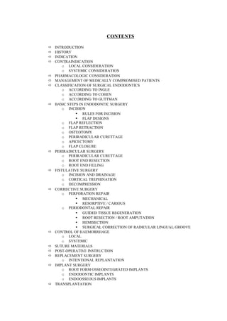
- 1. CONTENTS INTRODUCTION HISTORY INDICATION CONTRAINDICATION o LOCAL CONSIDERATION o SYSTEMIC CONSIDERATION PHARMACOLOGIC CONSIDERATION MANAGEMENT OF MEDICALLY COMPROMISED PATIENTS CLASSIFICATION OF SURGICAL ENDODONTICS o ACCORDING TO INGLE o ACCORDING TO COHEN o ACCORDING TO GUTTMAN BASIC STEPS IN ENDODONTIC SURGERY o INCISION RULES FOR INCISION FLAP DESIGNS o FLAP REFLECTION o FLAP RETRACTION o OSTEOTOMY o PERIRADICULAR CURETTAGE o APICECTOMY o FLAP CLOSURE PERIRADICULAR SURGERY o PERIRADICULAR CURETTAGE o ROOT END RESECTION o ROOT END FILLING FISTULATIVE SURGERY o INCISION AND DRAINAGE o CORTICAL TREPHINATION o DECOMPRESSION CORRECTIVE SURGERY o PERFORATION REPAIR MECHANICAL RESORPTIVE / CARIOUS o PERIODONTAL REPAIR GUIDED TISSUE REGENERATION ROOT RESECTION / ROOT AMPUTATION HEMISECTION SURGICAL CORRECTION OF RADICULAR LINGUAL GROOVE CONTROL OF HAEMORRHAGE o LOCAL o SYSTEMIC SUTURE MATERIALS POST-OPERATIVE INSTRUCTION REPLACEMENT SURGERY o INTENTIONAL REPLANTATION IMPLANT SURGERY o ROOT FORM OSSEOINTEGRATED IMPLANTS o ENDODONTIC IMPLANTS o ENDOOSSEOUS IMPLANTS TRANSPLANTATION
- 2. CAUSES OF FAILURE IN ENDODONTIC SURGERY CONCLUSION REFERENCES 2
- 3. INTRODUCTION : Not all dental problems are treatable by conventional procedures. A number of conditions, particularly those following trauma and disease, demand innovation and ingenuity. Endodontic surgery is a facet of comprehensive root canal treatment, which can manage problems that cannot be eliminated by non surgical techniques. HISTORY : Pre – 1900 Intentional Replantation Abulcasis (11th Century) Pare (1561) Fauchard (1712) Pfaff (1956) Berdmore (1768) Hunter (1778) Incision and drainage were managed by persons other than dentists and physicians. The need to manage chronic sinus tracts by either opening them, cleaning them out or burning them (Lorentz). The use of lancet or sharp pointed knife to puncture swelling (Harris). Surgical Trephination (Hullihen). Application of carbolic crystals to the gums followed by resection to alleviate pain (Bronson). Surgical treatment for management of alveolar abscess (Rhein) First root end resection (Smith) 1900-1939 (FOCAL INFECTION THEORY) 3
- 4. All root tissues located within the diseased tissue around apex was considered contaminated. 1940-1959 Single visit RCT followed by Surgical curettage (Jones) Open window method for apical curettage (Weaver) Use of root end preparations and root end fill with amalgam (Garvin, Luks, Guerny) Development of specific amalgam carrier for placement of material in apical third (Messing). 1960-1990 This period in the history of surgical endodontics represents what we know and practice today. INDICATIONS OF ENDODONTIC SURGERY: 1) Any condition or obstruction that prevents direct access to the apical third of the canal such as ; a. Anatomic – Calcifications, Curvatures, dens n dente, pulp stones. b. Iatrogenic – Ledging, broken instruments, old root canal fillings 2) Periradicular disease associated with a foreign body: over filled canals, excessive cement in the periodontium. 3) Apical perforations 4) Incomplete apexogenesis with blunderbuss that does not respond to apexification. 5) Horizontal fracture root tip with periradicular disease. 6) Failure to heal using non-surgical endodontic treatment. 7) Persistent and recurring exacerbations during non-surgical treatment or persistent unexplainable pain. 8) Treatment of any tooth and a suspicious lesion requiring a diagnostic biopsy. 9) Excessively large lesions. 10)Destruction of apical constricture of root canal due to uncontrolled instrumentation. 4
- 5. 11)Fenestration / Dehiscence 12)Lack of time CONTRAINDICATIONS : General Considerations : a) Medically compromised patients. b) Emotionally unstable patient c) Limitation in surgical skill and experience of the operator LOCAL CONSIDERATIONS : 1) Localized acute inflammation – Treatment is incision and drainage 2) Anatomic considerations. Procedures that penetrate the mandibular canal, maxillary sinus, mental foramen, floor of the nares should be avoided whenever possible. 3) Inaccessible surgical sites – inaccessible position and location of root apices, especially in posterior teeth and need to gain access to the surgical site through dense layer of bone may preclude a successful result. 4) Teeth with poor prognosis – Short rooted teeth, teeth with advanced periodontal disease, vertically fracture teeth. 5) Surgery should not be considered cure-all to compensate for inadequate technique that resulted in failure to heal. Esthetic Considerations : 1) Scarring – Any incision will leave a scar. The maxillary anterior gingiva is the most visible gingiva of the mouth and is exposed in varying degrees when laughing or talking. Since this segment has the greatest number of surgical endodontic entries, the normal lip line, smile line should be presurgically observed. When the labial gingiva is a major part of the smile, hiding the horizontal incision in gingival crevice is recommended as in reducing the 5
- 6. length and number of vertical releasing incisions. Semilunar incision produces the worst scar. 2) Amalgam Tattoo – A problem that exists with the use of the best and most frequently used reverse – fill and root-repair material is the tattoo. However when the material is confined to the root preparation, tattooing is minimized, if not eliminated. Many tattoos are associated with failing amalgam reverse fills, suggesting that percolation enhances the tattooing. If a tattoo is caused by reverse fill failure and re-entry for curettage an a new reverse fill are planned, a flap may be designed to excise the tattoo. Accessibility of Surgical Site : 1) Limitation of mouth opening – TMJ problems resulting from surgery, trauma, congenital defects like microstomia. Chemical or electrical burns etc. 2) Shallow mucobuccal fold – when the mucobuccal fold is shallow, the surgeon must incise and elevate thicker soft tissues, this procedure produces excessive capillary bleeding. If muscles represent in the fold area, such as mentalis, associated with lower incisions, the soft tissues of the lip or cheek may limit access and view. 3) Short alveolar process – In the case of incomplete development, or in mature individuals with small jaws, the root apexes maybe much closer to important intraosseous anatomic structures. Eg. Floor of the nose, maxillary sinus, mandibular canal and mental canal and foramen. 4) Extremely long roots : In long rooted teeth, the primary problem is that, their apexes are located in the widest portion of the alveolar process, which is narrowest at the crest and increasing in width apically. A secondary problem is the greater likelihood of their proximity to intraossoeus structures. A third consideration is the greater soft tissue thickness involved in the incision and elevation when the apex of such a tooth extends beyond the mucobuccal fold. 6
- 7. 5) Bony tori or exostoses : The most common alveolar process, tori occurs lingual to mandibular bicuspids often extending posteriorly to the molars. There are bony exostosis which occurs on the buccal cortical plate of maxillary molars and premolars. Their presence sometimes prohibits the normal buccal approach to maxillary posterior unless the exostoses are removed first. 6) Flaring or wide alveolar process : Some individuals have wider alveolar processes than others. Wide processes are more prevalent among men than women, among large boned compared to small boned individuals. The significance of this occurrence is the potential for greater bone thickness over the apexes of teeth. Mandibular teeth are most affected by flared processes. Dental Appliances : Access to certain areas may be slightly limited to fixed partial dentures and orthodontic appliances. Such prosthesis may possibly affect flap design, some instrumentation during apical surgery and suturing. Periodontal Considerations : 1) Periodontal pockets : It is important to know if periodontal pockets exists when considering flap design for surgery. Two choices are available when pockets are present. a) Avoid placing the incisions near the defects. b) Include the defects in a therapeutic manner. 2) Tissue Dehiscence : Soft tissue clefting, seen most frequently over mandibular incisors, affects flap design, when clefts are present, probing should be carried out to determine if any attached gingiva remains apical to the base of the cleft. 3) Lack of attached gingiva : If a flap is made apical to uncorrected mucogingival defect, chances are good for primary or secondary breakdown of the incision line. 7
- 8. Prosthetic Considerations : 1) Crown margins : When surgery is planned for an area with a pre- existing FPD or crown, several factors need to be considered. If the marginal gingiva is healthy and margins of the prosthesis are subgingival, avoiding a scalloped horizontal crevicular incision may prevent problems. If the margins are already exposed or if the crown / bridge is to be replaced, then a crevicular or inverse bevel incision is acceptable. 2) Crown-root ratio : When an apicocectomy is to be performed, the surgeon must consider, if the root that will be left is of sufficient length and diameter for the tooth to continue to stay in function and remain stable, especially, when the tooth is used as an abutment. Anatomic Considerations : Neurovascular bundles : The nasopalatine nerve, greater palatine nerve, inferior alveolar nerve, readily unites if severed, as the flap is easy to reposition and immobilize. The mental nerve has the poorest chance of regenerating, if severed as the flap is very difficult to reposition over the foramen and also nearly impossible to immobilize the flap because of cheek and lip movements. Maxillary Sinus : If the sinus is inadvertently entered and symptoms of an acute sinusitis develop, the patient should be referred to an ear, nose and throat surgeon for treatment. Floor of Nose : It is not uncommon for apical pathosis associated with maxillary incisors to extend superiorly and thin or perforate the cortex of the nasal fossa floor. Curettage of such lesions may lead to perforation of soft tissue lining of the external nasal fossa. 8
- 9. The potential for such a problem emphases the need for properly angulated, interpretable pre-operative radiographs. Frenums : The maxillary labial frenum is the most prominent frenum to be encountered in endodontic surgery. In cases where the frenum is unaesthetic or is suspected of occurrence of diastema, a concomitant frenectomy is considered. The other large frenum encountered is the mandibular lingual frenum, but rarely are mandibular lingual flaps needed. SYSTEMIC CONSIDERATIONS : Medically compromised patients comprise an ever increasing percentage of the population because of the rapid advances in medicine which have dramatically increased the survival rates associated with most diseases. Therefore it is essential to be aware of each patient’s systemic disorder and associated drug therapy, so that proper patient management procedures can be employed during dental treatment. A thorough case history has to be taken which includes the medical history and physical evaluation. Physical evaluation involves an assessment of patient’s health status, emotional stability. Evaluation of B.P., Pulse, Gait, Stature, Age, respiration, and any other signs (swollen ankles, obesity, clubbed fingers) which may indicate abnormalities. When any doubt exist, a medical consultation is obtained. Endodontic surgery routinely requires three categories of drugs : local anesthesia, vasoconstrictors and analgesics. Antibiotics may be indicated in patients susceptible to bacteremias. The stress reduction protocol is considered for compromised patients who are particularly susceptible to pain, anxiety, stress complex and require endodontic surgery. 9
- 10. a) Instill professional confidence. b) Avoid or minimize pain c) Administering preoperative sedation d) Administer intraoperative sedation e) Schedule surgery for mornings. Mid morning surgery appointments are best because of the patients functional reserves are at peak level. f) Accomplish surgical procedure expeditiously g) Avoid surprises Pre-warn the patient of all procedures which will produce some type of sensation. h) Ensure post surgical pain, anxiety and stress control. i) Schedule a post surgical evaluation appointment. MANAGEMENT OF MEDICALLY COMPROMISED PATIENTS : Systemic Hypertension : • Prescribe 5 mg diazepam, night before surgery. • Limit anaesthetic injections to three cartridges if possible. • Use 1:100000 or lower concentration for nerve black anaesthesia. • Use 1:50,000 epinephrine for hemostasis. Coronary Arterosclerosis, Angina, Myocardial Infarction : • Prescribe Ibuprofen or codeine (alone) for analgesic therapy. • Ensure availability of nitroglycerine tablets at all appointments. • Confirm bleeding time and clotting time • Avoid aspirin, acetoaminophen • Avoid antisialogogues • Consider prophylactic antibiotic coverage. Congestive Heart Failure - Infective Endocarditis : - Prophylactic antibiotic coverage 10
- 11. Under Local Anaesthesia - Amoxycillin 3 g orally 1 hour before - Clindamycin 600 mg orally 1 hour before Under General Anaesthesia – Amoxycillin 1g IV + 0.5 g aminoxycillin orally 6 hours later. - Vancomycin 1 g i.v. infusion for 1 ½ hours. PULMONARY SYSTEM : Chronic Bronchitis and Emphysema : • Treat in an upright sitting or slightly reclining position. • Avoid supine position • Avoid narcotics / barbiturates • Avoid antihistamines and anticholinergics • If patient is on corticosteroids, alterations of dosage level Asthma : • Acetoaminophen should be used for analgesic therapy. • Instruct patient to bring inhaler to each appointment. • Avoid exercise, infection and known allergens. Neurologic System : Cerebrovascular Accident • Prescribe ibuprofen or codeine for analgesic therapy. • Prothrombin time should be less than 35 seconds. • Bleeding time should be les than 6 minutes. • Limit anaesthetic / vasoconstrictor injections to 6 ml. • Avoid aspirin and acetoaminophen in patients on coumarin. Epilepsy: • Protect the patient from self injury by passive restraints. • Ensure patient has taken prescribed anticonvulsive drug. 11
- 12. • Determine bleeding time if patient is taking valproic acid. • Ensure staff is familiar with seizure control measures. ENDOCRINE SYSTEM : Diabetes Mellitus : • Prophylactic antibiotic coverage • Have source of glucose available • Schedule morning appointments • Instruct patient to inform immediately if early symptoms of insulin shock are detected. • Strict diet after surgery • If patient is on oral hypoglycaemic drugs, avoid salicylates. Adrenal Insufficiency : • If patient is taking less than 20 mg cortisol daily, steroid supplementation not required. • If patient is taking 20-40 mg cortisol, double or triple maintenance dose on morning of surgery, followed by normal maintenance dose in the second pre-operative day. • If more than 40 mg cortisol, steroid supplement not required. • Prophylactic antibiotic coverage. Hyperthyroidism : • Avoid all elective dental procedures • If emergency care is required o Prescribe NSAID for pain. o Prescribe antibiotics for infection. 12
- 13. • Avoid injection of vasopressor amines with local anaesthesia • Refer immediately for medical treatment HEPATIC SYSTEM : • Use minimal effective dosage levels of local anaesthetics. • Obtain presurgical prothrombin time and bleeding time. • Use strict infection control RENAL SYSTEM : • Prophylactic antibiotic therapy • Schedule surgery for morning after dialysis • Transplant patients are usually given steroids and cytotoxic drugs PREGNANCY : • Contraindicated in first and third trimester. • During these periods, only emergency treatment is indicated to alleviate pain of endodontic origin. PHARMACOLOGICAL CONSIDERATIONS : Analgesics : Non-Narcotic Analgesics : 1) Salicylates - Aspirin 650 mg every 4 hours. It is administered just prior to surgical procedure so that it is at maximum effectiveness as the local anaesthesia begins to wear off. Side effect – Reverse placebo effect. Diffusal : 1000 mg initially followed at 8-12 hours interval with 500 mg. 2) Paraaminophenol Derivative : - Acetoaminophen – 650 mg every 4 hours for several days following surgery. 3) Proprionic acid derivatives 13
- 14. - Ibuprofen – 400-600 mg every 4-6 hours following surgery. 4) Naproxen - 500 mg initially followed by 250 mg every 6-8 hours. Narcotic Analgesics : 1) Codeine 2) Oxycodone 3) Hydrocodone – 5-10 mg every 4 to 6 hours 4) Dihydrocodeine 5) Pentazozine Antibiotics : • Penicillin V 1000 mg followed by 500 mg 6 hourly. • Erythromycin 1000 mg followed by 500 mg 6 hourly • Clindamycin 300 mg followed by 300 mg 6 hourly • Cephalosporins 500 mg followed by 500 mg 6 hourly CLASSIFICATION OF SURGICAL ENDODONTICS : According to Ingle : 1. Surgical Drainage a) Incision and drainage b) Cortical trephination 2. Periradicular surgery a) Curettage b) Biopsy c) Root end resection d) Root end preparation and filling 3. Corrective surgery a) Perforation repair i) Mechanical ii) Resorptive 14
- 15. b) Root resection c) Hemisection 4. Replacement surgery 5. Implant surgery i) Endodontic Implants ii) Root form osseointegrated implants According to Cohen : 1. Class A : Absence of periapical lesion 2. Class B : Presence of small periapical lesion with no periodontal pocket 3. Class C : Presence of large periapical lesion with no periodontal pocket 4. Class D : Presence of large periapical lesion with periodontal pocket 5. Class E : Presence of periapical lesion with an endodontic and periodontal communication but no root treatment. 6. Class F : Tooth with an apical lesion and complete denudement of the buccal plate. According to Gutmann : 1) Periradicular surgery a) Curettage b) Root end resection c) Root end preparation and filling 2) Fistulative surgery a) Incision and drainage b) Cortical trephination c) Decompression 3) Corrective surgery a) Perforative repair i. Resorptive and carious ii. Mechanical 15
- 16. b) Periodontal management c) Intentional replantation Endodontic surgery consists of 7 basic steps. 1) Incision : In order to gain access to bone, soft tissue must be incised. This is accomplished with sharp sterile scalpel (#15 B.P. blade). Cardinal Rule I : The incision must be made with a firm, continuous stroke. Pen grasp is the most preferred hand position. Types of Incision : - Partial thickness - Full thickness Partial Thickness : Cut is made through mucosa and submucosa and separates superficial tissue form deep tissue. Full thickness : Made through mucosa, connective tissue and periosteum until bone is felt. Cardinal Rule II : - An incision should not cross an underlying bony defect that existed prior to surgery or produced by surgery. Cardinal Rule III : - Vertical incisions should be made in the concavities between bony eminences (tissue in thickness and more blood supply) Cardinal Rule IV : The termination of the vertical incision at the gingival crest must be at the line angle of a tooth. This will provide firm, attached tissue for suturing, will not split the papilla and will minimize the chances of causing a tissue cleft. 16
- 17. Cardinal rule V : The vertical incision should not extend into the mucobuccal fold. It an result in severe bleeding. To avoid incising through the fold, the vertical incision line should create an obtuse angle with the horizontal incision line. Cardinal Rule VI : The base of the flap must always be wider than the width of the free edge. This will maximize the blood supply. Cardinal VII : The periosteum must be reflected as an integral part of the flap. Cardinal Rule VIII : Any tissue retractor must rest on bone and not impinge on soft tissues. Cardinal Rule IX : All suturing begins by insertion of needle through the unattached tissue to the attached tissue. FLAP DESIGN : 1) Semilunar flap : curved, horizontal incision, convex position towards to gingival crest. Not recommended for surgical endodontic procedure as it shrinks form blood loss and follows on obvious, unsightly collagen scar. 2) Triangular flap / Single vertical flap : - Horizontal gingival crest incision formed by single vertical relaxing incision. - It allows for visualization of complete length of root, option of performing minor periodontal surgeries, and retains excellent blood supply. Indicated in short roots. 17
- 18. Disadvantage : Difficult to suture interproximally , gingival stripping can occur, oral hygiene is a problem. 3) Trapezoidal / Double vertical flap. Can be rectangular or trapezoidal in shape. It allows more complete surgical access than single vertical flap. Advantages : Retrofilling long roots, curettage of large periradicular lesions, repairing lateral root defects. Disadvantages : Chances of more haemorrhage and flap shrinkage. NB : Rectangular flap indicated when there is bony fenestration. 4) Leuke Ochsenbein flap / scalloped flap – modified double vertical flap combining the excellent visual assess and tissue relaxation of double vertical flap and also over coming the reparative defects in the interdental papilla. - Indicated in areas of veneered crowns and esthetics. - Preferred in maxillary anteriors Contraindications : Deep periodontal pockets on the labial surface of teeth. NB : the horizontal incision should be 3 mm from the free gingival groove. Disadvantages : Severe haemorrhage, Shrinkage. Mini Vertical Flap : - Indicate d in Trephination and minor curettage. Consist of sheet oblique incision to a proximal side of the apex of the involved tooth. Advantages : Simple, easy to suture, heals faster, less scoring, contraindication in large lesion. Gingival Flap : Extended horizontal incision along the gingival crest. Advantages : 18
- 19. - Gingivectomy procedures - Gingival levels can be change d in any position Disadvantages : - Difficult to retract - Tension on flap is excessive - Haemorrhage - Access and visualization - Oral hygiene - No relaxing incisions – Leaving of tissue 2) Flap Reflection : Periosteal elevator placed between periosteum and bone. It should begin in the vertical incision and then causing the horizontal component. 3) Flap Retraction : Holding in position of reflected flap during surgery. The size and shape of the retractor must be sufficiently longer to the size of the flap. Too small a retractor allows the tissue to flap over whereas large retractor will traumatize the surrounding tissue. The retractor must be positioned on solid bone and the soft tissue is not pinched. 4) Osteotomy : When bone is thin, cortical plate has been destroyed, the underlying inflammation is seen. However when the bone is intact, a window is created with a NO.6 or NO.8 carbide bur in a straight hand piece with copious sterile saline irrigation. Large round bur is preferred d in wiping motion away form bone over an apex. 5) Periradicular Curettage : - To eliminate the zone of irritation and contamination. - To take the specimen for histologic examination. 19
- 20. Curettage done with sharp curettes : Those scaler is an excellent instrument to sharply cantle the adhering inflammatory tissue. (NB) control of heamorrhage. 6) Apicoectomy : A large round bur is excellent for apical research. Round burs prevent gauging and formation of sharp line angles. Indication : Removes the untreated apical portion of root and enables the operator to determine the cause of failure. Provides a flat surface to prepare root end cavity and pack it with root end filling material. 7) Flap closure : The bone wax, ferric subsulphate is removed and surgical site is thoroughly irrigated to remove any loose particles, bone, filling materials. Thread Gauge : Based on thickness of tissues to be sutured. Most oral tissue : 4 – 0 silk Lip, tongue : 5 – 0 silk Palate : 3 – 0 silk Needles : Straight to 3/8 curve, ½, even a full curve. Needle length is relative to specific working area, which can vary form 3/8” to 5/8”. A suture needle should enter and exit both segments atleast 2 mm from the incision. Because bacteria collect on the surface of all suture material, it is recommended that all sutures be cleaned and disinfected with a germicidal mouthwash prior to their removal. 20
- 21. Probably, the finest innovation in suturing has been the development of the preswaged atraumatic needle. Suturing Technique : Sutures may be tied by hand or by instruments. The instruments used are straight haemostat, curved mosquito forceps and needle holder. The needle should be gripped atleast 2/3rd of the way back from the tip to allow room for needle to rotate through both edges of tissue. - Sling suspensory, Continuous mattress, Blanket techniques PERIRADICULAR SURGERY PERIRADICULAR CURETTAGE (APICAL CURETTAGE, PERIAPICAL CURETTAGE) : Definition : It is a surgical procedure to remove diseased tissue from the alveolar bone in the apical or lateral region surrounding a pulp less tooth. Indications : - To remove the infected tissues from the alveolar bone surrounding the root. - To remove necrotic cementum - To gain access to the root for additional surgical procedures - To remove excessive, overextended root canal filling materials - To assist in rapid healing and repair of periradicular tissues. - To remove septic lesions as convenience to the patient. - To remove a long standing, persistent lesion especially when a cyst is suspected. - To eliminate a persistent sinus tract - To remove external resorptive processes - To release confined inflammatory exudates - To manage wide open apex with necrotic pulp and apical lesions 21
- 22. - To obtain a specimen for biopsy Periradicular Curettage Versus Root End Resection : Over the years, various authors have advocated periradicular curettage as definitive treatment for a periradicular lesion, without subsequent root-end resection to maintain the root length for stability. However, no studies are available which address loss of tooth stability due to root-end resection. ROOT END RESECTION ( ROOT END AMPUTATION, APICOECTOMY) Definition : The ablation of the apical portion of the root and attached soft tissue. Indications : Removal of pathologic processes. They include resorptive processes, fracture root tips, contaminated root apices. - Removal of anatomic variations. They include accessory canals, apical canal bifurcations, apical deltas, severe curves, lateral canals and calcifications. - Removal of iatrogenic errors. They include broken instruments, ledges, perforations etc. - Removal of soft tissue lesions - Access to the canal system. In cases where major canal systems are blocked e.g. postcore restorations. - When non-surgical pretreatment is impractical. - Creation of an apical seal. - Evaluation for aberrant canals and root fracture. - Reduction of fenestrated root apices. 22
- 23. This situation is common in maxillary premolars and molars. In order to eliminate the inflammation in the periosteum and provide an osseous lowering for the roots, apical segment is removed or sculpted. Cleaning and Drying the Apical Preparation : The Stropko air/water syringe tip permits the controlled introduction of air, water, or saline into the apical preparation, so it can be rinsed and dried easily and effectively. The option of two Stropko irrigators and dryers, one for rinsing only and the second for drying only, eliminates this minor problem and adds versatility and precision to the surgical armamentarium. The irrigator and drier replaces the standard three-way tip on most air and water syringes and accepts most Luer-Loc needle attachments. For example, the Stropko air/water syringe tip is compatible with the following ; Ultradent tips, Monojet endodontic irrigating needles. A common finding of resected root surfaces of posterior teeth is an isthmus, which is a narrow connection between two root canals usually containing pulp tissue. It can be either complete or incomplete. At 3 mm from the apex, isthmuses are often found to merge two canals in one root. Thus the isthmus is a part of the canal system and not a separate entity; accordingly, it must be cleaned, shaped, and resealed. Ultrasonic tip preparation is the only way to carve an isthmus. This requires a careful and delicate approach, because the isthmus is located in the thinner portion of the root, which can easily be perforated or stripped. The ultrasonic tip with a diameter of less than 0.2 mm is the best tip to treat the isthmus without causing procedural mishaps. Ultrasonic Apical Preparation : First ultrasonic tips for endodontics and endodontic surgery were the CT tips made of stainless steel and designed by Dr.Gary Carr. The advantages of ultrasonic tips over microhead burs are ; 23
- 24. • Better access, especially in difficult-to-reach areas (e.g. a lingual apex). • More thorough debridement of tissue debris • Conservative preparations tracing the long axis at a precise depth of 3 mm. • Precise isthmus preparations with parallel canal walls for better retention of filling materials. • Bevel angle of the root tip was 0 to 10o when compared with burs which was 45o . ROOT END PREPARATION (RETROGRADE FILL, RETROFILL, REVERSE FILL). Definition : A method of sealing the apical extend of the root canal system through cavity preparation in the resected root end and placement of a restorative material. SURGICAL ACCESS: HARD TISSUE MANAGEMENT : Once the endodontic surgical flap is reflected and retracted, surgical access is made through the cortical bone to the roots of h teeth. This is one of the primary objectives of periradicular surgery. A knowledgeable approach to the proper management of osseous tissue will prevent tissue damage and enhance osseous healing, while at the same time effect a surgical access conducive for visibility and the management of the roots. The control of environmental and technical factors, such as heat generation, pressure placed on the cutting instrument, type of cutting instrument, speed of cutting, air and water coolants, flushing, manner of cutting, depth of cutting, and time of cutting, is necessary to ensure a normal, uneventful course of healing and to reduce postoperative sequelae. Speed of Cutting : Early studies in orthopedics focused on slow speeds of bone drilling to reduce heat levels generated and subsequent aseptic necrosis of bone. 24
- 25. Recommendations were in the range of 500 rpm. Initial studies on the removal of bone for dental purposes focused on speeds in the range of 8,000 rpm, claiming that, in dentistry the majority of bone is removed with slow speed rotary instruments. Histologically, the cutting of bone with or without a coolant or with a mixture of blood and saliva or water produced very comparable tissue changes at this speed. Type of Bur : The shape of the bur and its cutting edges play a significant role in ultimate osseous healing. Studies comparing the use of fissure bus with diamond burs and round burs favor the use of round burs to remove bone. This was verified in the positive osseous tissue response seen on moderate to high speed usage with #2 round burs, #4 round burs, #6 round burs, and #8 round burs. Cutting with round burs produced less inflammation, smoother cut edges, and earlier, more rapid healing of experimental surgical sites. Cutting osseous structure with a diamond stone was the most inefficient, with defects healing at an extremely slow rate. Use of Coolant : A major factor which contributes to frictional heat in adjacent bone during cutting stems form the clogging of the bur flutes with bone chips and coagulated tissue, especially when deep cuts are made. This leads to an increased torque and specific energy release with increasing bone cavity depth. The chips and tissue which clog the flutes of the bur exert a pressure against the internal surface of the cavity, reducing the bur’s efficacy, which enhances friction generated increased temperatures. The use of coolant, especially with high speed rotary instruments, can cause a backsplash effect and potentially contaminate a sterile field and the 25
- 26. operator. Various approaches have been suggested to offset these problems by minimizing the bacterial populations in the patient’s mouth prior to treatment, in water lines, in handpiece and in dental units. It is imperative that the endodontic surgeon employ the necessary procedures to control bacterial populations prior to surgery. Based on these studies, it is recommended that ; 1. All patients rinse thoroughly with a mouth wash for 1 minute prior to initiating the surgical procedures. 2. All handpiece be appropriately sterilized before use and/or flushed with atleast 100 ml of water or hypochlorite solution; 3. All water lines leading to the handpiece should be flushed prior to use with atleast 1 liter of water or hypochlorite solution and; 4. Devices which prevent water retraction and aspiration of contaminated fluids (check valves) should be placed in all dental units. Pressure and Time During Cutting Procedures : High speed cutting (250,000 to 300,000 rpm) required a minimal amount of hand pressure to effect a cut. This reduced time factor, along with light pressure, can be achieved by employing the technique of a “brush stroke” cut or the superficial shaving method of Tetsch. Technique of Bone removal : 1. Good anesthesia and hemostasis must always be obtained. If additional hemostasis is necessary, injection directly into cortical bone of an anesthetic solution with a vasoconstrictor (1:50,000) may be indicated. 2. Handpieces must be sterile with coolant lines flushed. In addition, water liens to the handpiece must be back flushed to reduce contamination. Check valves should be placed to prevent water retraction. 26
- 27. 3. Use high speed handpiece which have multiple apertures for coolant, and ensure the direction of the spray is towards the cutting edges of the bur. 4. Use sharp, sterile round burs to cut bone. 5. Light amounts of pressure are used when cutting bone. 6. Cut bone in a shaving or brush stroke manner, avoiding long contact of the bur with the osseous surface. 7. Use good aspiration, properly positioned to evacuate the coolant and debris form the cutting area rather than removing the coolant before it an contact the cutting edges of the bur. 8. Avoid deep penetration (3-5 mm) with the bur head during cutting. Deep penetration enhances frictional heat generation, especially in confined cutting areas. 9. Always position the handpiece, bur, and aspirator tip to facilitate full vision of the operative field. 10.Periodic irrigation of the surgical site with sterile saline or sterile water during osseous entry will help to remove debris form the site, enhance visibility, and dilute any bacterial contaminants present. Principles of Surgical Access to Root Structure : Once the tissue flap is properly retracted, the surface of the bone is examined for texture, contour, and integrity. Often fenestrations or dehiscences may be present on the root(s) anticipated for surgery. Also root eminences in the cortical plate will be visible long with areas of thin cortical bone through which the yellowish color of the root is often visible. These anatomic landmarks help to visualize the relationship of the roots to the crown and the angel of the roots in the alveolar process. No Periradicular lesion – Intact Cortical Bone : Surgical endodontics may be indicated when there is no apparent radiolucent lesion around the root. A common situation occurs when an 27
- 28. endodontic instrument separates in the canal, the canal has not been cleaned and shaped, the patient is experiencing discomfort, and all attempt to nonsurgically remove the segment have failed. However, the mere fact that no periradicular changes are evident radiographically does not dismiss the fact that a soft tissue lesion may be present. Centrally placed lesions in the alveolar process will often go undetected because they have not encroached upon the internal wall at the junction of the cancelous and cortical bone. Considerations should be given to exposing radiographic films form both a mesial and distal angulation, in addition to a straight on view. Common situations include lesions at the apices of the buccal root of the maxillary first premolar or mesial buccal root of the maxillary first molar which are superimposed directly over the palatal roots in straight on radiographs. Tooth length and position of the apex can be estimated by measuring an accurate radiograph and transposing this length along the tooth and bone with the use of a premeasured sterile file or millimeter ruler. The bone in the region of the root apex can be forcibly probed with the sharp tip of a DG-16 endodontic explorer, #23 or #5 explorer, Stewart probe, or sharp straight curette to determine if a small defect may be present. When an accurate location of the root apex has been determined, the bone is carefully cut away in a shaving motion, with light pressure and copious irrigation. During the removal of bone, the root surface can be distinguished form the surrounding osseous tissue in four ways. The root structure generally has a yellowish color, it does not bled when probed, its texture is smooth and hard as opposed to granular or porous, and it is surrounded by the periodontal ligament. Periradicular lesion – Intact Cortical Bone : When a periradicular lesion is present, it is often possible to penetrate the cortical plate with the sharp endodontic explorer or probe, or the sharp tip of a periosteal elevator or small straight curette. IN many cases the cortical 28
- 29. plate will be very thin and can be peeled off, exposing the soft tissue lesion. The undermined bone can be removed along the borders with a rongeuer forceps, hemostat, or sharp bone curette. Ultimately a round bur may have to be used to clearly define the extent of the lesion and expose its osseous borders. In cases where initial penetration of the cortical bone with a probe does not expose the soft tissue lesion, the bone is shaved off the surface. Once the position of the lesion is identified, the bony cortex around the borders of the lesion is carefully removed in a laterally cutting fashion, as opposed to penetrating into the soft tissue with the bur. If the lesion extends laterally towards adjacent roots, removal of bone should terminate before removing the cortical plate which covers roots not involved in the surgical procedure. Generally, an opening in the cortical plate, a few millimeters on either side of the root, will be sufficient. Periradicular Lesion – Fenestrated Cortical Bone : When fenestrations of cortical bone exist, whether due to thin bone, a perforating sinus tract, or perforating lesion, removal of the remaining cortical plate over the root apex. The brooders of the bony window being established are extended slowly, without disturbing the underlying soft tissue. If the soft tissue is hemorrhagic and vision in the surgical area is being obscured, injection of the soft tissue mass with an anesthetic solution containing vasoconstrictor (1:50,000) is indicated to control hemorrhage at the surgical site. During the removal of osseous tissue around the borders of the lesion, a shelf of bone is left, if possible, apical to the lesion on which to place the periosteal tissue retractor. Failure to do this may require a greater soft tissue reflection than anticipated or desired, or may result in constant impingement on the soft tissue flap with the retracting instrument. 29
- 30. Once the root or soft tissue lesion is properly exposed for surgical access and visibility, curettement, root end resection, and the establishment of an apical seal can be accomplished. Instrumentation and Technique : Once the osseous tissue overlying a periradicular lesion has been surgically removed, the soft tissue located around or adjacent to the root is curetted. Generally, both straight and angled sharp bone curettes are necessary along with multiple, variously angled periodontal curettes. Proper use of the curette will often facilitate the removal of soft tissue mass in one piece. Prior to curettage, the soft tissue mass may be injected with an anesthetic solution containing a vasoconstrictor (1:50,000). This will ensure comfort during the curettage and control haemorrhage in the surgical site. Efficient and rapid root end resection is accomplished with a high speed handpiece ( 45o or 90o angled head). Use of low speed fissure bur showed the smoothest root surface and the least disruption of gutta percha. The technique of root end resection employs a lingual to labial bevel, angled to the coronal aspect of the tooth. Suggested angles for root bevels ranges from 30o to 45o . Factors Influencing Degree of Removal of Root Tips : 1. Accessibility and visibility 2. Position and anatomy of root to the alveolar bone 3. Presence of any periodontal defects, root fracture, perforations. Root End Cavity Preparations : Indicated when the apical seal is inadequate. A small enlargement of the canal opening is made with a small round bur in a straight hand piece. The depth of preparation should be 2-3 mm. 30
- 31. An ultrasonic instruments offers advantages over round burs : - Control and ease of use - Permits less apical root beveling - Uniform depth of preparation - Follow the direction of canal - Clean the surface better the bur Root End Filling Materials : Seal the apical portion of root canal. Haemorrhage has o controlled prior to filling the cavity. 1/50000 epinephrine, bone wax. Retrofilling Materials : Zinc free amalgam, high cu amalgam, IRM, EBA, GIC, Cavit, Silastics, Composite resins, Varnishes, Dentin bonding agents, MTA, CO2, Nd YAG, Calcium phosphate cement. FISTULATIVE SURGERY Fistulative surgery involves procedures to establish by surgical means a communication between the oral cavity and submucoperiosteal or periradicular intraosseous areas of pathosis of endodontic origin. These procedures include incision and drainage, cortical trephination, decompression. Incision and drainage and cortical trephination are emergency procedures designed only to relieve acute pain so that definitive endodontic therapy can be completed after acute symptoms are solved. Patients requiring surgical emergency treatment can be categorized in patients with moderate to severe pain and a) Intraoral swelling only b) Intraoral and extra oral swelling c) No intraoral or extra oral swelling 31
- 32. In category I patients, respond immediately to incision and drainage. In category II patients, because of extra oral swelling, antibiotic therapy as well as analgesic therapy should be initiated immediately. Drainage is essential in these cases. In Category III patients, the involved tooth should be accessed, chemo mechanically debrided and apically trephinated. If drainage cannot be established, the emergency treatment of choice is cortical trephination. The preparation of the resected root to improve the seal of the root canal system is a multiphasic procedure consisting of haemorrhage control, root isolation and preparation of the root end to receive a restorative material. Blood hemostasis is initially achieved through proper anaesthetic techniques prior to tissue incision and flap reflection, local haemostasis is by injection into the soft tissue lesion. If haemorrhage still exits, attempts can be made to inject directly into the bony cavity or flush the area with anaesthetic solution. Incision and drainage and cortical trephination are extremely effective in alleviating most acute conditions caused by periradicular tissue inflammation and infection. However, they are not adequate in treating cases presenting with signs and symptoms of overwhelming infections which may rapidly progress to life threatening condition. INCISION AND DRAINAGE : It involves an incision in the inferior border of an intraoral swelling, followed by probing into the swollen tissues with a mosquito hemostat, curette to release exudates trapped in the tissue compartment. Analgesic therapy should be initiated before the procedure to allow the drug to reach peak serum level at the earliest possible time. 32
- 33. In patients with large, fluctuant intraoral swelling, another type of pain phenomenon can occur which may alarm both patients and practitioners. As this blades passes into the target area, there is sudden outpouring of purulent exudates, moments later, the patient experiences excruciating pain, far more severe, than the pain that led the patient to seek treatment. This lasts for 5-7 minutes and results from severe change in tissue pressure gradients causing excitory stimulation of sensory nerves. The incision should be horizontal, not vertical and made along the dependent base of the swelling, so that the release of exudates is gravity assisted. Probing into the incisional wound to release exudates entrapped in tissue compartments completes the incisions and drainage procedure (No.11, 12, 15 scalpel blade). CORTICAL TREPHINATION : Patient who complains of pain with no swelling, the treatment of choice is apical trephination but they are limited in certain cases (post and core, fractured instruments, ledging). Cortical trephination is the only option. Trephination is a surgical technique used to all eviate acute pain caused by accumulation of purulent material when drainage through root canal is impossible. The cortical trephination procedure involves an incision through mucoperiosteal tissues, exposure of the surface of cortical bone, penetration of the cortex with a rotary (round bur) instrument and creating a pathway through cancellous bone to the vicinity of involved periradicular tissues. Cortical trephination is always initiated from a buccal approach, never lingual or palatal. Thus palatal roots of maxillary molars are rarely amenable to this emergency procedure. Trephination only relieves the symptoms, it is 33
- 34. not always a definitive treatment. If it does not relieve symptoms, then antibiotic coverage is required. DECOMPRESSION : This procedure is designed to reduce the size of the lesion so that surgical intervention is not necessary, or if necessary will be limited to immediate periradicular tissues of the involved teeth. The procedure disrupts the integrity of the lesion wall, eliminates the internal osmotic pressure differential and results in healing by osseous regeneration. Other terms used to describe decompression include marsupialization, Partsch operation etc. Indications : A periapical lesion involving one or more non vital teeth. A lesion greater than 200 mm2 in size. Lesion seen radiographically as a circumscribed, ell defined lesion bounded by thin radiopaque line. Lesion that produces a straw colored fluid upon aspiration. Decompression Technique : After clinical and radiographic examination, the site of penetration through the mucoperiosteum and cortical plate is selected. The route must allow penetration of a cannula into the deepest extend of the lesion and should be between the roots of adjacent teeth (in interdental bone). The selected site should be as coronal as possible. After initiating at analgesic therapy and obtaining sound local anesthesia, a small vertical incision is made in the mucoperiosteal tissues to allow separation with a periosteal elevator and exposure of cortical bone. Using a rotary instrument (6 or 8 round bur) in high speed handpiece with a water coolant, the cortical bone is removed. After aspiration, the cavity is thoroughly irrigated with normal saline. 34
- 35. It is necessary to insert a cannula to maintain the patency of the surgical opening otherwise, it will close. Metal tubes, acrylic tubes, rubber dam and other materials have been used. Creating flange on the cannula to prevent its displacement into the cavity lesion by warming the cannula over the flare. The patient is instructed on proper irrigation technique. A 10 ml disposable Luer-Lok syringe irrigating needle is prescribed. The patient is asked to return after 48 hours for suture removal, irrigation of the lesion and adjustment of the cannula for tissue comfort. Recall after 3 months. (NB : Ideally endo therapy should be completed after tube placement. It prevents occurrence of root treatment flare up). Procedure : An incision is made in a No.11 blade directed into center of the lesion. A vertical incision is made through the periosteum to bone and usually penetrates into the defect on occasion the underlying bone may require perforation with a round bur before the tube can be placed. Initial drainage usually occurs at this time. The beveled end of the tube is inserted using a slight rotary motion until a resistance is felt at the posterior wall of the defect. Any adjustment is done at the beveled end. The other end is made into a button by gently touching with warm spatula. CORRECTIVE SURGERY Involves the correction of defects in the body of the root other than the apex. Corrective surgical procedures may be necessary as a result of procedural accidents, resorption (internal or external) root caries, root fracture and periodontal disease. 35
- 36. Corrective surgery may involve periradicular surgery, root resection (removal of an entire root from multirooted teeth leaving clinical crown intact), hemisection or intentional replantation. Reparative defects of the root and associated procedures are classified as; 1) Perforation repair a) Mechanical b) Resorptive / caries 2) Periodontal repair a) Guided Tissue Regeneration b) Root Resection / Hemisection c) Surgical Correction of radicular lingual groove Perforation Repair : Mechanical : Are procedural accident that can occur during root canal or post space preparation common sites are pulp chamber floor of molars and distal aspect of mesial root of mandibular molars and distal aspect of mesial root of mandibular molars and mesiobuccal roots of maxillary molars. Perforations can be caused by two errors : 1) Creating a ledge in the canal wall during initial instrumentation and perforating through the side of the root at the point of canal obstruction or root curvature. 2) Using too large or too long an instrument and either perforating directly through the apical foramen or creating a hole in the lateral surface of the root by over instrumentation (canal stipping). Cervical Canal Perforations : The cervical portion of the canal is most often perforated during the process of locating and widening the canal orifice or inappropriate use of Gates-Glidden burs. 36
- 37. Diagnosis : Sudden appearance of blood. Correction includes both internal and external. Small perforations may be sealed from inside the tooth. If the perforation is large, it may be necessary to seal first from the inside and then surgically expose the external aspect of the tooth and repair the damaged tooth structure. MTA has been used with excellent results. Mid Root Perforations : They occur mostly in curved canals during instrumentation. It can be recognized as sudden appearance of bleeding. Access to midroot perforation is most often difficult and repair is not predictable. Two step method is used wherein the root canal are first obturated and then the defect is repaired surgically. MTA has been used. Prevention : In lower first molars at 1.5 mm below the bifurcation, they found the dentin of the root to be 1.5 – 1.3 mm thick from the canal to the cementum. The mesiobuccal canal is in most danger of being striped. Anticurvature filing - maintaining mesial pressure on enlarging instruments to avoid delicate danger zone of distal wall where root is very thin. Stripping can be prevented by exercising caution : Careful use of rotary instruments inside the root canal. Apical Perforations : Cause : The instrument not negotiating a curved canal, or not establishing accurate working length and over instrumentation. It is as a result of ledging, apical transportation, apical zipping. 37
- 38. The term apical zip is defined as an elliptical shape that may be formed in the apical foramen during preparation of a curved canal and subsequently transports the apical foramen to the outer wall. Sites are mesiobuccal and palatal roots of maxillary molars and mesial roots of mandibular molars. Diagnosis : Patient may complains of pain during treatment and bleeding is seen. Treatment : If apical zip is created, there will be two foramen : One natural and other is iatrogenic. In this case, obturation of both these foramin and main body of the canal requires the vertical compacting techniques with heat softened gutta percha, often surgery is necessary if a lesion is present apically. If the perforation is caused by over instrumentation, treatment includes re-establishing tooth length short of original length and then enlarging the canal with larger instruments to that length. Creating an apical barrier is another technique that can be used to prevent over extensions during root canal filling, materials used to develop such barriers include dentin chips, calcium hydroxide powder, proplast, MTA. Post Space Perforation : A well done root canal procedure can be destroyed in a few seconds by a misdirected post space preparation. Treatment : The use of resin composite bonded to adjacent root dentin with a bonding agent. Preparing the space at the time the root canal is obturated reduces the risk of perforating. It is safer to do so with a hot instrument or a file than with a round bur or an end cutting bur. Gates Glidden and Peeso drills are not likely to be at risk in causing perforations. Resorption (External / Internal) : 38
- 39. Repair of a defect on root surface depends on whether there is a communication between resorptive defect and the oral cavity / pulp space. When there is communication between resorptive defect and oral cavity, surgical procedure is required. When the defect communicates with pulp space, there is excessive bleeding present. Treatment : Before repairing the defect, gutta percha point may be placed in root canal to prevent the repair material from obstructing the root canal. The gutta percha point acts as an internal matrix. The pulp space can either be obturated at the same appointment or at a later date. If the resorptive defect has extended into the gingival sulcus, then mucoperiosteal flap should be raised and defect is established and repaired and flap is repositioned and stabilized with sutures. IF resorptive defects are on lingual / palatal surface accessibility is difficult. Treatment intentional replantation or extraction should be considered. Periodontal Repair : Guided Tissue Regeneration In the past, extensive periodontal defects required extraction or root amputation. Today with techniques of guided bone regeneration and demineralized freeze-dried bone allografts, many teeth that were previously untreatable can be saved. Can be resorbable and non resorbable type. Resorbable type : Collagen membrane, polylactic acid, polyglycolic acid. Non resorbable type : Latex, Expanded polytetrafluoroethane. ROOT AMPUTATION : Root amputation are procedures to eliminate a weak, diseased root to allow the stronger root to survive, whenever if retained together would 39
- 40. collectively fail. Selected root removal allows improved access of home care and plaque control with resultant bone formation and reduced pocket depth. Indications : 1) Existence of periodontal bone loss to extend that periodontal therapy and patient maintenance do not sufficiently improve the condition. 2) Destruction of a root through resorptive process, caries or mechanical perforations. 3) Surgically inoperable roots that are calcified, contain broken instruments. 4) Fracture of one root that does not involve the other. Contraindications : 1) Lack of necessary osseous support for remaining root. 2) Fused roots or roots in unfavorable proximity to each other. 3) Remaining roots not favourable endodontically 4) Lack of patient motivation 5) Poor crown : root ratio AMPUTATION TECHNIQUE FOR MAXILLARY MOLARS : Maxillary molars typically have mesiobuccal roots that are broad buccolingually, narrow mesiodistally and extend 2/3 of the distance to lingual root. Distobuccal roots are more conical in shape. Palatal canal imparts stability. Amputation is performed with 701 XL bur because of its length. Especially in mesiobuccal roots. Preshaping crown with a bur so that large crown structure is removed over the root to be extracted. To resect the root involving abutment, it is horizontally resected at oblique angle. Osseous recontouring is done. Finally reshaping of crown is done with diamond stones. 40
- 41. Morphologic Factors : The length, width and contour of roots are important factors in determining where the resective cut is made and strength of remaining tooth structure. Two different approaches to resection are available. One approach is to amputate horizontally or obliquely the involved root at the point where it joins the crown, a process called root amputation. The other approach is to cut vertically the entire tooth in half from mesial to distal of crown in maxillary molars, and form buccal to lingual of crown in mandibular molars – removing in either case the pathologic root. This procedure is called hemisection. INDICATIONS FOR HEMISECTION : 1. Furcal invasion by inflammatory periodontal disease not amenable to correction by root planning, oral hygiene procedures. 2. A carious lesion involving one root of a multirooted tooth. 3. Fracture of a single root of multirooted tooth. 4. Perforation of a root during endo treatment. 5. Partial calcification of root canal not amenable to conventional endodontic treatment 6. Severe dilacerations CONTRAINDICATION : 1. Extensive bone loss 2. Pronounced pre-operative mobility 3. Fusion of roots at or near the apices 4. Inoperable canals 5. Ineffective oral hygiene 41
- 42. 6. Furcal involvements where the furcation is far apical to CEJ that gaining surgical access is difficult with loss of osseous support. HEMISECTION : The radiograph is examined to determine that the fusion of the roots is not present. Endodontic treatment is completed on the root to be retained. The chamber and root to be resected is condensed with amalgam. The area is anaesthetized and the coronal segment of the tooth is sectioned with a fissure bur. The bur is placed in the bifurcation and moved in the buccal and lingual direction until the entire crown is severed. The bur is then move dint eh apical direction to severe the furca. When the crown and furca are severed, a periosteal elevator is used to release the periodontal attachment and luxate the root. Extraction forceps are used to grasp and luxate the section to be removed. Smoothen the sharp, furca with a blunt tapered diamond. A radiograph is taken to determine all of the overhanging furca is removed. The buccal and lingual plates are compressed with finger pressure. Sutures are not usually required. Tooth is restored with a crown. Bisection / Bicuspidization – Refers to a division of the crown that leaves the two halves and their respective roots. They are designed to form a favourable position for the remaining segments that leaves them easier to clean and maintain a good oral hygiene. Procedure : - Endodontic treatment should be carried on the root to be retained. - The occlusion should be adjusted to eliminate the trauma of lateral excursion - After endodontic treatment, post endodontic restoration has to be placed. SOFT TISSUE MANAGEMENT CONTROL OF HAEMORRHAGE LOCAL AND SYSTEMIC 42
- 43. LOCAL CONTROL : A) Topical a) - Adrenaline - Nor-adrenaline b) Absorbable Agents Cellulose – Oxidized Cellulose (Oxycel) Oxidized regenerated cellulose Absorbable gelatin sponge Human fibrin foam Calcium alginate c) Thromboplastin agents Thrombin – Human and Bovine Russel Viper Venom – Powder and solvent d) Chemical agents Tannic Acid (2-5%) Tin ferric chloride (15%) Zinc chloride (6-10%) Alum (5%) H2O2 (Dilute) e) Socket plugs Ethicon bone wax Horpley’s wax White head varnish Bismuth iodophor paste ZnO and Eugenol on cotton wool f) Electrocautery – When tissues are touched with cautery, it causes precipitation of protein elements in the end of the wound resulting in sealing of the vessel. g) Cold compressors – Ice application h) Mechanical measures 43
- 44. Pressure (Use of wet gauze) Splints i) Ligation and Sutures Atraumatic needles and silk Absorbable suture Bone punch – Occlude the orifice of bone canal for intraosseous bleeding. SYSTEMIC CONTROL : 1. Adrenochrome monosemicarbozone dehydrate (available in Tab / Inj) 2. Prenarin (conjugated estrogen) 20-40 mg. 3. Vitamin K – 5 mg tab. TDs, 2 mg/iv 4. Vitamin C + rutin 5. Calcium Gluconate 6. Ehtamsylate 7. Styptobion tab / IM 8. Fresh frozen plasma (20-30o C storage) 9. Freezed dried human AHG 10.Epsilon Amino Caproic Acid 11.Cyelocapron Classification of Topical Hemostatic Agents : 1) Mechanical agents (Non Resorbable) a) Bone wax 2) Chemical agents a) Vasoconstrictors : Epinephrine b) Ferric sulphate 3) Biologic agents Thrombin 4) Absorbable hemostatic agents a) Mechanical agents i) Calcium sulphate 44
- 45. b) Intrinsic action agents i) Gelfoam ii) Absorbable collagen : collatape iii) Microfibrillar collagen hemostats : Avitene c) Extrinsic action agents i) Surgical Bone Wax : Composition : Beewax + Softening agent + Conditioning agent. Mechanism of action : when placed under pressure, lugs the vascular openings. Drawback : Presence of persistent inflammation vasoconstrictors. Epinephrine is commonly used. Cotton pellets containing racemic epinephrine (1.9 mg). Ferric Sulphate (Monsel’s Solution) : Mechanism of Action : Agglutination of blood proteins and acidic pH of the solution. Advantages : Easy to apply, does not require pressure. Disadvantages : Known to be cytotoxic and cause necrosis and tattooing. Thrombin : Mechanism of Action : Initiate the extrinsic and intrinsic clotting pathways. (NB) It is designed for topical application only. Disadvantages : - Difficulty in handling - Cost factor Calcium Sulphate : Consist of powder and liquid component which is mixed into a thick putty like consistency and placed in bony walls. Mechanism of Action : It plugs the vascular channel of action. 45
- 46. Advantages : - It resorbs in 2-4 weeks - It is porous which allows fluid exchange and does not allow flap necrosis. - In expensive. Gelfoam and Spongostan : Hard, gelatin based sponge that are water insoluble and resorbable. Mechanism of Action : They promote the disintegration of clot causing subsequent release of thromboplastin. Collagen : Mechanism of Action : 1) Stimulation of platelet adhesion, aggregation and release reaction 2) Activation of Factor VIII 3) Mechanical tamponode action 4) Release of serotonin Applied directly to heeding site while using pressure. Haemostasis achieved in 2-5 minutes. Microfibrillar Collagen Hemostat : Avitene and Instat are 2 popular forms. It is derived form purified bovine dermal collagen, shredded into fibrils and converted into an insoluble partial hydrochloric acid salt. Mechanism of Action : It functions through topical hemostasis, providing collagen framework for platelet adhesion, which initiates platelet plug formation. Disadvantages : - Difficult to apply to wet surface - Inactivated by autoclaving 46
- 47. - Their contamination may enhance infection - Expensive Surgical : Prepared by oxidation of regenerated cellulose, spun into threads and woven into a gauze that is sterilized with formaldehyde. Mechanism : Act as a barrier to blood and then as an artificial coagulum as plug. Suturing : To secure the flap in its original position should not pull or stretch the tissue and should not compromise circulation. Suture kit : Thread, needles, scissors, haemostat, cotton tips. Suture Materials : a) Silk Non resorbable Twisted or braided Increased strength Easy to handle Least expensive - Disadvantage : Oral hygiene b) Catgut suture : Collagen made form submucous layer of small intestine of sheep. Resorbable. - Disadvantages : Takes atleast 24 days to resorb, so oral hygiene is a problem. c) Monofilament (Polyester). Advantages : Strength Disadvantages : Can tear the tissue like wire cuts cheese. Complications : 47
- 48. - Haemotoma formation - Ecchymosis - Stichabscess Removal of suture done after 7 days. 1) Incision : In order to gain access to bone soft tissue must be incised (No.15 BP Blade). 2) Elevation : A most fracture 9 periosteal elevator is used to reflect or elevate tissue. A total exposure of the lesion is the most important objective in elevation. 3) Retraction : to hold the soft tissue away form the surgical area without impinging upon the circularly system of the flap. 4) Ostectomy: Removal of the overlying bone at the root apex of the offending teeth. For greatest efficiency in cutting bone, carbide should be used with a heavy water spray to cool or cleanse the bone and bur. For endodontic surgery, assorted round burs ranging from size #2 to #8 and the round ended tapered fissure burs sizes #402 and #458 efficiently accomplish most entries into bone. The bony window should be large enough to permit access to all dimensions of the lesion and facilitate total nucleation. 5) Curettage : The purpose of removing the pathologic tissue is to eliminate the zone of irritation and contamination and take the specimen for histologic examination. The Allis forceps is an excellent instrument to remove the tissue without damaging it. Three efficient curettes - #4, #50C, #11. 6) Apicectomy: 48
- 49. 7) Closure : Before re-attaching the reflected tissue, it is advisable to examine the surgical site radiographically. Haemorrhage has to be controlled before patient is dismissed. Various techniques has been employed. - Reinject L.A. solutions containing 1:50,000 epinephrine and suture the flap. - Pack the bony cavity wit iodoform gauze and apply pressure for 5-10 minutes. - Pack the bony cavity with bone wax for 5-10 minutes, remove the wax and suture the flap. Pack the bony cavity with artificial clotting agents. The most commonly used preparations are Gelfoam and surgical. Gelfoam is a specially treated sterile porous gelatin sponge. It is inert and when packed in bone and it is absorbed in 4-6 weeks. Overfilling is avoided as the material expand an contact with blood. Prophylactic antibiotic session is recommended to reduce the potential for postoperative sensitivity. Surgical, a modified carboxymethyl cellulose, an oxidized, regenerated cellulose and is also effective in controlling bleeding from bony cavities. Once in contact with blood, the cellulose develops and products which can cause an artificial clot. This material is not absorbed and its low pH has been suspected to be cause of postoperative pain. Suturing : A standardized suture kit should contain thread, needles, scissors and a hemostat or needle holder, cotton tips. Suture Materials : 3 types - Catgut suture : Collagen made form submucous layer of small intestine of sheep. - Advantages : Absorbability i.e. suture removal is not necessary. - Disadvantages : It takes atleast 24 days to resorb, so oral hygiene is a problem. Monofilament (Polyester) (Plastic) Advantages : Strength 49
- 50. Disadvantages : Can tear the tissue like a wire cuts cheese. The cut ends are sharp and can lacerate the lips and cheeks. POST-OPERATIVE INSTRUCTIONS 1. It is normal for blood to seep from site of a surgical procedure for several hours after the operation. Moth rinses are avoided, as they stimulate bleeding. Cleanse the mouth with ½ teaspoon of table salt and ½ glass of hot water. Try to locate the bleeding spot and apply pressure with a piece of gauze. If this fails to stop, call the surgeon’s office. 2. Swelling may be seen which may last form few hours to several days. Following surgery, an icepack should be applied to swollen area, at 10 minutes interval for first 6-8 hours and heating pad should be applied for 30-60 minutes 4 times a day on second, third and fourth days. 3. Pain may be present following surgery using ice pack and taking two aspirin tablets every 3-4 hours will help reduce this pain. 4. Careful brushing is desired and promotes healing. 5. Post-operative infection may be seen. Hot saline rinses are taken. 6. Patient may experience loss of appetite as the teeth may be tender and certain foods may be difficult to chew. Treatment – high protein diet as well as multivitamin capsule. 7. If one or more stitches loosens and hangs free, it should be trimmed with scissors. REPLACEMENT SURGERY INTENTIONAL REPLANTATION Replantation is defined as the replacing of a tooth in its socket following deliberate or traumatic avulsion. The only true indication for intentional replantation is when there is absolutely no other treatment available to maintain a strategic tooth. 50
- 51. The major contraindications include unrestorable, fracture or non strategic teeth, presence of periodontal disease, irregular root structure and lack of patient compliance. Technique : - Ensure proper removal of all debris and irritating substances form the tooth. - Root canals should be cleaned, shaped and obturated to the extent possible. - It is advisable to fill the endodontic access opening with an amalgam to strengthen the crown, so it can withstand the forces of extraction. - Patient premedication with antibiotics and anti-inflammatory agents. - After anesthesia, the tooth to be extracted is isolated and surrounding area is cleaned with an antiseptic solution. Elevators should be carefully used to loosen the tooth, minimizing injuries to the investing tissues and root surface. Extraction forceps should make minimal contact with tooth structure. - The tooth is gently lifted form its socket and base of socket is carefully curetted to remove any foreign debris. Under no circumstances, should the walls of socket be curetted. - Socket can then be covered with a sterile gauze. - Examine the tooth for fracture, extra roots or foramina or any unusual configurations such as C-shaped roots. - When the tooth is ready to be replaced in its socket, the walls should be gently flashed with saline to remove the blood clot. The tooth is carefully and slowly placed into its socket allowing for a slow escape of blood which has build up in the socket. Slight pressure is applied to the buccal and lingual cortical plates to ensure adaptation. - The occlusion is re-checked and a splint is placed if necessary. Splints are removed within 5-7 days. 51
- 52. - Analgesics are used as necessary, with NSAID’s being the drug of choice. INDICATIONS : 1. Inadequate interocclusal space is perform non surgical endodontic treatment. 2. Canal obstruction 3. Surgical approach not possible 4. Non-surgical and surgical treatment have failed 5. Visual access is inadequate to perform root end resection. 6. Root defects (resorption, perforation). Factors Affecting the Outcome : 1) Keeping the out of socket time as short as possible. 2) Keeping the periodontal ligament cells on root surface moist with saliva. 3) Minimizing damage to cementum and periodontal ligament cells. Contraindications : 1. Periodontal involvement with excessive mobility of tooth. 2. Buccal or lingual palate that is destroyed or missing. 3. Septal bone at bifurcation and trifurcation that is destroyed or missing. 4. Extraction may cause fracture of crown. IMPLANT SURGERY Can be divided into root form osseointegrated implants and endodontic implants. ROOT FORM OSSEOINTEGRATED IMPLANTS : Osseointegration is defined as the direct structural and functional connection between ordered, lining bone and surface of load carrying implant. Biomechanical as well as bacterial factors have long been recognized to play a substantial role in osseointegration maintenance. 52
- 53. Immediate Implant Placement : Implant placement is done immediately following tooth extraction. Advantages : - Incorporation of two procedures into one appointment. - The expediency of total treatment time. - Minimization of osseous collapse as well as resorption and maintenance of soft tissue architecture. Indications for Root Form Osseointegrated Implants : 1. Vertical root fracture 2. Horizontal root fracture in coronal 1/3 to ½ of the root. 3. Non-repairable resorption 4. Non-Treatable endo-perio lesions 5. Non treatable endodontic failures 6. Non-treatable retained primary teeth 7. Gross post perforations 8. Non-restorable teeth Contraindications : 1. Lack of special training by surgeon 2. Uncontrolled diabetes mellitus 3. Psychiatric factors 4. Post menopausal women on thyroid medication and without estrogen replacement therapy Procedure : The tooth should be extracted with as little trauma as possible. It is important to retain the cortical bone buccal and lingual to the extraction socket. 53
- 54. The implant apex should be stabilized in atleast 3 to 4 mm of bone and implant head should be positioned to confirm to either central fossa, in posterior teeth or cingulum in anterior teeth, for screw retained prosthesis. Bone graft materials. Soft tissue closure and supportive therapy. ENDOSSEOUS IMPLANTS : Partially enclosed and submerged within the bone. They are in varying designs and composition. Shapes : Blades, spirals, screws, hollow cylinders, cones or cylinders with porous surfaces. Endosseous implants with polymeric inserts, which acts s shock absorber. Coated with ceramic such as hydroxyapatite or Ti-plasma stage to enhance their interaction to biological environment. Indications : To replace a removable prosthesis with fixed prosthesis in posterior edentulous area of mandible. To lend support to middle of a long span bridge. To support either a usually complete denture or fixed prosthesis in a totally edentulous mandible. Types : a) Subperiosteal implants : Is a framework specially fabricated to fit on top of supporting areas in mandible or maxilla under mucoperiosteum with perimucousal extension for support and attachment of a prosthesis. b) Transosteal implants – are inserted through an extraoral incision below the chin with series of projection that penetrate the mandible from its inferior border and connected by a bone plate that rest on inferior border of mandible. c) Endosteal implants – Surgically placed within alveolar and basal bone, they are further divided into root form and blade form implants. 54
- 55. Requirements of an Implant : 1. The implant must be fabricated from an alloplastic biocompatible material such as titanium, titanium alloy or hydroxyapatite. 2. The preparation of bony socket must be done with a gentle surgical technique. Electric handpiece revolve at slow RPM’s are designed specifically for implant surgery. 3. The implant must closely fit the precise bony preparation throughout its length. 4. The implant must be mechanically fixed to bone by either threads or a roughened surface on the implant. 5. The implant must remain unloaded during healing phase of 3 to 9 months. 6. The implant must be properly restored with an even distribution of occlusal forces. Factors Affecting the Placement : - Severity of initial infection - Location of root relative to the alveolus - Residual bone buccolingually and coronally apically - Vascularity of residual bone - Density of residual bone - Quality of cancellous marrow spaces - Availability of bony walls to contain the bone graft material - Volume of bone regeneration necessary - Soft tissue available for closure - Experience of the operator ENDODONTIC IMPLANTS Definition : Is a metallic extension of the root with the object of increasing the root- to-crown ratio, to give the tooth better stability in the arch. 55
- 56. Indications : 1. Transverse root fracture 2. Internal resorption involving apex 3. Pathologic resorption involving apex 4. Periodontally weak tooth 5. When there is less root length compared to crown Contraindications : 1. Recently erupted tooth 2. Impacted teeth 3. Cyst/tumors of maxilla and mandible 4. Complete lingual spiny ridge 5. Carcinoma of oral tissues 6. Drug addictions 7. On going radiation therapy 8. Extensive neuralgic diseases 9. Pregnancy 10.Alcoholism 11.Blood diseases Procedure The equipment needed for endodontic implantation is the same as for endodontic treatment, with the addition of a series of extra-long reamers, 40 mm, in sequential sizes and implants of corresponding size. First, anesthetize the tooth and involved area with a local anesthetic. Next, with the rubber dam in place, aseptically complete the usual treatment of access preparation, enlargement, and irrigation of the root canal. The access preparation should differ from the usual in that it must be larger and wider in the clinical crown, to accommodate the placement of a rigid implant that requires “straight-line” insertion into the canal. In addition, the root canal must 56
- 57. be enlarged to at least the size of a No.60 instrument. In a tooth with multiple canals or an apical root curvature, the canals should be filled and sealed with gutta percha and cement in the usual manner. Unfilled canals or a partially unfilled canal in a curved root tip, as occurs with penetration of the osseous layer form the root canal in a “straight line” preparation, will result in failure and formation of subsequent periapical rarefaction. A marker is then set on the 40 mm reamers at a level equivalent to the length of the tooth plus the number of millimeters the implant will extend beyond the root apex. The first 40 mm reamer used to perforate the root apex should be several sizes smaller than the last sized instrument used to complete the preparation of the root canal. The last 40 mm reamer used should be at least equivalent in size to the last endodontic instrument used in the root canal alone and the bone is reamed to the desired length. Irrigate the root canal with anesthetic solution or sterile saline solution rather than sodium hypochlorite, which can irritate the periapical tissue. Irrigation of the canals debrides as well as controls hemorrhage within a few minutes. Dry the canal with sterile absorbent points. Select an implant of equivalent size to the last instrument used, score it lightly to indicate the desired length, that is, form the occlusal tip through the root canal to the exact length cut into the cancellous bone, and insert it into the root and bone. The implant must fit tightly and must penetrate the bone to the prepared length. If necessary, enlarge the root canal a little more to accommodate the implant, but the implant must fit at the apical foramen. Dry the root canal again. Shorten the implant at its apical tip by 1 mm, to ensure that will seat snuggly and will not bind in the cut osseous bed. Insert a plugger into the access opening until it binds, and measure the exact length it can be inserted unimpeded into the canal. This plugger will be used to seat the implant during cementation; because the butt end of a Vitallium or chrome 57
- 58. cobalt implant must be cut off prior to insertion into the tooth because of the hardness of the metal. Using a diamond or carborundum disc, cut the butt end of the fitted implant and remove a length equivalent to that measurement obtained by inserting the plugger blade into the root anal. Insert cement into the dried canal (Grossman suggests a polycarboxylate cement; Frank suggests AH 26 cement), and try to avoid cement extrusion beyond the canal. Using a hemostat to hold the sterilized implant, insert the implant slowly not the canal and bone. Seat the implant by pressing the plugger blade firmly against the butt end of the implant until it binds completely in the canal. When a post type crown is to be made, seat the implant to the level corresponding to the midroot and leave sufficient space to cement a post-core crown afterward. TRANSPLANTATION It is not as successful as intentional replantation because of the immunologic factors. Autotransplantation is a transplantation of a tooth in the same individual. It is a method of choice. Allotransplantation is from one person to another is still considered as experimental procedure. VALID CAUSES FOR SURGICAL FAILURE : 1) Failure to debride the root canal space thoroughly. 2) Failure to seal the root canal space three – dimensionally. 3) Tissue irritation from toxic root – canal fillings or root end fillings. 4) Failure to manage root canal or root end filling materials properly. 5) Superimposition of periodontal disease 6) Vertical fractures 7) Recurrent cystic lesion 8) Improper management of the supporting periodontium. QUESTIONABLE CAUSES FOR SURGICAL FAILURE. 58
- 59. 1) Infected dentinal tubules and surgical site. 2) Antibiotics 3) Accessory and lateral canals 4) Loss of alveolar bone : The loss of alveolar bone, especially the buccal cortical plate, has been shown to render a poor postoperative prognosis following periradicular surgery. 5) Root resorption 6) Timing of root canal obturation : Always put in the root filling first, because if you cut off the root tip first it will be difficult to make a successful root filling; but if you fill the root canal first and then cut off the root tip you will heave a clean, oval surface. On the other hand, a better prognosis has been identified by many authors when the root is filled prior to surgery. Studies conducted using ultrasonics for canal debridement during surgical treatment have shown a greater reduction in bacterial populations. Anatomic Factors : 1) Fenestration – Dehiscence 2) Aberrant root anatomy or canal space 3) Root proximity in the alveolar bone 4) Maxillary sinus 5) Focal myositis 6) Loss of osseous structure Technical Factors : 1) Quality of canal cleaning and obturation 2) Quality of root end resection. a) Root apices which are not completely resected, exhibiting a smooth flat root face, may serve as a source of irritation to the surrounding periradicular tissues. 59
- 60. b) Roots or root systems not completely resected from buccal to lingual may hide obscure roots canals, canal extensions, anastomoses, or coronal canal exiting. c) Extensive resections without regard for adjacent teeth may cause needless pulp devitalization or damage to adjacent root structure resulting in possible resorptive defects. 3) Quality of root end cavity preparation. a) Failure to include an isthmus or portion of a canal extension in the preparation. b) Failure to achieve a minimum or ideal depth of cavity preparation based upon the particular anatomic situation. c) Lack of retention in the root end preparation d) A palatal, lingual or lateral perforation of the root. e) Choosing an improper cavity preparation design for a particular root anatomy without considering all the anatomic factors, e.g. a long axis prepared in a deeply platally placed root of a maxillary first premolar, or a slot preparation when the buccal crystal bone is already compromised and f) Inadvertent preparations of a cavity in bone which resembles root structure. Quality of the Root End Filling Materials and Techniques : 1) Poor adaptation and condensation. 2) The biologic breakdown of root end filling materials 3) Excess material left in the surgical site, either as overhangs, overcontours, or scattered debris, has also been implicated in case failure. 4) Cytotoxic root filling materials which have been expressed beyond the apex or which are left or placed s the root end filling material can cause 60
- 61. damage to the surrounding vital structures (antrum, alveolar nerve) and periradicular surgery will invariably not be able to correct the damage because of the irreversible tissue destruction. Improper Soft Tissue Management : Failures in periradicular surgery can also be attributed to improper flap design, tissue reflection, retraction and repositioning, and suturing. 61
- 62. CONCLUSION Improvement in conventional therapy in last 5-10 years has dramatically reduced the number of cases requiring surgery. This fact must be attributed to better endodontic instruments and materials, increased knowledge of canal anatomy, refinement of operative skills and added understanding of basic medical sciences. Yet even with the improvement, there still exist a need for surgical procedures to manage problems that cannot be eliminated by non-surgical techniques. 62
- 64. COLLEGE OF DENTAL SCIENCES DEPARTMENT OF CONSERVATIVE DENTISTRY AND ENDODONTICS SEMINAR ON “ENDODONTIC SURGERY” Presented By : -
- 65. Dr. Sharno Mathai Varghese 65