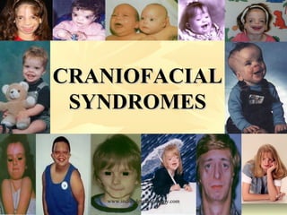
Craniofacial syndromes /certified fixed orthodontic courses by Indian dental academy
•
202 gefällt mir•34,757 views

The document discusses various craniofacial syndromes and anomalies. It begins by introducing craniofacial anomalies and their significance. It then defines key terms like syndrome, anomaly, sequence, and association. It describes the four types of congenital anomalies: malformation, disruption, deformation, and dysplasia. It also discusses classifications of craniofacial syndromes and some specific syndromes associated with midfacial deficiencies like Apert syndrome, Crouzon syndrome, and Pfeiffer syndrome. It provides details on the etiology, clinical features, dental anomalies, and treatment of these conditions.

Empfohlen
Empfohlen
Weitere ähnliche Inhalte
Was ist angesagt?
Was ist angesagt? (20)
Ähnlich wie Craniofacial syndromes /certified fixed orthodontic courses by Indian dental academy
Ähnlich wie Craniofacial syndromes /certified fixed orthodontic courses by Indian dental academy (20)
Mehr von Indian dental academy
Mehr von Indian dental academy (20)
Kürzlich hochgeladen
Kürzlich hochgeladen (20)
Craniofacial syndromes /certified fixed orthodontic courses by Indian dental academy
- 2. INDIAN DENTAL ACADEMY Leader in continuing dental education www.indiandentalacademy.com www.indiandentalacademy.com
- 3. INTRODUCTION It is small wonder that anomalies of the craniofacial complex are of major significance not only to dental health but also to mental health and social wellbeing. The anomalies of development of the craniofacial complex are extremely vast and varied. For most patients, it is difficult or impossible to describe a specific etiologic cause. In an adult, the etiologic factors that caused the problem to develop during growth may no longer have an effect even if they are still present. And yet, some thought must be given to etiology when treatment is planned because this is a major factor in estimatingwww.indiandentalacademy.com results, the stability of particularly in children but also in adults.
- 5. •SYNDROME: syn= together, dromos= a running The aggregate of signs & symptoms associated with any morbid process & constituting together the picture of the disease & related to each other anatomically, biochemically or physiologically. •An anomaly is a deviation from the average or norm, anything that is structurally unusual or irregular or contrary to a general rule. •A sequence is a pattern of multiple anomalies derived from a single known or presumed structural defect or mechanical factor. •An association is a nonrandom occurrence in two or more individuals of multiple anomalies not known to be a polytopic field defect, sequence, or syndrome •Dysmorphology is an area of clinical genetics that is concerned with the diagnosis and interpretation of patterns of structural defects. Recurrent patterns of birth defects are the hallmarks of syndrome recognition. www.indiandentalacademy.com
- 6. There are four clinically significant types of congenital anomaly Malformation Disruption Deformation and Dysplasia MALFORMATION :- A morphological defect of an organ, part of an organ, or larger region of the body that results from an intrinsically abnormal developmental process Intrinsic implies that the developmental potential of the primordium is abnormal from the beginning, such as a chromosomal abnormality of a gamete at fertilization. Most malformations are considered to be a defect of a morphogenetic or developmental field "which responds as a coordinated unit to embryonic interaction and results in complex or multiple malformations www.indiandentalacademy.com
- 7. DISRUPTION A morphological defect of an organ, part of an organ, or a larger region of the body that results from the extrinsic breakdown of, or an interference with, an originally normal developmental process. Thus, morphological alterations following exposure to teratogens —agents such as drugs and viruses — should be considered as disruptions. A disruption cannot be inherited, but "inherited factors can predispose to and influence the development of a disruption." www.indiandentalacademy.com
- 8. DEFORMATION An abnormal form, shape, or position of a part of the body that results from mechanical forces. Intrauterine compression that results from oligohydramnios — insufficient amount of amniotic fluid — produces a clubfoot an example of a deformation produced by extrinsic forces. Some central nervous system defects, such as meningomyelocele — a severe type of spina bifida produce intrinsic functional disturbances that also cause fetal deformation. www.indiandentalacademy.com
- 9. DYSPLASIA An abnormal organization of cells into tissue (s) and its morphological result (s). Dysplasia is the process and the consequence of dyshistogenesis (abnormal tissue formation). All abnormalities relating to histogenesis are therefore classified as dysplasias, e.g., congenital ectodermal dysplasia www.indiandentalacademy.com
- 10. TYPES OF PROBLEMS IN MORPHOGENESIS POOR TISSUE FORMATION UNUSUAL FORCES ON NORMAL TISSUE MALFORMATION DEFORMATION BREAKDOWN OF NORMAL TISSUE DISRUPTION www.indiandentalacademy.com ABNORMAL ORGNZN OF CELLS IN TISSUE DYSPLASIA
- 11. ANOMALAD: Term coined by Dr. F. Clarke Frase To denote a pattern of morphologic defects stemming from a single localized structural anomaly, which result in a cascade of consequent defects. Ending “ad” is meant to imply more than one defect (as in diad, triad) So anomalad connotes the initiating anomaly and its derived secondary defects. The cause for initiating structural defect may be heterogeneous, so anomalad does not connote a particular mode of etiology. As severity of initial morphologic anomaly may vary, so the range of severity & extent of anomalad may also be variable. Eq. Robin Anomalad www.indiandentalacademy.com
- 13. 1) Richard & Goodman classified multiple malformation disorders as : i) Syndrome a) Chromosomal abnormality b) Gene abnormality c) Environmental factor ii) Anomalad iii) Association iv) Combination of malformations not assigned to any of above categories. www.indiandentalacademy.com
- 14. 2) Embryonic or histologic classification: i) Single germ layer involved ii) All the three germ layers involved ex. Gardner syndrome 3) Monothetic syndrome classification: Syndromes that share single common feature are grouped together. ex. Syndromes with cleft palate Syndromes with polydactyly This aids in differential diagnosis 4) Polythetic classification: Syndromes sharing a large proportion of their principal anomalies are grouped together.They are even better aid in www.indiandentalacademy.com differential diagnosis.
- 15. 5) Syndrome prototype: By Herrmann & Opitz 1974 & Cohen 1982 i) Dysmetabolic syndrome (Metabolism) iii) Malformation syndrome (Organ or Feild) • • • • • • Normal at birth, Uniform clinical features, No congenital malformations, Biochemically defined, Recessive mode of inheritance eg. Hurler Syndrome Tay- Sachs Disease • Noncontiguous Malformation • Embryonic Pleiotropy • Lack of biochemical definition • eg. Trisomy 13 syndrome Rubenstein Tyabi Syndrome iv) Dyshistogenetic syndrome (Tissue) 1) Simple • Involvement of only 1 germ layer ii) Deformation syndrome (Region) • Inheritance- dominant or recessive • Changes in shape of previously normal • eg. Achondroplasia structures • Lack of movement- mechanical , 2) Hamartoneoplastic Sydrome • Hamartomas or neoplasias functional • • More than 1 germ layer Commonly affects musculoskeletal • Commonly dominant system • eg. Peutz Jeghers Syndrome www.indiandentalacademy.com Gardners Syndrome
- 16. Graphic illustration of the causes of human birth defects Unknown Etiology Multifactorial Inheritance Chromosomal Abnormalities Mutant Gene Environmental Agents www.indiandentalacademy.com
- 17. 1. Syndrome Associated With Midfacial Deficiency 2. Syndromes Associated With Midfacial Excess 3. Syndromes Associated With Mandibular Deficiency 4. Syndromes Associated With Mandibular Prognathism 5. Syndromes Associated With Problems Of Facial Height 6. Syndromes Associated With Facial Asymmetry 7. Syndromes Associated With Ocular Hypertelorism 8. Syndromes Associated With Chromosomal Disorders 9. Environmental Induced Syndromes 10.Other Common Syndromes www.indiandentalacademy.com
- 18. Syndrome Associated With Midfacial Deficiency •Achondroplasia •Apert Syndrome •Crouzon Syndrome •Pfeiffer Syndrome •Stickler Syndrome •Cleidocranial Dysplasia •Ectodermal Dysplasia •Marfan Syndrome •Treacher Collins Syndrome •Hemifacial Microsomia( Goldenhar Syndrome) •Gorlin Syndrome •Waardenberg Syndrome •Larsen Syndrome •Oral – Facial – Digital Syndrome www.indiandentalacademy.com •Frontonasal Dysplasia
- 19. ACHONDROPLASIA The term Achondroplasia was first used by Parrot in 1978 to describe short limbed dwarfism associated with enlarged head. AETIOLOGY:•Autosomal dominant, about 90 percent of the cases represent a fresh gene mutation. •Older paternal age has been a contributing factor in these cases. • It is a disturbance of Endochondral bone formation. •The most common chondrodysplasia, true achondroplasia, occurs with a frequency of about 1 in 15,000. CLINICAL FINDINGS:•Small stature. Mean adult height in males is 131 ± 5.6 cm and in females is 124± 5.9 cm. •Megalocephaly •Small foramen magnum •Short cranial base with early spheno occipital closure. •Low nasal bridge with prominent forehead. www.indiandentalacademy.com •Mild midfacial hypoplasia with narrow nasal passages
- 20. •Short Limbed dwarfism •Waddling gait with exaggerated lordotic lumbar spine • Prominent Buttocks • Protuberant abdomen •Short trident hand, fingers being similar in length, with short proximal and mid phalanges. •Short femoral neck; incomplete extension of elbow. •The metacarpals and phalanges are shortened www.indiandentalacademy.com
- 21. LeFort III osteotomy TREATMENT •Correction of class III malocclusion and midface deficiency in achondroplasia ideally is accomplished by a LeFort III osteotomy to move the entire midface forward. •Unfortunately, no analogous surgical procedure exists to deal with the limb deficiencies although progress has been made recently toward lengthening the short limbs www.indiandentalacademy.com Pre- treatment Post- treatment
- 22. CRANIOSYNOSTOSIS SYNDROMES These constitute a group of conditions each characterized by premature fusion of the cranial sutures resulting in disproportionate growth of the cranial bones and as a sequence the growth of the facial bones. TYPES OF CRANIOSYNOSTOSIS •Scaphocephaly (Sagital Suture) •Trigonocephaly (Metopic suture) •Turricephaly (Bilateral Coronal •Suture) •Plagiocephaly •Frontal Plagiocephaly ( Unilateral Coronal Suture) •Occipital Plagiocephaly (Unilateral Lambdoid Suture) www.indiandentalacademy.com •Parallelism ( Frontal Bone/Contralateral Occipital Lobe)
- 23. APERT SYNDROME •The condition was reported by Wheaton in 1894. • In 1906, Apert summarized nine cases, and in 1920, Park and Powers published an exceptional essay on this entity. Numerous cases have been reported. AETIOLOGY:•Autosomal dominant •Most cases sporadic •Estimated in 1 in 160,000 live births •Apert FGFR2 mutation lead to an increase in the number of precursor cells that enter the osteogenic pathway,leading ultimately to increased subperiosteal bone matrix formation and premature calvaria ossification during fetal development www.indiandentalacademy.com
- 24. Clinical Features Craniofacial. Short anteroposterior diameter with high, full forehead and flat occiput. Irregular craniosynostosis, especially of coronal suture. Fontanels may be large and late in closure. www.indiandentalacademy.com
- 25. Flat facies, Supraorbital horizontal groove, Shallow orbits, Hypertelorism, Strabismus, Down-slanting of palpebral fissures, Small nose, . Lips frequently assume a trapezoidal cofiguration in the relaxed state. www.indiandentalacademy.com
- 26. Maxillary hypoplasia The maxillary dental arch may be V-shaped with severely crowded teeth and bulging alveolar ridges.. Narrow palate with median groove, with or without cleft palate or bifid uvula. www.indiandentalacademy.com
- 27. DENTAL ANOMALIES Delayed and/or ectopic eruption and shaped incisors. . Supernumerary teeth have been observed. Class III malocclusion is usually present, with anterior open bite or cross bite and unilateral or bilateral posterior crossbite. www.indiandentalacademy.com shovel-
- 28. OTHER CLINICAL FINDINGS Syndactyly in varying degrees generally affecting second to fourth fingers and usually all toes. Considerable range of mental deficiency noted although normal intelligence occasionally found. Numerous skeletal abnormalities of elbow, shoulder, lip, and cervical spine. Various cardiac and gastrointestinal malformations are noted. Acne vulgaris about forearms common Hearing loss. www.indiandentalacademy.com
- 29. TREATMENT Goals of surgery -Increase intracranial space in the anterior cranial vault for the brain. -To increase the orbital volume. -To improve the morphology of the forehead and upper orbits. Thus it requires -Bicoronal suture release with decompression -simultaneous cranial vault and upper orbital Osteotomies with reshaping and advancement. Usually done at 9 to 11 months PRE of age unless signs of increased TREATMENT intracranial pressure are identified earlier in life. www.indiandentalacademy.com POST
- 30. CROUZON DISEASE In 1912 Crouzon first described a woman and her son with this disorder. In 1915 he reported a family in which seven of twenty one members affected and thus stressed the genetic aspects of the syndrome ETIOLOGY Autosomal dominant Wide range of expressivity About one third cases arise as a new mutation CLINICAL FEATURES •Premature closure, especially of coronal suture, occasionally lambdoidal. •Variable cranial form depending on order and rate of progression of suture closure • Optic nerve damage www.indiandentalacademy.com
- 31. FACIAL FEATURE Exophthalmos Divergent strabismus Hypertelorism Short upper lip Hypoplasia of maxilla with relative mandibular prognathism Nose may appear beaked because of midfacial hypoplasia High arched, short palate V-shaped maxillary dental arch. Crowding of the upper teeth. Class III Malocclusion Bilateral Atresia of Auditory meatus noted occasionally www.indiandentalacademy.com
- 32. PFEIFFER SYNDROME • Described by Pfeiffer in 1963 in eight members of one family •Autosomal dominant inheritance pattern. ETIOLOGY: • It is heterogeneous because it is caused by a single recurring mutation(Pro 252 Arg) of the FGFR1 gene and by several different mutations affecting FGFR2. •Type I •Type II •Type III www.indiandentalacademy.com
- 33. PFEIFFER SYNDROME ·Generally compatible with life • Tower Skull · Normal or near normal intelligence · Bilateral coronal synostosis · Midface growth often normal. · Minimal Ocular proptosis.. · Hydrocephalus is absent. · Supraorbital ridge recessed. · Anterior cranial base short and wide • Orbits are shallow,eyes are proptopic with a degree of hypertelorism •Syndactyly • Great toe deviated medially www.indiandentalacademy.com
- 34. TREATMENT • Primary cranio-orbital decompression and reshaping in infancy at 9-11 months of age. •Management of total midface deformity. •A frontofacial(monobloc) osteotomy with horizontal advancement and need for additional segmentation of the orbits to normalize the dysmorphology is generally required. •Le Fort III osteotomy. •Age of treatment:5 to 7 years of age. •Management of orthognathic deformity in adolescents. •Mandible has a normal basic growth pattern, as the growth of maxilla may be limited, Angles class III malocclusion with anterior open bite often results •A Le Fort I Osteotomy with horizontal advancement Segmentation in combination with an oblique osteotomy of the www.indiandentalacademy.com chin(genioplasty) to correct lower face deformity.
- 35. STICKLER SYNDROME (1965) •Autosomal dominant, high degree of penetrance •Flattening of midface and nasal bridge •Facial Asymmetry •Micrognathia •Submucous palatal cleft •Epicanthal folds •Myopia •Hearing Loss •Bony enlargement of joints •Joint hypermobility •Hip joint Deformity www.indiandentalacademy.com •Occasional Mental Retardation
- 36. CLEIDOCRANIAL DYSPLASIA Syn. : Marie Sainton Disease, Cleidocranial dysostosis In 1765 Martin first reported the clavicular findings In 1897 Marie and Sainton noted the genetic transmission and coined the name cleidocranial dysostosis In 1975 Goodman and co-workers described an autosomal recessive form of the disease Autosomal dominant with wide variability in expression, but usually showing penetrance. Autosomal Recessive disease is usually more severe www.indiandentalacademy.com
- 37. CLINICAL FEATURES: Growth. Slight to moderate shortness of stature. Craniofacial. Brachycephaly with bossing of frontal, parietal, and occipital bones; Late closure of fontanels and mineralization of sutures; Late or incomplete development of accessory sinuses and mastoid air cells; Wormian bones Small sphenoid bones. Calvarial thickening. Midfacial hypoplasia with low nasal bridge, narrow high-arched palate. Hypertelorism. www.indiandentalacademy.com
- 38. DENTITION Late eruption, Malformed roots, Retention cysts, Enamel Hypoplasia, Caries, Supernumerary teeth. CLAVICLE AND CHEST •Partial to complete aplasia of clavicle with associated muscle defects, •small thorax with short www.indiandentalacademy.com oblique ribs.
- 39. MARFAN SYNDROME A hereditary disease transmitted as an autosomal dominant trait. It is basically a disease of connective tissue related to a defective organization of collagen. Here collagen is abnormally soluble. An outstanding feature is excessive length of tubular bones resulting in : Dolichostenomelia disproportionately long thin extremities Arachnodactyly spidery fingers. Shape of face and skull is long and narrow Hyperextensibility of joints, habitual dislocations, Kyphosis or scoliosis, flat foot. www.indiandentalacademy.com
- 40. Myopia is usually present Cardiovascular complications – aortic aneurysm and aortic regurgitation, valvular defects, enlargement of the heart. High arched palatal vault is very prevalent and may be a constant finding. Odontogenic cysts of maxilla and mandible TMJ dysarthrosis www.indiandentalacademy.com
- 41. TREACHER COLLINS SYNDROME • • • • • • Autosomal dominant condition with variable expressivity. Bilateraly symmetrical abnormalities of structures within the first and second branchial arches. Incidence:1 in 25,000 to 1 in 50,000 live births. Pathogenesis:- Theories include failure of differenciation of the branchial arch mesoderm, defective facial bone ossification, and tissue ischemia resulting from stapedial artery hypoplasia. Behrents, Mcnamara, and Avery pointed out that all major aspects of MFD are fully expressed by the 15th week of embryonic development. Current research suggest that the abnormality may occur early as developmental defects of the neural crest cells www.indiandentalacademy.com
- 42. CLINICAL FEATURES CRANIUM •Flat parieto-occipital Bone • • • • • • • • • • • • • MIDFACE Antimongoloid palpebral Fissures Colobomas of lower eyelid Eyelash(or Follicle) Malformations Malar defects Atresiaof lacrimal puncta Macrostomia Auricular defects External auditory canal defects Preauricular hair displacement High arched palate Nasal deformity Preauricular sinuses www.indiandentalacademy.com Malocclusion
- 43. · MANDIBLE Mandibular defects • Micrognathia • Open bite • OTHER FEATURES Fish like facial Appearance • Deafness frequent • Mental retardation Infrequent • www.indiandentalacademy.com
- 44. TREATMENT • • • • • • Zygomatic and orbital reconstruction. Maxillomandibular reconstruction. Nasal reconstruction. Soft tissue reconstruction External ear reconstruction. External auditory canal and middle ear reconstruction www.indiandentalacademy.com
- 45. HEMIFACIAL MICROSOMIA Syn.:- Goldenhar syndrome, Occulo Vertebral Dysplasia The term Hemifacial microsomia was issued by Gorlin to describe patients with unilateral microtia , macrostomia and failure of formation of the mandibular ramus and condyle. •Gorlin suggested that the Goldenhar syndrome was a variant of this complex characterized in addition by vertebral anomalies and epibular dermoids AETIOLOGY :•The mode of inheritance is not established with rare familial aggregation •Multifactorial, Drugs- Thalidomide, Triazene •Poswillo, using animal model of hemifacial microsomia, was able to show that destruction of differentiating tissues in the region of the ear and jaw by an expanding hematoma produced a branchial arch dysplasia. •Thus a vascular abnormality can disrupt the normal embryologic www.indiandentalacademy.com development of first and second branchial arches
- 46. CLINICAL FEATURES •Prominent fore head •Midfacial Asymmetry •Varying malformation of the mandible •Flattened gonial angle and narrowed maxilla on the involved side •Maxillary, temporal and malar bones on the involved side are reduced in size •Muscles of palate, mastication, tongue, face may be hypoplastic or paralyzed •Malformation of the external ear may vary from displaced pinna to complete aplasia •Macrostomia of mild degree and crowding of teeth •Palpebral fissures slant downwards www.indiandentalacademy.com www.indiandentalacademy.com
- 47. OTHER CLINICAL FEATURES •Mild mental retardation occasionally •Bony anomalies, especially of the vertebral column •Congenital heart diseases like VSD and hypoplasia of the lung TREATMENT •Mandibular osteotomies to reposition the mandible. -Unilateral mandibular osteotomy. -Bilateral mandibular osteotomy. -Le fort I osteotomy to correct the maxilla. •Functional orthopedic appliance therapy. www.indiandentalacademy.com •Orthodontic therapy after combined surgical correction
- 48. FRONTONASAL DYSPLASIA Syn.:- Median Cleft Face Syndrome AETIOLOGY :•The basic defect is unknown but thought to result from failure of flow of ectomesenchyme allowing brain vesicle to fill space normally occupied by the nasal capsule CLINICAL FEATURES:•Facial alteration graded from mild to severe •Anterior encephalocoele in some •Occular hypertelorism •Microopthalmia •Nose often flattened with widely spaced nostrils •Cleft lip •Mental Retardation www.indiandentalacademy.com
- 49. Syndromes Associated With Mandibular Deficiency •Robin Anomalad •Nager Acrofacial Dysostosis •Wilder Vanek – Smith Syndrome •Moebius Syndrome •Hallermann- Streiff Syndrome •Treacher Collins Syndrome www.indiandentalacademy.com
- 50. PEIRRE ROBIN ANOMALAD In 1923 Robin reported this condition, In 1975 the term anomalad was joined AETIOLOGY •May be isolated or in association with other syndromes eg. Stickler syndrome, Fetal Alcohol Syndrome, drug induced syndromes. •The primary defect lies in arrested development and ensuing hypoplasia of the mandible, ultimately producing the characteristic “bird facies”. •This in turn prevents the normal descent of the tongue between the palatal shelves, resulting in cleft palate. Because of this mechanism a cleft lip is not found in conjunction with the cleft palate. www.indiandentalacademy.com •Incidence is 1 in 39,000 live births
- 51. CLINICAL FEATURES •The primary components are cleft palate micrognathia and glossoptosis. •Occasional hydrocephaly or microcephaly •U shaped cleft palate • Mandibular hypoplasia •Eye anomalies such as congenital cataract •Deafness •Mental retardation •Sternal Anomalies •Congenital heart defects •Jaw malformation may result in respiratory difficulty as the tongue may www.indiandentalacademy.com fall back and obstruct the epiglottis.
- 52. NAGER ACROFACIAL DYSOSTOSIS •Possibly autosomal Recessive inheritance CLINICAL FEATURES •Malar Hypoplasia •Down slanting palpebral fissures •Absent Eyelashes •Cup shaped low set ears •Cleft palate •Micrognathia •Missing or hypoplastic thumbs •Syndactyly www.indiandentalacademy.com •Abnormal Radius or Ulna
- 53. MOEBIUS SYNDROME Syn.:- Congenital Facial Diplegia •Basic defect is unknown but nuclear hypoplasia accounting for cranial nerve paralysis has been documented CLINICAL FEATURES •Both 6th and 7th nerve palsy. •Bilateral facial paralysis •Convergent Strabismus •Protruding malformed ears •Micrognathia, high broad nasal bridge •Atrophy of tongue due to lack of hypoglossal innervation •Drooping angles of the mouth •Mental retardation •Limb reduction defects www.indiandentalacademy.com
- 54. Syndromes Associated With Mandibular Prognathism •Klienfelter Syndrome •Osteogenesis Imperfecta •Gorlin Syndrome •Marfan Syndrome •Waardenberg Syndrome www.indiandentalacademy.com
- 55. KLINEFELTER SYNDROME • Due to non disjunction of the X chromosome during meiosis • Commonly affects males and the have an extra X chromosome XXY, XXXY CLINICAL FEATURES • Skeletal disproportion • Mandibular prognathism • Hypertelorism, Strabismus • Upward slant of the Palpebral fissure • Depressed Nasal bridge • Cleft Palate • Taurodontism • Mental Deficiency www.indiandentalacademy.com
- 56. OSTEOGENESIS IMPERFECTA Syn.:- Brittle bones disease •Van der Hoeve in 1918 •Disturbance of mesodermal tissues especially calcified tissues CLINICAL FEATURES •Extreme fragility and porosity of bones with increased proneness to fracture •Blue Sclera, D/D Ehler Danlos syndrome, Marfan syndrome •Dentinogenesis Imperfecta •Triangular facies with craniofacial disproportion •Ears displaced/ Deafness •Mandibular prognathism •Short stature due to repeated fractures •Loose ligaments- dislocation of the TMJ www.indiandentalacademy.com
- 57. GORLIN AND GOLTZ SYNDROME (Jaw cyst basal cell nevus bifid rib syndrome) 1st described by Binkley and Johnson in 1951, thoroughly reviewed by Gorlin et al. It is transmitted as an autosomal dominant trait CLINICAL FEATURES •Cutaneous anomalies : Basal cell carcinoma, other benign dermal cysts and tumors. •Palmar pitting, palmar and plantar keratosis and dermal Basal Cell Carcinoma calcinosis. • Dental and osseous lesions : Odontogenic keratocyst (often multiple), mandibular prognathism. •Bifid ribs, vertebral anomalies, www.indiandentalacademy.com
- 58. Eyes Hypertelorism with wide nasal bridge,congenital blindness Neurologic anomalies Mental retardation, dural calcification, agenesis of corpus callosum, congenital hydrocephalus Sexual abnormality Hypogonadism in males, ovarian tumors Oral findings odontogenic keratocysts As they often develop early in life, deformity and displacement of developing teeth may occur. TREATMENT •Several cases of ameloblastoma have developed in cysts of this syndrome •This emphasizing the importance of surgery of normal cysts and and their histologic examination www.indiandentalacademy.com
- 59. Syndromes Associated With Problems Of Facial Height •Amelogenesis Imperfecta •Beckwith – Weidmann Syndrome www.indiandentalacademy.com
- 60. BECKWITH WIEDMANN SYNDROME •Uncertain mode of inheritance although familial occurrence has been observed CLINICAL FEATURES •Macroglossia •Anterior open bite •Mandibular prognathism •Earlobe grooves •Depression on posterior rim of ear •Exopthalmos •Infra orbital hypoplasia •Midfacial recession •Mild mental retardation •Neonatal hypoglycemia www.indiandentalacademy.com
- 61. Syndromes Associated With Facial Asymmetry •Hemi Hypertrophy •Facial Hemiatrophy •Neurofibromatosis •Hemifacial MicrosomIa www.indiandentalacademy.com
- 62. FACIAL HEMIHYPERTROPHY AETIOLOGY •Condition has been ascribed to hormonal imbalance, chromosomal abnormalities, Localized alteration on intrauterine development, lymphatic abnormalities vascular abnormalities CLINICAL FEATURES •Enlargement of one half of the head •Dentition on the affected side of abnormal shape and size •Both bone and soft tissue is involved •Tongue involvement is common •Mandibular hemihypertrophy •May be associated with Wilm’s tumor of the kidney and adrenal cortical carcinoma www.indiandentalacademy.com
- 63. FACIAL HEMEATROPHY Syn.:- Parry Romberg Syndrome AETIOLOGY • Exact cause is unknown but suggested factors are 1. Trophic malformation of the cervical sympathetic system 2. Trauma 3. Infection 4. Heredity 5. Peripheral Trigeminal Neuritis 6. Form of localized Scleroderma • Onset is noticed in the 1st or 2nd decade of life as a white line, furrow on one side of the face or brow near the midline • This initial lesion extends to include atrophy of the skin, subcutaneous tissue muscle and bone depending on the severity www.indiandentalacademy.com of the atrophy
- 64. CLINICAL FEATURES •Unilateral facial involvement •Trigeminal neuralgia or parasthesia •Vitiligo •Atrophy of half of tongue •Delay in dental eruption on the affected side •Eye involvement •Migraine headaches or epilepsy •Affected skin becomes darkly pigmented •Loss of facial hair •Predilection for involvement of left side www.indiandentalacademy.com of face
- 65. NEUROFIBROMATOSIS Syn.:- Von Recklinghausen’s disease •Simple autosomal dominant inheritance with variable penetrance •Incidence of 1 in 3000 live births •Two types- superficial nodules, deeper diffuse lesions CLINICAL FEATURES •Café- au-lait spots •Neurofibromas of tongue skin •Skeletal defectswhen tumor is located within bone •Endocrinedisturbances •Macrocephaly •Mandibular asymmetry •Malignant transformation of lesions in 15% www.indiandentalacademy.com •Associated congenital defect mental retardation and ocular
- 66. Syndromes Associated With Ocular Hypertelorism •Apert Syndrome •Basal Cell Nevus Syndrome •Crouzon Syndrome •Frontonasal dysplasia •Fetal Face Syndrome •Hypertelorism- Hypospadias Syndrome •Noonan Syndrome •Oro facial Digital Syndrome www.indiandentalacademy.com
- 67. FETAL FACE SYNDROME •Syn.:- Robinow Syndrome •Both autosomal dominant and recessive inheritance CLINICAL FEATURES •Facial resemblance to a fetus of 8 weeks •Frontal bossing •Hypertelorism •Short upturned nose •Macrocephaly •Triangular mouth with downturned corners •Dental malalignment •Vertebral anomalies www.indiandentalacademy.com
- 68. NOONAN SYNDROME CLINICAL FEATURES •Broad forehead •Coarse hair •Flat nasal bridge •Occular hypertelorism •Small receding chin •Sad facial expression •High arched palate •Dental anomaliess with malocclusioncleft uvula •Mental retardation •Webbing of neck •Congenital heart disease www.indiandentalacademy.com
- 69. ORO FACIAL DIGITAL SYNDROME In 1954 Papillon described a syndrome characterized by congenital anomalies of the face, oral cavity, and digits X-linked dominant Possibly 1 in 250,000 CLINICAL FEATURES Frontal bossing Alopecia or dryness of scalp Pseudocleft of mid-upper lip Cleft or defect of hard palate Malposition of teeth Supernumerary teeth Hypoplasia of malar bones Broad nasal root www.indiandentalacademy.com
- 70. Cleft tongue Ankyloglossia Absence of lower lateral incisors Thick fibrous bands (frenula) in upper and lower mucobuccal fold mental retardation Abnormalities of hair and sebaceous glands Digital anomalies include syndactyly, clinodactyly, www.indiandentalacademy.com
- 71. Syndromes Associated With Chromosomal Disorders Down Syndrome •Trisomy 18 •Trisomy 13 •Turner Syndrome • www.indiandentalacademy.com
- 72. DOWN SYNDROME In 1866 Langdon Down first adequately described this syndrome. This disease is the most common chromosomal abnormality to occur in man. 3 forms of down syndrome: a) Typical trisomy 21 with 47 chromosomes (95% of cases) b) Translocation type- Here the extra chromosome material of number 21 is translocated to another chromosomed of G or D group (3%) c) Chromosomal mosaicism (about 2% of cases) The risk of having an affected child of the typical trisomy 21 type is one is 2000 live births in women under 30 years of age Rises dramatically to one is 50 live births is women over 45 years of age. Overall incidence is 1 in 600 live births www.indiandentalacademy.com
- 73. CLINICAL FEATURES Brachycephalic skull Open metopic suture, late closure of fontanels Hypoplastic maxilla and nasal bones Flattening of nasal bridge, orbital ridges, and maxilla Ocular hypotelorism Short hard palate Cleft lip/palate about 3 times frequency in normal population Protruding fissured tongue with hypertrophy of papillae Dentition delayed, increased periodontal disease, and reduced dental caries www.indiandentalacademy.com
- 74. Oblique palpebral fissures Epicanthal folds Spotting of iris Cataracts Strabismus Small ear Excessive skin on nape Reduced Height Delay in skeletal maturation Short Broad Neck Hypermobility of Joints. Cardiac abnormalities IQ range from 25 to 70 and decreases with age. Simian crease and clinodactyly www.indiandentalacademy.com
- 75. TRISOMY 18 – EDWARDS SYNDROME •Triplication of chromosome 18, most common cause is non disjunction . CLINICAL FEATURES •Prominent occiput •Micrognathia •Small oral opening •Short upper lip •Cleft lip and palate • narrow palatal arch •Short palpebral fissures •Corneal opacities •Microopthalmos •Low set malformed ears •Short neck www.indiandentalacademy.com
- 76. TRISOMY 13 – PATAU SYNDROME •Trisomy 13 stated account for 15 % of the spontaneous abortions and there is a substantial tendency for recurrence CLINICAL FEATURES •Microcephaly •Port wine stain in glabellar area •Scalp defects in parietoccipital area •Absent eye brows •Shallow supraorbital ridges •Micropthalmos with multiple eye defects •Deafness •Cleft lip and palate •Micrognathia •Congenital abnormalities involving heart, kidney, brain and GIT www.indiandentalacademy.com
- 77. TURNER SYNDROME About 1% of monosomy X female embryos survive. The incidence of 45, X or Turner syndrome in newborn females is approximately 1 in 8000 live births. It accounts for about 18% of all abortions caused by chromosome abnormalities. Half the affected individuals have 45, X; the other half have a variety of abnormalities of a sex chromosome The phenotype of Turner syndrome is female. Secondary sexual characteristics do not develop in 90% of affected girls and hormonal www.indiandentalacademy.com replacement
- 78. CLINICAL FEATURES 1. Facies -shows premature aging 2. Face frequently heart shaped 3. Multiple eye findings including cataract, strabismus, blue sclera. 4. Color Blindness 5. Depressed Corners of mouth 6. High arched palate 7. Dental malocclusion 8. Micrognathia 9. Prominent ears www.indiandentalacademy.com
- 79. OTHER CLINICAL FINDINGS Short stature, average adult height 140 cm Webbed neck Shield-like chest Undeveloped breasts www.indiandentalacademy.com
- 80. Low hairline on posterior neck Infant often presents with lymphedema of hands and feet Short fourth metacarpal bone in some patients Hypoplastic toenails www.indiandentalacademy.com
- 81. Environmental Induced Syndromes An environmental agent which is demonstrated to have a statistically significant, nonrandom association with a particular congenital malformation or set of malformation is termed as environmental teratogenic agent or teratogen. •Potter Syndrome •Fetal Alcohol Syndrome •Fetal Hydantoin Syndroem www.indiandentalacademy.com
- 82. POTTER SYNDROME AETIOLOGY :- Oligohydraminos- reduced amniotic fluid CLINICAL FEATURES •Occular hypertelorism •Prominent skin fold from the inner canthus to the cheek •Flattened nose with downward turned nasal tip •Large flattened ears •Micrognathia •Mandibular asymmetry •Congenital sternomastoid torticollis •Clefts of lip and palate •Micropthalmia www.indiandentalacademy.com
- 83. FETAL ALCOHOL SYNDROME Infants born to chronic alcoholic mothers exhibit a specific pattern of defects, including prenatal and postnatal growth deficiency, mental retardation, and other anomalies. Exposure to high level of ethanol at an early stage of fetal development produces fetal alcohol syndrome [FAS] www.indiandentalacademy.com
- 84. Deficiencies of midline tissue of the neural plate very early in embryonic development give rise to a sires of malformation collectively known as the holoprosencephalies which are characterized by a failure of the first three ventricles of the brain to separate The olfactory placodes which are partly derived from the anterior neural plates, are too close together, and this causes deficient development of the median and nasal prominences. The result is a spectrum of facial deformities ranging from total absence of the nose and related structures to an intact but moderately www.indiandentalacademy.com underdeveloped midface.
- 85. FETAL HYDANT01N SYNDROME The fetal hydantoin syndrome occurs in 5 to 10% of children born to mothers treated with phenytoins or hydantoin anticonvulsants. GENERAL CLINICAL FEATURES Short neck, Rib anomalies, Umbilical and inguinal hernias, Coarse profuse scalp hair, hirsutism, Low-set hairline, Abnormal palmar crease. Strabismus. www.indiandentalacademy.com
- 86. CLINICAL FEATURES Microcephaly Wide anterior fontanel Metopic ridging Ocular hypertelorism Broad, depressed nasal bridge Short nose with bowed upper lip Broad alveolar ridge Cleft lip and palate www.indiandentalacademy.com
- 87. Limbs Hypoplasia of distal phalanges with small nails, especially postaxial digits; Low arch dermal ridge patterning of hypoplastic fingertips; Digitalized thumb; Dislocation of hip. www.indiandentalacademy.com
- 88. Other Common Syndromes • Binder Syndrome • Ehlers Danlos Syndrome www.indiandentalacademy.com
- 89. BINDER SYNDROME Syn. :- Maxillonasal Dysostosis •Affects primarily the anterior part of the maxilla and nasal complex •Initially described by Noyes in 1939, but clinically defined as a syndrome by Binder in 1962 CLINICAL FEATURES •Failure of development in the premaxillary area, with associated deformities of the nasal skeleton •Rudimentary nasal spine •Mandibular prognathism •Increased gonial angle •Typical Class III occlusion with marked proclination of the upper incisors •Poorly developed philtrum •Half moon appearance of external nares inferiorly www.indiandentalacademy.com
- 90. EHLERS – DANLOS SYNDROME (CUTIS HYPERELASTICA) Originally described by Van Meekeren in 1682 Further classified by Ehler in 1901 & Danlos in 1908 Ten distinct forms of syndrome have now been delineated EDS I, EDS II, EDS III are more common. Inherited as autosomal dominant EDS V has X-linked inheritance. It is a hereditary disorder of connective tissue with abnormal collagen www.indiandentalacademy.com
- 91. •CLINICAL FEATURES •Hyperelasticity of skin, hyperextensibility of joints and fragility of skin and blood vessels resulting in excessive bruising as well as defective healing of skin wounds. Auricles are hyper mobile with tendency towards “lop ears”. Mitral valve prolapse with or without tricuspid valve prolapse. Oral mucosa is of normal color but excessively fragile and bruised easily. Healing of mucosa is slightly retarded and no remarkable hyper extensibility of mucosa can be demonstrated. Gingival bleeding after brushing Hyper mobility of TMJ, repeated dislocations www.indiandentalacademy.com
- 92. Accurate diagnosis of a specific syndromes among the 0.7% of babies born with multiple malformations is a necessary pre requisite of providing a prognosis and plan of management for the affected infant, as well as genetic counseling for the parents. Until recent years, treatment of dentofacial deformity concentrated on the dentition, orthodontic treatment was the accepted approach to these problems. However the situation has changed with explosive rapidity and new treatment options involving comprehensive intervention have enhanced the management of craniofacial syndromes. When dealing with craniofacial syndromes and the individuals affected by them it is worthwhile to keep in mind the words of James Paget “We ought not to set them aside with idle thoughts or idle words about "curiosities" or "chances." Not one of them is www.indiandentalacademy.com without meaning; there is not one that might not become the
- 93. REFERENCES • Oral & maxillofacial pathology, 2nd edi, Neville. • Text book of oral pathology, 4th edi, Shafer. • Atlas of “The face in genetic disorder” Richard & Goodman 2nd edi. • Syndromology : an updated conceptual overview Int. J. Oral Maxillofacial Surg. 1989 • Surgical Correction of Dentofacial Deformities, Bell Proffit and White www.indiandentalacademy.com • J.W. Fergnson and RPJ Thomson EJO 7:145