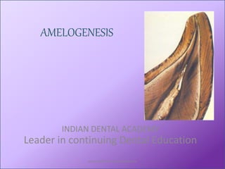
Amelogenisis / dental implant courses by Indian dental academy
•
37 gefällt mir•3,632 views

The Indian Dental Academy is the Leader in continuing dental education , training dentists in all aspects of dentistry and offering a wide range of dental certified courses in different formats.for more details please visit www.indiandentalacademy.com

Empfohlen
Weitere ähnliche Inhalte
Was ist angesagt?
Was ist angesagt? (20)
Andere mochten auch
Andere mochten auch (6)
Ähnlich wie Amelogenisis / dental implant courses by Indian dental academy
Ähnlich wie Amelogenisis / dental implant courses by Indian dental academy (20)
Mehr von Indian dental academy
Mehr von Indian dental academy (20)
Kürzlich hochgeladen
Kürzlich hochgeladen (20)
Amelogenisis / dental implant courses by Indian dental academy
- 1. AMELOGENESIS INDIAN DENTAL ACADEMY Leader in continuing Dental Education www.indiandentalacademy.com
- 2. • Tencate’s oral histology and embryology-8th edition • Oral histology-Berkovitz,3rd edition • Orban’s oral histology,13th edition • Oral development & histology ,James K. Avery 2nd edition. • Dental enamel, John wiley www.indiandentalacademy.com
- 3. Learning objectives • At the end of the seminar learner should be able to understand : – Various stages of amelogenesis – Morphology of ameloblast in various stages of amelogenesis – Modification of the matrix and mineralization – Clinical considerations www.indiandentalacademy.com
- 4. • Enamel is an ectodermally originated tissue covering the anatomical crown of a tooth. • It is formed by the enamel organ . • Enamel organ is derived from a localized proliferation of the oral epithelium. www.indiandentalacademy.com
- 5. EPITHELIAL ENAMEL ORGAN At the stage preceding the formation of hard structures, the enamel organ, originating from the stratified epithelium of the primitive oral cavity, consist of the four distinct layers – • Outer enamel epithelium • Stellate reticulum • Stratum intermedium • Inner enamel epithelium (ameloblastic layer) www.indiandentalacademy.com
- 6. • Before enamel formation begins cells of IEE assume a columnar shape and differentiate into ameloblast that produces organic matrix. www.indiandentalacademy.com
- 7. • Ameloblasts are 4-5 μ in diameter and 40μ high, attached to one other by junctional complexes. • Amelogenesis i.e.formation of enamel by ameloblast is divided into 3 functional stages. www.indiandentalacademy.com
- 8. Functional stages in the life cycle of ameloblast Presecretory stage Morphogenetic phase Differentiation phase Secretory stage Maturation stage Transitional phase Maturation proper www.indiandentalacademy.com
- 10. MORPHOGENETIC STAGE:- • Before the ameloblasts are fully differentiated and produce enamel, they interact with the adjacent mesenchymal cells, determining the shape of the dentinoenamel junction and the crown. www.indiandentalacademy.com
- 11. • The cells are cuboidal or low columnar, with large oval nuclei that almost fill the cell body. • The Golgi apparatus and the centrioles- proximal end of the cells . • Distal end (dentinal ) • Proximal /Basal end (Str. Intermedium) • Mitochondria are evenly dispersed throughout the cytoplasm. www.indiandentalacademy.com
- 13. Differentiation stage: • As cell of IEE differentiate into ameloblasts they elongate and their nuclei shift proximally toward the stratum intermedium. • Golgi complex increase in volume and migrates distally. • Mitochondria cluster in proximal region www.indiandentalacademy.com
- 14. • Specialized attachment which encircle distal and proximal extremities of cells. • Fine actin filament from these complexes radiates into cytoplasm of cells and can be distinguish as terminal webs. • Their function is to hold ameloblast together and regulate the substance to enter and leave the enamel. www.indiandentalacademy.com
- 16. Secretory stage: • At this stage ameloblast reflects their intense synthetic and secretory activity. • Golgi complex and RER is present extensively. • Protein synthesized is processed in golgi complex and packed into secretory granules • These granules migrate into Tomes’ process. • They are released against newly formed mantle dentin . www.indiandentalacademy.com
- 17. • When enamel formation begins Tomes’ process comprises only of proximal portion. www.indiandentalacademy.com
- 18. • Ameloblast moves away from the dentin as enamel is formed and develops distal portion of Tomes’ process . www.indiandentalacademy.com
- 19. Secretion of enamel protein confine to 2 sites Proximal part of Tomes’ process forms interrod enamel Distal part of Tomes’ process forms enamel rod www.indiandentalacademy.com
- 22. • The interrod and rod growth sites are associated with membrane infoldings on proximal and distal portion of Tomes’ process . • These infoldings represent the site where secretory granules release enamel proteins extracellularly. • It leads to increase in length of crystals and thickness of enamel layer. www.indiandentalacademy.com
- 23. • The distal portion of Tomes’ process lengthen as enamel thickens & disappear . • Creating a narrow space between rod and interrod forming rod sheath. www.indiandentalacademy.com
- 24. • The enamel layer is composed of rod containing layer sandwiched between rodless initial and final enamel. • It is because initial and final enamel is formed by proximal portion of Tomes’ process and rods are in relation to distal portion of Tomes’ process.www.indiandentalacademy.com
- 27. • Enamel maturation (full mineralization) occurs after most of the thickness of enamel matrix has been formed in the occlusal or incised area. • In the cervical parts of the crown, enamel matrix formation is still progressing at this time. • During enamel maturation the ameloblasts are slightly reduced in length and are closely attached to enamel matrix. MATURATION STAGE:- www.indiandentalacademy.com
- 28. Transition stage • After immature enamel is formed ameloblasts undergo significant morphologic changes in preparation for their next role of maturing enamel. The changes seen during transitional stage are : •Reduction in height of ameloblasts •Cessation of enamel secretion www.indiandentalacademy.com
- 29. •50% ameloblasts undergo apoptosis •Organelles involved in protein synthesis undergo autophagocytosis •Development of ruffle border in the apical cytoplasm www.indiandentalacademy.com
- 30. Maturation proper • In this phase there is bulk removal of water and organic substance from enamel , to allow introduction of inorganic material. www.indiandentalacademy.com
- 31. Modulation • Modulation is very dramatic phenomenon shown by ameloblasts. • It is cyclic creation, loss & recurrence of highly invaginated ruffle ended apical surface and alternate with smooth bordered cells. www.indiandentalacademy.com
- 32. • It waves across the crown from least matured region to most matured region i.e. from the cervical region to incisal /occlusal region. • Ameloblast modulate rapidly – once every 8 hrs – thereby yielding 3 complete modulations per day. www.indiandentalacademy.com
- 34. Smooth & ruffle ended ameloblasts • Ruffle ended ameloblasts possess proximal junction that are leaky and distal junctions that are tight, whereas most smooth ended ameloblasts have proximal junction tight & the distal ones are leaky. www.indiandentalacademy.com
- 35. • Ruffle ended ameloblasts show considerable endocytic activity & contain numerous lysosomes, calcium binding protein & membrane-associated calcium ATPases that appear to promote the pumping Ca ions into the maturing enamel. www.indiandentalacademy.com
- 36. • Smooth –ended ameloblast leak small proteins ,show little endocytic activity , no ATPase activity . • Interstitial fluids may leak into maturing enamel and contribute to neutralization of pH of enamel matrix. www.indiandentalacademy.com
- 38. • Ca+ pass actively through the ruffle ended ameloblasts & passively through the sides of smooth ended ameloblasts to the mineralizing front. • Ruffle ended ameloblasts secrete bicarbonate ion to keep the mineralizing front alkaline, prevent acidification & helps to keep the mineralization process to continue. www.indiandentalacademy.com
- 39. • Loss of organic matrix is observed during enamel maturation. • It is due to enzymes that degrade extracellular matrix protein into polypeptide fragments. www.indiandentalacademy.com
- 40. • As ameloblast begin first series of modulation cycle , they deposit basal lamina. • It adheres to enamel surface by hemidesmosomes. • Basal lamina contains laminin-332 , which is essential for formation of hemidesmosomes. • Its deficiency leads to focal enamel hypoplasia.www.indiandentalacademy.com
- 41. • Amelotin(ameloblast secretory product) is also associated with basal lamina. • Basal lamina rich in glycoconjugates which helps in adhesion and also regulate the movement of substance into and out of enamel layer. www.indiandentalacademy.com
- 42. PROTECTIVE PHASE:- • When formation of full thickness of enamel is completed , ameloblast reduces in size. • Cells of stratum intermedium, stellate reticulum and OEE reorganizes such that their differentiation is not possible. • Blood vessels invaginate deeply into these cells to form a convoluted structure known as papillary layer. www.indiandentalacademy.com
- 43. Reduced enamel epithelium • When fully matured enamel is formed, ameloblast and papillary layer regresses and form reduced enamel epithelium. • Which protects the Enamel until the tooth erupts. www.indiandentalacademy.com
- 44. DESMOLYTIC STAGE • The reduced enamel epithelium proliferates and induce atrophy of the connective tissue separating it from the oral epithelium, so that fusions of the two epithelia can occur. www.indiandentalacademy.com
- 45. • It is probable that the epithelial cells elaborate enzymes that are able to destroy connective tissue fibers by desmolysis. • Outer layer of REE and cells of oral epithelium proliferate into degenerated connective tissue. • It forms a mass of cells over the erupting tooth called epithelial plug. www.indiandentalacademy.com
- 46. • Cell death in the middle of the epithelial plug leads to formation of epithelial lined canal through which tooth erupts without haemorrhage. • Premature degeneration of the reduced enamel epithelium may prevent the eruption of a tooth. www.indiandentalacademy.com
- 48. • On the basis of ultrastructure & composition two processes are involved in amelogenesis: Organic matrix formation Mineralization www.indiandentalacademy.com
- 49. FORMATION OF ORGANIC MATRIX • Secretory activity begins after first layer of dentin is laid down . • Islands of enamel matrix deposited along the predentin. • Dentinoenamel membrane : a thin continuous layer of enamel formed along the dentin, which is aprismatic. www.indiandentalacademy.com
- 51. • Amelogenin : • Major enamel matrix protein. • It undergoes extracellular degradation by proteolytic enzyme like MMPs into smaller low molecular weight fragments. • Many isofoms of amelogenin are produced with similar terminal amino acid sequence but with differing sequence at central portion. www.indiandentalacademy.com
- 52. • The genes coding for amelogenin is present on both X and Y chromosomes. •Most of the secreted amelogenin is removed during maturation. www.indiandentalacademy.com
- 53. • Amelogenin form thixotrophic gels , they can be easily squeezed out by pressure from growing crystals. • It forms minute nanospheres between which enamel crystals are formed.Thus it maintains space between crystals. • Its absence leads to formation of hypoplastic teeth. www.indiandentalacademy.com
- 54. • Ameloblastin and enamelin are non- amelogenin . • They also undergo extracellular degradation by proteolytic enzymes but at a rapid rate. • They help in nucleation and crystal growth. www.indiandentalacademy.com
- 55. • Tuftelin is localized to DEJ and involved in cell signaling. • Recently new protein amelotin suggestive to help in enamel formation. www.indiandentalacademy.com
- 56. • Proteinases are involved in extracellular degradation of enamel protein. • Enamelysin (MMP20) , an enzyme from matrix metalloprotinase family (MMP) is involved in this. • Another enzyme kallikrein 4 functions as bulk digestive enzyme during maturation stage. • Loss of function of these enzymes hypomineralized enamel. www.indiandentalacademy.com
- 57. • The organic component & water are lost in mineralization. • Over 90% of initially secreted protein is lost. • Remaining protein forms envelops around individual crystals. www.indiandentalacademy.com
- 58. MINERALIZATION • Two stages : Immediate partial mineralization (30%) Completion of mineralization www.indiandentalacademy.com
- 59. Immediate mineralization occurs in enamel matrix as it is laid down. Nucleation is initiated by the apatite crystallites of dentin on which enamel is laid down. Initial mineral formed is octacalcium phosphate which is unstable and converted into hydroxyapatite. www.indiandentalacademy.com
- 60. Gradual completion of mineralization starts from height of the crown & progresses cervically It begins at dentinal ends of rods www.indiandentalacademy.com
- 61. Each rod mineralizes from the depth to the surface. The sequence of mineralization of rods is from cusps or incisal edge toward the cervical line. Rate of enamel formation is 4 μ/day and it is more in permanent teeth. www.indiandentalacademy.com
- 63. • Characterized by growth of crystals • Ribbon shaped crystals increase in thickness. • Gradually the organic matrix becomes thinned & more widely spaced to make room for growing crystals • Crystals increase in size from 1.5 µm to 25 µm • Crystal sizes increase further after tooth eruption due to ionic exchange with saliva www.indiandentalacademy.com
- 64. • Ca reaches the matrix via enamel organ. • It happens by transcellular route. www.indiandentalacademy.com
- 65. Intracellular movement of Ca in ameloblast . & Transcellular route of Ca transportation. www.indiandentalacademy.com
- 66. • Ca transport may be active by carriers or passive by simple flow of Ca across the cell . • The layer of ameloblasts has limited but controlled permeability to ions like calcium & fluoride. • Initial nucleation occurs in dentin, which cross the DEJ. • No matrix vesicles have been demonstrated in enamel mineralization. www.indiandentalacademy.com
- 67. • Nanospheres [which are self assembled amelogenin fragments] are formed between the 1st crystals of enamel. • Through this organization amelogenins control initial enamel biomineralisation. • Initially crystals grow by fusion of nucleation sites but once prismatic structure is achieved crystals enlarge preferentially in length than in width. www.indiandentalacademy.com
- 68. The matrix can control crystal growth by 2 mechanisms • By breaking down in a controlled pattern to provide space for new crystal deposition • By modulating the effect of directly inhibiting molecules. • 1st Could be achieved by formation of appropriately oriented microchannels with the same dimensions as crystals & crystal form at growing end of the prisms. www.indiandentalacademy.com
- 69. The degraded matrix proteins accumulate in extracellular space around the ameloblasts & by inhibiting further activity, control & limit the thickness of enamel deposited. www.indiandentalacademy.com
- 70. Development of prismatic structure • The 1st formed enamel is laid by proximal process of tomes’ process of ameloblasts. • It is prismless & contains small crystals (15nm x1.6nm). • As the ameloblasts form pyramidal process tomes' process at distal end & the enamel formed thereafter is prismatic. www.indiandentalacademy.com
- 71. • The crystals in head of prisms are approximately parallel to long axis of the prism but in tail deviate 60 o . www.indiandentalacademy.com
- 72. Incremental lines • Enamel is formed incrementally with periods of alternating activity & quiescence. • This leads in structural markings known as Incremental Lines. • They are of two types- – Short periods: Cross striation (diurnal) – Long periods: Enamel striae (weekly) www.indiandentalacademy.com
- 73. CROSS STRIATIONS • They are seen as lines traversing the enamel at right angle to their long axis. • They reflect diurnal rhythm, relating to rate of secretion by ameloblasts www.indiandentalacademy.com
- 74. • At low power cross striations appears lines 2.5-6μm apart , being closer together near DEJ . • Cross striations result as subtle changes in the nature of the organic matrix &/or crystallite orientation . • The prisms are seem to narrow at cross striation. www.indiandentalacademy.com
- 75. ENAMEL STRIAE • These are the structural lines appear as brownish bands in ground section of enamel. • These represent the incremental pattern of the enamel & are known as Incremental lines of Retzius. www.indiandentalacademy.com
- 76. • In transverse sections they run in circumferential pattern like rings of tree. www.indiandentalacademy.com
- 77. • In logitudinal section they surround the tip of the dentin. • In the middle & cervical part they run obliquely. • The striae overlying the cusp & incisal edge don't reach the surface. www.indiandentalacademy.com
- 78. • The term incremental lines –reflect variations in structure & mineralization, either hypomineralization or hypermineralization www.indiandentalacademy.com
- 79. • The rhythmic alteration of periods of enamel matrix formation & rest can be upset by metabolic disturbances . • Prolonged rest period -broadening of incremental lines, rendering them more prominent. www.indiandentalacademy.com
- 80. • In human tooth there are about 7-10 cross striations between adjacent striae in any individual. • So the striae are formed at about weekly interval. • Average distance between adjacent cross striations is 4μm & between enamel striae in middle part of tooth is 25-35μm. www.indiandentalacademy.com
- 81. • In cervical areas enamel is formed slowly& cross striation may be only about 2μm apart & enamel striae 15- 20μm apart. www.indiandentalacademy.com
- 83. HYPOPLASIA : • Defective matrix formation : pitting , furrowing ,even total absence of enamel www.indiandentalacademy.com
- 84. Causes : 1. Systemic: nutritional deficiencies, endocrinopathies, febrile diseases & chemical intoxication . www.indiandentalacademy.com
- 85. 2. Local :Single tooth • Infection of pulp which causes infection of periapical tissues of a deciduous tooth that subsequently causes defect in enamel in permanent successor. 3. Genetic : Transmitted as a mendelian dominant character. • Entire enamel of both deciduous as well as permanent is affected. • The surface of crown is yellow-brown , smooth , glossy and hard. www.indiandentalacademy.com
- 86. HYPOCALCIFICATION • Defects in matrix structure and mineral deposition: opaque or chalky areas on normally contoured enamel surface . • Enamel matrix : soft & acid soluble . www.indiandentalacademy.com
- 87. Causes: 1.Systemic hypocalcification : Mottled enamel.(White patch of hypominerelized & altered enamel) It is endemic and limited in areas where fluoride content in water is more than 1.5 PPM . 1-1.2 PPM of fluoride reduces susceptibility to dental caries. www.indiandentalacademy.com
- 88. 2.Local :Injury during maturation stage. 3.Genetic : normal enamel matrix but defective maturation . www.indiandentalacademy.com
- 89. • Discolouration of teeth due to tetracycline . www.indiandentalacademy.com
- 90. CONCLUSION: • The process of amelogenesis involves cells that secrete enamel proteins • Which immediately participate in mineralization (30%). • Once the entire thickness of enamel is formed , it then acquires additional mineral coincident with bulk removal of enamel protein and water www.indiandentalacademy.com
- 91. • Yield a unique layer of 95% minerals. • This process is under cellular control . • The associate cells undergo significant morphological changes throughout amelogenesis, reflecting their evolving physiological activity . www.indiandentalacademy.com