
Empfohlen
Weitere ähnliche Inhalte
Was ist angesagt?
Was ist angesagt? (20)
Andere mochten auch
Andere mochten auch (13)
Ähnlich wie Drug Hypersensitivity Types and Mechanisms
Ähnlich wie Drug Hypersensitivity Types and Mechanisms (20)
Kürzlich hochgeladen
Kürzlich hochgeladen (20)
Drug Hypersensitivity Types and Mechanisms
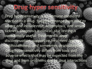
- 1. Drug hypersensitivity differs from toxic and adverse effects that may be expected from the drug and from problems due to drug interactions Drug hypersensitivity is an immune-mediated reaction to a drug. Symptoms range from mild to severe and include rash, anaphylaxis, and serum sickness. Diagnosis is clinical; skin testing is occasionally useful. Treatment is drug discontinuation, supportive treatment (eg, with antihistamines), and sometimes desensitization.
- 2. DRUG HYPERSENSITIVITY TYPE 1 (IMMEDIATE HYPERSENSITIVITY) TYPE III HYPERSENSITIVITY TYPE 4(DELAYED HYPERSENSITIVITY) TYPE 2 (CYTOTOXIC HYPERSENSITIVITY) TYPE 5 HYPERSENSITI VITY
- 3. In type 1 hypersensitivity, an antigen is presented to cells specific to the antigen that stimulate B-cell production of IgE antibodies also specific to the antigen. The difference between a normal infectious immune response and a type 1 hypersensitivity response is that in type 1 hypersensitivity the antibody is IgE instead of IgA, IgG, or IgM. During sensitisation, the IgE antibodies bind to Fcε receptors on the surface of tissue mast cells and blood basophils. Mast cells and basophils coated by IgE antibodies are "sensitised." Later exposure to the same allergen cross-links the bound IgE on sensitised cells, resulting in degranulation and the secretion of pharmacologically active mediators such as histamine, leukotriene (LTC4 and LTD4), and prostaglandin that act on the surrounding tissues. The principal effects of these products are vasodilation and smooth-muscle contraction. Type 1 hypersensitivity can be further classified into an immediate and late-phase reaction. The immediate hypersensitivity reaction occurs minutes after exposure and includes release of vasoactive amines and lipid mediators, whereas the late-phase reaction occurs 2–4 hours after exposure and includes the release of cytokines. Type I hypersensitivity (or immediate hypersensitivity) is an allergic reaction provoked by reexposure to a specific type of antigen referred to as an allergen.[1] Type I is not to be confused with Type II, Type III, or Type IV hypersensitivities. Exposure may be by ingestion, inhalation, injection, or direct contact.
- 4. In type II hypersensitivity (or cytotoxic hypersensitivity) the antibodies produced by the immune response bind to antigens on the patient's own cell surfaces. The antigens recognized in this way may either be intrinsic ("self" antigen, innately part of the patient's cells) or extrinsic (adsorbed onto the cells during exposure to some foreign antigen, possibly as part of infection with a pathogen). These cells are recognized by macrophages or dendritic cells, which act as antigen-presenting cells. This causes a B cell response, wherein antibodies are produced against the foreign antigen. An example of type II hypersensitivity is the reaction to penicillin wherein the drug can bind to red blood cells, causing them to be recognized as different; B cell proliferation will take place and antibodies to the drug are produced. IgG and IgM antibodies bind to these antigens to form complexes that activate the classical pathway of complement activation to eliminate cells presenting foreign antigens (which are usually, but not in this case, pathogens). That is, mediators of acute inflammation are generated at the site and membrane attack complexes cause cell lysis and death. The reaction takes hours to a day. Type II reactions can affect healthy cells. Examples include Red blood cells in Haemolytic Anaemia, Acetylcholine receptors in Myasthenia Gravis, and TSH receptors in Grave's Disease. Another example of type II hypersensitivity reaction is Goodpasture's syndrome where the basement membrane(containing collagen type IV) in the lung and kidney is attacked by one's own antibodies.
- 5. Type III hypersensitivity occurs when there is an excess of antigen, leading to small immune complexes being formed that do not fix complement and are not cleared from the circulation. It is characterized by solvent antigens that are not bound to cell surfaces (which is the case in type II hypersensitivity). When these antigens bind antibodies, immune complexes of different sizes form. Large complexes can be cleared by macrophages but macrophages have difficulty in the disposal of small immune complexes. These immune complexes insert themselves into small blood vessels, joints, and glomeruli, causing symptoms. Unlike the free variant, a small immune complex bound to sites of deposition (like blood vessel walls) are far more capable of interacting with complement; these medium-sized complexes, formed in the slight excess of antigen, are viewed as being highly pathogenic. Such depositions in tissues often induce an inflammatory response, and can cause damage wherever they precipitate. The cause of damage is as a result of the action of cleaved complement anaphylotoxins C3a and C5a, which, respectively, mediate the induction of granule release from mast cells (from which histamine can cause urticaria), and recruitment of inflammatory cells into the tissue (mainly those with lysosomal action, leading to tissue damage through frustrated phagocytosis by PMNs and macrophages). Type III hypersensitivity occurs when antigen-antibody complexes that are not adequately cleared by innate immune cells accumulate, giving rise to an inflammatory response and attraction of leukocytes.
- 6. Type 4 hypersensitivity is often called delayed type hypersensitivity as the reaction takes two to three days to develop. Unlike the other types, it is not antibody mediated but rather is a type of cell-mediated response. CD4+ helper T cells recognize antigen in a complex with Class 2 major histocompatibility complex. The antigen-presenting cells in this case are macrophages that secrete IL-12, which stimulates the proliferation of further CD4+ Th1 cells. CD4+ T cells secrete IL- 2 and interferon gamma, further inducing the release of other Th1 cytokines, thus mediating the immune response. Activated CD8+ T cells destroy target cells on contact, whereas activated macrophages produce hydrolytic enzymes and, on presentation with certain intracellular pathogens, transform into multinucleated giant cells.
- 7. This is an additional type that is sometimes (often in the UK) used as a distinction from Type 2. Instead of binding to cell surface components, the antibodies recognise and bind to the cell surface receptors, which either prevents the intended ligand binding with the receptor or mimics the effects of the ligand, thus impairing cell signaling. Some clinical examples: Graves' disease Myasthenia gravis The use of Type 5 is rare. These conditions are more frequently classified as Type 2, though sometimes they are specifically segregated into their own subcategory of Type 2.