![Diagnostico diferencial ,[object Object],[object Object],[object Object],[object Object],[object Object],[object Object],[object Object]](data:image/gif;base64,R0lGODlhAQABAIAAAAAAAP///yH5BAEAAAAALAAAAAABAAEAAAIBRAA7)
Empfohlen
Weitere ähnliche Inhalte
Was ist angesagt?
Was ist angesagt? (20)
Ähnlich wie Sca
Ähnlich wie Sca (20)
Sca
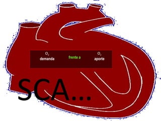
- 1. SCA… O 2 demanda O 2 aporte frente a
- 14. Síndromes Coronarios Agudos Braunwald E et al. J Am Coll Cardiol 2000;36:970-1062 Sin elevación del ST Con elevación del ST Síndrome coronario agudo Angina inestable Infarto de miocardio IM sin onda Q IM con onda Q IM sin elevación del ST
- 15. Sindromes Coronarios Agudos (SCA) SCA sin elevación persistente del segmento ST SCA con elevación persistente l segmento ST Troponina incrementada Angina Inestable IM Sin Onda Q IM Con Onda Q CPK-MB
- 16. No Elevación ST Elevación ST Sindrome Coronario Agudo Angina Inestable IMNQ IMA Q IMANEST Infarto Miocárdico Agudo Dolor tipo isquémico Presentación Enfoque Dx ECG Marcadores Bioquímicos Dx Final Curr Cardiol 2006,31:769-817
- 20. Estudio Angina Estable (+) Test de Esfuerzo Cuadro clínico ECG Test de Provocación Cintigrama Mioc Eco Stress Coronariografía .
- 22. Estudio Sindrome Coronario Agudo IAM Q IAM NO Q Cuadro clínico ECG Coronariografía Normal SD ST Enzimas Cardiacas Angina Inestable Infarto Miocárdico (+) (-) ID ST Test de Provocación
Hinweis der Redaktion
- Figure 1. Endothelial Dysfunction in Atherosclerosis. The earliest changes that precede the formation of lesions of atherosclerosis take place in the endothelium. These changes include increased endothelial permeability to lipoproteins and plasma constituents, which is mediated by nitric oxide, prostacyclin, platelet-derived growth factor, angiotensin II, and endothelin; up-regulation of leukocyte adhesion molecules, including L-selectin, integrins, and platelet–endothelial-cell adhesion molecule 1, and the up-regulation of endothelial adhesion molecules, which include E-selectin, P-selectin, intercellular adhesion molecule 1, and vascular-cell adhesion molecule 1; and migration of leukocytes into the artery wall, which is mediated by oxidized low-density lipoprotein, monocyte chemotactic protein 1, interleukin-8, platelet-derived growth factor, macrophage colony-stimulating factor, and osteopontin.
- Figure 2. Fatty-Streak Formation in Atherosclerosis. Fatty streaks initially consist of lipid-laden monocytes and macrophages (foam cells) together with T lymphocytes. Later they are joined by various numbers of smooth-muscle cells. The steps involved in this process include smooth-muscle migration, which is stimulated by platelet-derived growth factor, fibroblast growth factor 2, and transforming growth factor b ; T-cell activation, which is mediated by tumor necrosis factor a , interleukin-2, and granulocyte–macrophage colony-stimulating factor; foamcell formation, which is mediated by oxidized low-density lipoprotein, macrophage colony-stimulating factor, tumor necrosis factor a , and interleukin-1; and platelet adherence and aggregation, which are stimulated by integrins, P-selectin, fibrin, thromboxane A 2 , tissue factor, and the factors described in Figure 1 as responsible for the adherence and migration of leukocytes.
- Figure 3. Formation of an Advanced, Complicated Lesion of Atherosclerosis. As fatty streaks progress to intermediate and advanced lesions, they tend to form a fibrous cap that walls off the lesion from the lumen. This represents a type of healing or fibrous response to the injury. The fibrous cap covers a mixture of leukocytes, lipid, and debris, which may form a necrotic core. These lesions expand at their shoulders by means of continued leukocyte adhesion and entry caused by the same factors as those listed in Figures 1 and 2. The principal factors associated with macrophage accumulation include macrophage colony-stimulating factor, monocyte chemotactic protein 1, and oxidized low-density lipoprotein. The necrotic core represents the results of apoptosis and necrosis, increased proteolytic activity, and lipid accumulation. The fibrous cap forms as a result of increased activity of platelet-derived growth factor, transforming growth factor b , interleukin-1, tumor necrosis factor a , and osteopontin and of decreased connective-tissue degradation.
- Figure 4. Unstable Fibrous Plaques in Atherosclerosis. Rupture of the fibrous cap or ulceration of the fibrous plaque can rapidly lead to thrombosis and usually occurs at sites of thinning of the fibrous cap that covers the advanced lesion. Thinning of the fibrous cap is apparently due to the continuing influx and activation of macrophages, which release metalloproteinases and other proteolytic enzymes at these sites. These enzymes cause degradation of the matrix, which can lead to hemorrhage from the vasa vasorum or from the lumen of the artery and can result in thrombus formation and occlusion of the artery.
- This slide shows the acute coronary syndrome spectrum. Patienst present with ischemic discomfort; the 12 lead ECG is at the center of the decision pathway to distinguish those patients presenting with ST elevation from those without ST elevation. Biochemical markers are used to confirm the diagnosis of myocardial infarction. Prompt reperfusion is critical for those patients presenting with ST elevation.