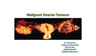
Malignant ovarian tumours Dr H.K.Cheema
- 2. 15-20% of all genital malignancies 20% ovarian tumours are malignant 5th most common cause of cancer deaths 1 Ca Breast 2.Ca cervix 3.Ca lung 4. Ca Colon 5. Ca Ovary Any Cancerous growth of ovaries is known as Ovarian Carcinoma 1.Primary Ovarian tumour 2.Secondary Ovarian Tumour
- 4. Socio-Epidemiological Features • Nulligravida & low parity • Repeated ovulation trauma • Excessive use of ovulation induction drugs • Early menarche • Late menopause • White race • Family history • Use Of Talcom Powder & asbestos • Use of coffee, tobacco, alcohol, dietary fat
- 5. • Age group 40-60 yrs • Nulliparity and low parity • Relative or absolute infertility • Family history of Breast, colon, endometrial, ovarian Cancer • Lynch II/ HNPCC (Hereditary non-polyposis colorectal cancer syndrome • .Post menopausal palpable ovaryvol.8cm3 • Obesity
- 10. Primary 80% Epithelial (80-90%) 1. Serous 2. Mucinous 3. Endometroid 4. Brenner 5. Undifferenciated Secondary 20% Krukenberg tumour Non-epithelial Germ cell tumours Sex chord stromal tumours
- 11. • 1. Benign • 2. Borderline • 3. Malignant
- 12. Classification Of Ovarian tumours Sister Meera Began Experiencing Cancer. Doctor Examined The Cancer. She Felt Good. Epithelial Tumours Serous Mucionous Brenner Endometriod Clear cell Sex chord stromal tumours Sertoli Leydig cell Fibroma Granulosa Theca cell Germ Cell tumours Dysgerminoma Endometrial sinus tu. Teratoma Chorio-carcinoma
- 13. Types Of Ovarian Carcinoma
- 15. • 90% of all primary ovarian carcinomas • any age but 60%-Post-menopausal, • 20% Pre-menopausal • Majority are not familial. • Include both cystic, solid & mixed types • Bilateral in 50% of cases
- 20. Clinical Presentation • Ovarian cancer is called “Silent Killer”. When signs & symptoms appear, it’s too late. • Patients remain asymptomatic for several months, even with early stage. • It is difficult to distinguish the symptoms and make decisive diagnosis of Ovarian cancer.
- 26. Ascites 1. Increased transudate 2. Obstruction of peritoneal fluid outflow from diaphragm. Right sided Pleural effusion More fluid in right sub- diaphargmatic space Left supra-clavicular lymph node enlarged Lymph nodes 1. Para-aortic 2. Superior gastric 3. Supra-clavicular
- 27. Signs • The presence of a fluid wave or less commonly, flank bulging suggests the presence of significant ascites. • In a woman with a pelvic mass and ascites, the diagnosis is ovarian cancer until proven otherwise. • However, ascites without an identifiable pelvic mass suggests the possibility of cirrhosis or other primary malignancies such as gastric or pancreatic cancers
- 31. Physical Examination • A pelvic or pelvic-abdominal mass is palpable in most patients with ovarian cancer. • In general, malignant tumors tend to be solid, nodular, and fixed, • To aid surgical planning, a rectovaginal examination also should be performed. Mass Feel-solid/ hetrogenous Mobility –restricted Tenderness-usually present Surface-irregular Margins-well defined lower border-not reachable Percussion-dull note
- 32. Physical Examination • In advanced disease, examination of the upper abdomen usually reveals a central mass signifying omental caking. • Auscultation of the chest is also important because patients with malignant pleural effusions may not be overtly symptomatic. The remainder of the examination should include palpation of the peripheral nodes in addition to a general physical assessment
- 33. Omental caking
- 34. • To confirm malignancy pre-operatively • To identify the extent of disease • To detect primary site
- 37. Ultra-sonography/TVS • In general, malignant tumors are multi-loculated, solid or echogenic, large (>5 cm), and have thick septa with areas of nodularity • Other features may include papillary projections or neo-vascularization.
- 38. Radiography • Every patient with suspected ovarian cancer should have a chest radiograph to detect pulmonary effusions or infrequently, pulmonary metastases. • Rarely, a barium enema is helpful clinically in excluding diverticular disease or colon cancer or in identifying involvement of the recto- sigmoid by ovarian cancer.
- 39. Computed Tomography Scanning CT-Scan • The main advantage of computed tomography (CT) scanning is in treatment planning of women with advanced ovarian cancer. • Preoperatively, it may detect disease in the liver, retroperitoneum, omentum, or elsewhere in the abdomen and thereby guide surgical, • However, CT scanning is not particularly reliable in detecting intra-peritoneal disease smaller than 1 to 2 cm in diameter. • Moreover, the accuracy of CT scanning is poor for differentiating a benign ovarian mass from a malignant tumor when disease is limited to the pelvis. In these cases, transvaginal sonography is superior. Positron Emission Tomography PET-Scan Differenciates normal from cancerous tissue. More sensitive than CT_Scan or MRI Helps identify recurrance
- 41. Paracentesis • A woman with a pelvic mass and ascites can be assumed to have ovarian cancer until proven otherwise surgically. • However, paracentesis may be indicated for patients with ascites and the absence of a pelvic mass.
- 52. Staging ovarian cancers Stages= Ovarian Carcinoma Stage I A One ovary involved Stage 1 B Both ovaries involved Stage 1 C One or both ovaries + Surface of ovary+ rupture of capsule+ Ascites/+ peritoneal washings
- 57. Stage II-Ovarian carcinoma • II A • Extension and/or metastases to the uterus and/or tubes • IIB • Extension to other pelvic tissues • II C • Tumor limited to the genital tract or other pelvic tissues, but with disease on the surface of one or both ovaries; or with capsule(s) ruptured; or with malignant ascites or positive peritoneal washings
- 61. Stage III-Ovarian carcinoma • III A • The cancer is present in one or both of the ovaries, and cancer cells are also present in small ranges in parts of the abdomen with this stage without nodular involvement. • III B • On this particular stage, the cancer is present in one or both of the ovaries, and cancer cells are also present in amounts less than 2 cm or 3/4″ in parts of the abdomen • III C • Abdominal implants at least 2 cm in diameter and/or positive pelvic, para-aortic, or inguinal nodes
- 66. Stage IV-Ovarian carcinoma • Stage IV • Distant metastasis including Pleural effusion or parenchymal liver metastasis
- 68. Stage of disease Histology of tumour Age of patient General condition
- 70. Staging Laprotomy • Aim • To stage the disease & resect as much tumour as possible. Steps 1. General anaesthesia 2. Liberal Vertical incision 3. Aspirate ascitic fluid/Peritoneal washings 4. Exam ovaries & pelvis 5.Systematic exploration of all organs 6.Multiple biopsies 7.TAH with B/L S O 8.Infra colic omentectomy 9.Pelvic & para-aortic lymphadenectomy
- 71. Management of Early-Stage Ovarian Cancer • When a malignancy appears clinically confined to the ovary, surgical removal and comprehensive staging should be performed • Fertility-Sparing Management :may be an option in selected patients when disease appears confined to one ovary in younger patients • Adjuvant Chemotherapy: In general, patients with stage IA or IB, tumors should be treated with three to six cycles of platinum based-combinations
- 92. • Average age=10-20yrs. • Rx=Primary treatment- surgery • Young patient-conservative surgery • (unilateral oophorectomy) • Adjuvant therapy with chemotherapy • All Germ cell tumours-highly chemo sensitive • Stage Ia Grade I—no need • Other stages- Bleomycin+etoposide+cisplatin
- 94. Tumour Markers LDH hCG Alpha-fetoproteins Young patient Staging laprotomy+Unilateral salpingo- oophorectomy+ Biopsy of other ovary + (Chaemotherapy/radiotherapy) BEP Bleomycin+Etoposide+Cisplatin VBP Vinblastin+bleomycin+cisplatin
- 96. Sex chord stromal carcinoma • Granulosa cell tumour • Thecoma • Fibroma • Sertoli-Leydig cell tumour • Gynadroblastoma • Unclassified
- 97. Most common Ostrogen producing Any age 10% pre- pubertal Coffee bean nuclei Unilateral salpingo- oophorecctomy+chaemoth arapy
- 104. Common Primary sites Stomach, colon, Gall bladder, Pancreas, Breast
