2. Neoplasm
Definition Is a new growth
0r abnormal mass of tissue ,the growth
of which exceed & uncoordinated with
that of normal tissue & persist in the
same excessive manner even after
cessation of stimuli which evoke the
changes
Tumor : (Greek ,swelling)
3. Tumor classify into
benign or malignant on the basis of:
• Histologic and cytologic features
• The biological behavior of tumor
All malignant tumor : are Cancer
4. Tumor basic component
All tumor compose of
1-proliferating neoplastic cells (parenchyma).
• The parenchyma determine the biologic
behavior & this component from which the
tumor derives its name
2-supportive stroma non neoplastic (c.t & bl.v).
• tumor growth and evolution is critically
dependent on their stroma
5.
6. Nomenclature of Tumors
• All have the suffix oma
Depending on histogenesis (cell or
tissue of origin ) tumor can broadly
divided into those derived from
epithelial or mesenchymal
7. EPITHELIAL NEOPLASMS
• Adenoma A benign epithelial neoplasm that
arises within a gland (eg, thyroid adenoma, colonic
adenoma)
• papilloma (Latin, papilla = nipple) when arising
from an epithelial surface.
Papillomas may arise from squamous, glandular, or
transitional epithelium .
• Carcinoma Malignant epithelial neoplasms
(adenocarcinomas if derived from glandular
epithelia;
squamous carcinoma
transitional cell carcinoma
8.
9. MESENCHYMAL NEOPLASMS
• Benign mesenchymal neoplasms are
named after the cell of origin followed
by the suffix -oma
• E.g fibroma
• Malignant mesenchymal neoplasms are
named after the cell of origin, to which
is added the suffix -sarcoma.
liposarcomas
13. Mixed tumorsMixed tumors
• Tumors with mixed differentiationTumors with mixed differentiation
– e.ge.g.. pleomorphic adenoma of salivarypleomorphic adenoma of salivary
glandgland
– carcinosarcomacarcinosarcoma
14. TeratomaTeratoma
• Tumor contain elements of all three
germ layers: endoderm, ectoderm, and
mesoderm. Thus, brain, respiratory and
intestinal mucosa, cartilage, bone, skin,
teeth, or hair may be seen in the
neoplasm.
• The constituent tissues are not limited
to those normally present in the area of
origin.
15. - Arise from totipotent cells (usually gonads)- Arise from totipotent cells (usually gonads)
testis or ovarytestis or ovary
- benign cystic teratoma of ovary is the most- benign cystic teratoma of ovary is the most
common teratomacommon teratoma
Teratomas are classified as
• mature (well-differentiated and composed
of adult-type tissues) benign.
• immature (made up of fetal-type tissues).
are malignant,
17. • The names of some malignant neoplasms
are formed by adding the suffix -oma to
the cell of origin, eg,
• lymphoma (lymphocyte),
• plasmacytoma (plasma cell),
• melanoma (melanocyte),
• Neoplasms of blood-forming organs are
called leukemias
18. some confusing terminology
Aberrant differentiation (not trueAberrant differentiation (not true
neoplasms)neoplasms)
• Hamartoma :is disorganized mass ofdisorganized mass of
tissue (malformation)tissue (malformation) compose of
mature cell normally present in that
tissue in haphazard arrangment
e,g Hamartoma of lung (contain
bronchial cell,bl.v., cartilage,lymphoid
tissue )
19. choristoma
• (ectopic normal tissue in abn.location )
e.g arrest of adrenal cell under kidney
capsule or small nodule containing
pancreatic tissue found in submucosa
of the stomach.
20. Character of
benign & malig.neoplasm
Differentiation by following feature
1.Differentiation & anaplasia.
2.Growth rate
3.Local invasion
4.metastasis
21. DifferentiationDifferentiation
Differentiation refers to the extent to whichDifferentiation refers to the extent to which
neoplastic parenchymal cells resemble theneoplastic parenchymal cells resemble the
corresponding normal parenchymal cells, bothcorresponding normal parenchymal cells, both
morphologically and functionallymorphologically and functionally
- Well differentiated- Well differentiated Resembles mature cells of tissue of originResembles mature cells of tissue of origin
- Poorly differentiated- Poorly differentiated neoplasmneoplasm
– Composed of primitive cells with little diffrerentiation.Composed of primitive cells with little diffrerentiation.
– In general all benign t.are well diff.
In contrast malig.t. range from well to poorly diff.
• Differentiation determine the tumor grade
• Lack of differentiation (UndifferentiatedLack of differentiation (Undifferentiated
tumor) are “anaplastic” tumortumor) are “anaplastic” tumor
22. The degree of anaplasia correlate with
aggressiveness of the tumor( biologicbiologic
behavior)behavior)
– Poorly differentiated malignant tumorsPoorly differentiated malignant tumors
usually have worse prognosisusually have worse prognosis
23.
24. • PleomorphismPleomorphism : tumor cell display variation in both: tumor cell display variation in both
(cytologic abn,)(cytologic abn,)
– SizeSize
– Shape (Bizzar tumor giant cell )Shape (Bizzar tumor giant cell )
• Abnormal nuclear morphologyAbnormal nuclear morphology
– HyperchromasiaHyperchromasia
– High nuclear cytoplasmic ratioHigh nuclear cytoplasmic ratio
– Chromatin clumpingChromatin clumping
– Prominent nucleoliProminent nucleoli
– nuclear-to-cytoplasm ratio may approach 1 : 1 insteadnuclear-to-cytoplasm ratio may approach 1 : 1 instead
of the normal 1 : 4 or 1 : 6of the normal 1 : 4 or 1 : 6 thethe
• MitosesMitoses
– Mitotic rateMitotic rate
– Type of mitoses (atypical )Type of mitoses (atypical )
• Loss of polarityLoss of polarity ,,
• presence large area of necrosispresence large area of necrosis
““ANAPLASIAANAPLASIA””
25.
26.
27. DysplasiaDysplasia
• DysplasiaDysplasia is a term that meansis a term that means
disordered growth.disordered growth.
• Dysplasia often occurs in metaplasticDysplasia often occurs in metaplastic
epitheliumepithelium
• Dysplasia is encounteredDysplasia is encountered in epitheliain epithelia
• Dysplastic cells exhibit considerableDysplastic cells exhibit considerable
pleomorphism and often contain largepleomorphism and often contain large
hyperchromatic nuclei with a high nuclear to-hyperchromatic nuclei with a high nuclear to-
cytoplasmic ratio.cytoplasmic ratio.
• The architecture of the tissue may beThe architecture of the tissue may be
disorderdisorder
28. DysplasiaDysplasia
Malignant transformation is a multistep processMalignant transformation is a multistep process
• In dysplasia some but not all of the features ofIn dysplasia some but not all of the features of
malignancy are present, microscopicallymalignancy are present, microscopically
• DysplasiaDysplasia maymay develop into malignancydevelop into malignancy
– Uterine cervixUterine cervix
– Colon polypsColon polyps
• Graded as low-grade or high-gradeGraded as low-grade or high-grade
• Dysplasia mayDysplasia may NOTNOT develop into malignancydevelop into malignancy
• HIGH grade often classified with carcinoma-in-situHIGH grade often classified with carcinoma-in-situ
29. High-grade dysplasiaHigh-grade dysplasia::
• The abnormal dysplastic cells involveThe abnormal dysplastic cells involve
whole thickness of epith.whole thickness of epith.
• The lesion remain confine by theThe lesion remain confine by the
basement membrane ,it is pre-invasivebasement membrane ,it is pre-invasive
or called (or called (carcinoma in situ)carcinoma in situ)
• Dysplasia does not necessarily progress toDysplasia does not necessarily progress to
cancercancer..
Mild to moderate changes that do not involveMild to moderate changes that do not involve
the entire thickness of epithelium may bethe entire thickness of epithelium may be
reversible, after removal of the inciting agentreversible, after removal of the inciting agent
34. Features of Malignant TumorsFeatures of Malignant Tumors
• Cellular featuresCellular features
• LocalLocal invasioninvasion
– CapsuleCapsule
– Basement membraneBasement membrane
• MetastasisMetastasis
– Unequivocal sign of malignancyUnequivocal sign of malignancy
– Seeding of body cavitiesSeeding of body cavities
– LymphaticLymphatic
• HematogenousHematogenous
35.
36. Invasion
• Invasiveness is the most reliable feature
differentiate malig.from benign t.
• Some cancer are in pre-invasive stage called
(ca.in situ) e.g ca.in situ of cervix without
invasion to basement memb.
• Metastasis tumor implant discontinues with
the primary tumor .
• It is unequivocal mark that the tumor malig with
the exception B.C.C. of skin& C.N.S. glioma,
both locally invasive but not give rise for distant
metastasis
37. • Benign t. :slowly growing tumor remain
localized, amenable to local surgical
removal
• Malignant: spread distant
38.
39. FeatureFeature BenignBenign MalignantMalignant
Rate of growthRate of growth Progressive butProgressive but
slow. Mitosesslow. Mitoses
few and normalfew and normal
Variable.Variable.
Mitoses moreMitoses more
frequent andfrequent and
may bemay be
abnormalabnormal
DifferentiationDifferentiation WellWell
differentiateddifferentiated
Some degree ofSome degree of
anaplasiaanaplasia
LOCALLOCAL
INVASIOINVASIO
NN
Cohesive growth.Cohesive growth.
Capsule & BMCapsule & BM
not breachednot breached
Poorly cohesivePoorly cohesive
and infiltrative.and infiltrative.
44. • Acqiured causes orAcqiured causes or
predispostionpredispostion
• Heredity form or predispostionHeredity form or predispostion
(5%-10%of all human(5%-10%of all human
cancer)which can be dividedcancer)which can be divided
into three categories)into three categories)
45. Genetic inherited
predispostion of cancer
can be divided into three categories :
1- Familial cancer syndromessyndromes
Example :
• BRCA1&BRCA2 ca.breast, ovary
Feature cc.familial cancer:
1-early age at onset
2-tumor arise in two or more close relatives
3-some time multiple & bilateral tumor
46. Inherited cancer synd
Inherited as A.D mutation (point mutation) in
t.supp.gene e.g
• RB-Familial retinoblastoma ,40% inherited
• APC- (familial adenomatous polyposis) FAP
of colon,germ line mutation of APC
• NF-Neurofibromatosis type 1& 2
• MEN(multiple endocrine neoplasia)
syndromes
• MSH2&MSH6-hereditary non polyposis
colonic cancer(HNPCC),inherit defective
copy of mismatch repair gene
• P53 :various t.
55. Molecular Basis of Cancer
NON-lethalNON-lethal genetic damagegenetic damage(or mutation)
may be
• Acquired by the action of environmental
agents, caused by exogenous agents such as
chemicals, radiation, or viruses
• or it may be inherited in the germ line.
• Not all mutations, are “environmentally”
induced. Some may be spontaneous , falling into
the category of bad luck.
56. Molecular Basis of Cance….cont.
• tumors are monoclonal formed
by the clonal expansion of a
single precursor cell that has
incurred genetic damage.
• Carcinogenesis is aCarcinogenesis is a multistepmultistep
processprocess
.
57. • The process which result in
transformation of normal cell to
neoplastic cell by causing permanent
genetic alteration.
• No single gene mutation is sufficient to
cause cancer
• Occur by accumulation of genetic
lesions that result in tumor
progression
58. MOLECULAR BASISMOLECULAR BASIS
of CANCERof CANCER
• Four classesFour classes of normal regulatoryof normal regulatory
genesgenes
– PROTO-oncogenesPROTO-oncogenes
– Growth inhibitor gene (cancer suppressor
gene)
– DNA repair genesDNA repair genes
– Apoptosis genesApoptosis genes
59. TRANSFORMATION &TRANSFORMATION &
PROGRESSIONPROGRESSION
• Self-sufficiency in growth signalsSelf-sufficiency in growth signals
• Insensitivity to growth-inhibiting signalsInsensitivity to growth-inhibiting signals
• Evasion of apoptosisEvasion of apoptosis
• Defects in DNA repair: “Spell checker”Defects in DNA repair: “Spell checker”
• Limitless replicative potential: TelomeraseLimitless replicative potential: Telomerase
• AngiogenesisAngiogenesis
• Invasive abilityInvasive ability
• Metastatic abilityMetastatic ability
60. ESSENTIAL ALTERATIONS FOR
MALIGNANT TRANSFORMATION
The seven key changes are the following
• Self-sufficiency in growth signals: Tumors
have the capacity to proliferate without external
stimuli, usually as a consequence of oncogene
activation.
• Insensitivity to growth-inhibitory signals:
Tumors may not respond to molecules that are
inhibitory to the proliferation of normal cells such
as transforming growth factor β (TGF-β) and
direct inhibitors of cyclin-dependent kinases
(CDKIs). •
61. • Evasion of apoptosis: Tumors may be
resistant to programmed cell death, as a
consequence of inactivation of p53 or
activation of anti-apoptotic genes.
• Limitless replicative potential: Tumor
cells have unrestricted proliferative
capacity, avoiding cellular senescence
and mitotic catastrophe.
62. LIMITLESS REPLICATIVELIMITLESS REPLICATIVE
POTENTIALPOTENTIAL
• TELOMERES determine theTELOMERES determine the
limited number of duplicationslimited number of duplications
• TELOMERASETELOMERASE, present in >90%, present in >90%
of human cancers, changesof human cancers, changes
telomeres so they will havetelomeres so they will have
UNLIMITED replicative potentialUNLIMITED replicative potential
63. • Sustained angiogenesis: Tumor cells,
like normal cells, are not able to grow
without formation of a vascular supply to
bring nutrients and oxygen and remove
waste products. Hence, tumors must
induce angiogenesis.
64. TUMOR ANGIOGENESISTUMOR ANGIOGENESIS
• Activation of VEGF and FGF-bActivation of VEGF and FGF-b
• Tumor size is regulated (allowed) byTumor size is regulated (allowed) by
angiogenesis/anti-angiogenesisangiogenesis/anti-angiogenesis
balancebalance
65. • Ability to invade and metastasize: Tumor
metastases are the cause of the vast majority of
cancer deaths and depend on processes that
are intrinsic to the cell or are initiated by signals
from the tissue environment.
• Defects in DNA repair: Tumors may fail to
repair DNA damage caused by carcinogens
during unregulated cellular proliferation, leading
to genomic instability
68. Invasion FactorsInvasion Factors
• DetachmentDetachment ("loosening up") of("loosening up") of
the tumor cells from each otherthe tumor cells from each other
• AttachmentAttachment to matrixto matrix
componentscomponents
• DegradationDegradation of ECM, e.g.,of ECM, e.g.,
collagenase, etc.collagenase, etc.
• MigrationMigration of tumor cellsof tumor cells
69. Invasion FactorsInvasion Factors
• 11stst
step:E-cadherin (keep n.cell together),-itstep:E-cadherin (keep n.cell together),-it
will bind to B-catenin .-lead to B cateninwill bind to B-catenin .-lead to B catenin
sequestation .in malig.cell E cadherin issequestation .in malig.cell E cadherin is
lost .lost .
• 2ns step degradation of b.m &ECM by2ns step degradation of b.m &ECM by
proteolytic enzy (MMP-9&cathespin –proteolytic enzy (MMP-9&cathespin –
D)which cleave type IV collgen.D)which cleave type IV collgen.
70.
71. Elaboration of variety of enzyme by
t.cell aid in degradation of ECM&
invasiveness .
Cathepsin D proteinase enzy.that
cleaves fibronectin & laminin
High level of this enzy are ass with
greater invasiveness.
73. oncogenes
• Gene promote autonomous cell growth
in cancer cell in the absence of normal
mitogenic signals.
• Their normal counterpart are
protooncogenes which involve in
physiologic regulation of cell
proliferation& differentiation
74. ONCOGENESONCOGENES
• Are MUTATIONS of NORMAL genesAre MUTATIONS of NORMAL genes
(PROTO-oncogenes)(PROTO-oncogenes)
– Growth FactorsGrowth Factors
– Growth Factor ReceptorsGrowth Factor Receptors
– Signal Transduction Proteins (RAS)Signal Transduction Proteins (RAS)
– Nuclear Regulatory ProteinsNuclear Regulatory Proteins
– Cell Cycle RegulatorsCell Cycle Regulators
75. oncogens & their function
Neoplastic cell prolif. self stimulation mediated by :
1-Over-expression of GF gene sis
2 - Receptor for epidermal growth factor HER2 –or
ERBB2 located at cell surface
3- signal transducer
Ras oncogen act on intracellular signaling (signal
transducer)
e.g: pancreatic adenoca ,cholangioca, ca.colon shows
point mutation
4- DNA Binding nuclear protein
myc oncogen :stimulate direct DNA synthesis
translocation t8:14 in Burkitt lymphoma.
5- cell cycle protein (regulator) Cyclin & CDK
6- Inhibitor of apoptosis (bcl-2).
76.
77. Most common type of nonrandom karyotypic
changes in tumor cell
e.g of balanced translocation
1-philadelphia chromosome in C.M.L.represent
transl.bet.chr.9&22
2- Burkitts lymphoma in 90% transl.bet chr 8 &14
e.g of deletion
–solid t. of non hematopoietic origin –embryonal t.of
childhood (retinoblastoma ,wilms t)
Loss of some normal cancer suppressor genes located
on chr.13 &11 in case of wilms t.
Gene amplification
e.g neuroblastoma the amplified gene is oncogen
called N-myc ,or in case of ca.breast.
78. Tumor suppressor gene
& their function
• P53 : nuclear protein
• TGFB: potent inhibitor for proliferation
mutation seen in ca.colon
• APC Inhibitor for transcription regulator
individual born with one mutant allel develop hundreds to
thousands of adenomatous polyp in the colon in10-20 yr.
• RB gene : in nucleus ,cell cycle inhibitor
(retinoblastoma,osteosarcoma)
• WT1 : location in nucleus
• NF1&NF2 : in plasma membrane ,inhibitor for signal
transducer
• BRCA 1& BRCA 2 (DNA repair)
79. P53 gene
The Guardian of the Genome
• The most common mutated gene in human cancer
• It is DNA binding protein ,regulate DNA repair & prevent
DNA transcription error
Function in normal cell (regulate cell proliferation) through
1-Activation of temporal cell cycle arrest (quiescence )
through p21
2- permanent cell cycle arrest (senescence)
3-promotes cell death through BAX gene which inhibits
BCL2 (Apoptotic inhibiting gene)
4- help in repair process (through GADD 45)
80. P53 &P53 & RASRAS
p53p53
• Activates DNAActivates DNA
repair proteinsrepair proteins
• Sentinel of G1/SSentinel of G1/S
transitiontransition
• Initiates apoptosisInitiates apoptosis
• Mutated in moreMutated in more
than 50% of allthan 50% of all
human cancershuman cancers
RASRAS
• H, N, K, etc.,H, N, K, etc.,
varietiesvarieties
• Single mostSingle most
commoncommon
abnormality ofabnormality of
dominantdominant
oncogenes inoncogenes in
human tumorshuman tumors
• Present in about 1/3Present in about 1/3
of all humanof all human
82. Function of P53 In normal cell
• Activates DNA repair proteinsActivates DNA repair proteins
• Sentinel of G1/S transitionSentinel of G1/S transition
• Initiates apoptosisInitiates apoptosis
activation of P53 occur by DNA damaging
agent or hypoxia lead to cell cycle arrest in
G1 phase & induction of DNA repair .
The mechanism of arrest is mediated by cell
cycle inhibitor protein P21
Successful repair of DNA allow cell to proceed
with cell cycle
If DNA repair fail ,then P53 trigger either
apoptosis or senescence.
83. • .
In mutant P53 :
DNA damage cell go through cell
cycle without DNA repair ,this
genetically abn.cell proliferate
give rise to malig.
84.
85. •Two hit hypothesis,LOH & cancer
• No.of syndromes result from germ line
mutation in various t.supp.gene .
Example :
• Li-Fraumeni synd.(inherited predispos to ca.)
due to germ line mutation of p53.
• Familial polyposis coli synd : APC gene
mutation ,also cause m.m.& ov.ca
• Hereditary wilms t.
• Von Hipple Lindau synd.
89. Chemical carcinogen
multistep process through four stages
•Initiation & promotion sequences
•Progression: stage in which tumor growth
become autonomus (independent of
carcinogen or the promoter result from
acc.mutation result in immotalize cell)
•Cancer: the end result of entire sequence
90. Initiation
• Is the event that induce lesion in cell
genome (DNA)
Result from exposure of cell to
• appropriate dose of carcinogenic agent .
• Initiator is mutagenic
• Initiated cell (altered ,transformed cell )
render it susceptible to give rise of tumor
91. • Initiation alone is not sufficient for
tumor formation.
• It give rise of tumor only after promotor
application
• initiation is rapid & irreversible
• the initiating carcinogen produce
permanent changes in DNA of the
target cell
92. Promotion
Is the event lead to clonal proliferation of
the initiated transformed cell .
• promotion can induce changes in the
initiated cell but they are not
tumorogenic by themselves
• tumor does not result when promotion
applied before rather than after the
initiating agent .
93. • promotion is reversible changes (not affect
DNA directly )
• promotion is dose-threshold" dependent
concentration of promoter below which
neoplasia will not occur.
• The altered cell remain dependent on the
continue presence of promoter stimuli which
may be (exogenous chemical ,physical or
endogenous like hormonal stimulation in
breast,endometrium ,prostate).
94.
95. Major chemical carcinogens
• Some chemical agent possess the
capability of both (initiation &
promotion ) called
• complete carcinogen like Alkyalting
agent ,anticancer drug ,busulphan
cyclophosphamide,chlorambucil.
• Incomplete carcinogen capable of
producing t. only after initiation
96. carcinogenic initiators
Examples
• Alkylating agents like cyclophosphamide.
• (PCAH) polycyclic aromatic hydrocarbons
found in smoked foods,cigarette smoke (have
broad range of target organs)produce cancer at
site of application
They are metabolized at cytochrome p450 which
will oxidases to electrophilic epoxides which in
turn react with cellular protein & nucleic acid
• Nitrosamines in pickled foods. gut ca.
• Preserved food contain nitrites which react with
other dietary component to form nitrosamines
which activated by hydroxylation to form reactive
ion .
97. • Aromatic amines or azo dyes used in food
coloring .
• Vinyl choride-liver angiosarcoma
• beta-naphthylamine-use in rubber industry–
bladder ca.
• microbial product
Fungi : aflatoxin b1 (produce by some strain
of aspergillus flavus )
cause hepatic cancer
Act as initiator
98. Examples of promoters
• hormones such as estrogen,
• drugs such as diethylstibesterol,
• chemicals such as cyclamates used as
sweeteners
• Chemical act as both initiator &
Promoter
• (cigarette smoking & Asbestoses )
100. (HPV)
• Anogenital cancer
• Sq.c.c. of cervix (HPVhigh risk gp.
(16-18,31,33,35,51)
• benign wart low risk gp.(HPV 6-11)
The oncogenic potential of HPV is related
to E6 & E7 viral protein which bind to
P53 & RB gene respectively so they
overcome cell cycle inhibitor
101. Role of EBV
• Infectious mononucleosis(a short lifed
lymphproliferative dis.)
• Oral hairy leukoplakia (in AIDS)
• Nasopharyngeal carcinoma
• African Burkitt's lymphoma
• Non-Hodgkin's lymphomas in AIDS
102. Effect of tumor on the host
I- Local effect according to tumor location
• Pressure effect e.g pituitary adenoma
when increase in size
• ulceration &obstruction e.g ca.colon
• secondary infection &bleeding
e.g malena in ca.colon or hematurea in
bladder ca
103. systemic effect
Cancer cachexia
It is waisting syndrome: cc by loss of
body fat .anemia
• there are several factors contribute to
malnutrition in cancer pat.
1-high prot.& fat turnover ,hypermetabolism
anorexia ,reduced food intake,,
2-Cachectin (TNF) - produced by tumor cell
¯ophages & it is a mediator of
waisting syndrome
104. other soluble factor produced by tumor cell .
3- PIF proteolysis inducing factor which
involved in abn.metabolism by increase
catabolism of muscle & adipose tissue,
4- Impair immune defenses –prone to
infection .
105. paraneoplastic syndrome
• It is clinical syndrome with symptom complexes
occur in cancer pat, (10% of advanced malig)
• Result from synthesis of bioactive sub. by tumor
cells.
symptom may be
1- endocrinopathies :elaboration of hormon from
tumor cell which are not of endocrine origin
(ectopic hr.production )
Example:
• Insulin release of by fibrosarcoma
• Erythrpoitein hr.production by renal cell.ca.
• Parathyroid like hr-sq.c.ca of lung
106. 2-Neuromyopathic paraneoplastic synd-
Myasthenia Gravis. like synd.in small ell
ca.of lung
3- cutaneous :Acanthosis nigrican cc by
grey black patches of hyperkeratotic skin
4- Vascular & hematological changes.
• DIC (hypercoagulability)-release of
PAF&procoagulant from tumor cell.
107. Tumor marker
Biochemical indicator of presence of tumor
• Aid in DX
• Determine response to therapy
• Indicate relapse in follow up
Hormones :
• HCG :Trophoblastic t,Non seminoma
testicular t.
• Calcitonine : Medullary ca.of thyroid
• Catecholamine : pheochromocytoma
• Ectopic hr.: paraneoplastic synd.
108. Tumor marker cont….
Isoenzy.:
• PAP:prostatic ca.
• NSE : small cell ca.of lung,neuroblastoma
Specific protein :
• Immunoglobulins : multiple myloma
• Prostatic specific Ag.:ca.prostate
109. Tumor marker cont….
Mucins
• CA-125 : ovary ca.
• CA-19-9 : colon, pancreas ca.
• CA-15.3 : Breast ca.
Oncofetal Ag.:
• (AFP)Alpha fetoprotein: liver ca,testicular t.
• CEA : ca. pancreas,colon,lung,stomach
New molecular marker :
• P53,APC,RAS in stool& serum ca.colon
110. Lab.Dx of cancer
1- Biopsy
2- Cytology
3- FNA
4- Immunocytochemistry
(by specific monoclonal Antibody for identification of cell product
or surface marker )
5- flow cytometry :
measure several cell character such as
1- DNA content like aneuploidy seems to be associated with poor
prognosis
2- Identification of cell-surface antigens
widely used in the classification of leukemias and lymphomas
111. The value of
immunohistochemistry
1-categorization of undiff.malig.t
(To determine line of differentiation )
2-categorization of leukemia/lymphoma
3- determination the site of origin of metastatic
tumor
like thyroglobulin marker for tumor of thyroid
origin
e.g Desmin –specific for muscle
4- Detection of molecule that of prognostic &
therapeutic significance
like ER,PR,erb B2 in ca.breast
Hinweis der Redaktion
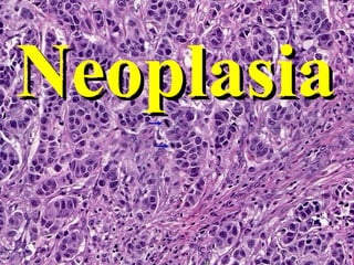
Poorly differentiated carcinoma of breast . Histopathology
Colonic polyp.(Adenoma )
Figure 7-4 A, Gross appearance of an opened cystic teratoma of the ovary. Note the presence of hair, sebaceous material, and tooth .
Dysplasia means potential PRE-cancer. Anaplasia means cancer. The three words: pleomorphism, hyperchromasia, and increased mitoses, are the three most widely used terms to describe malignant tumors on pathology reports .
Anaplastic tumor
Do you remember from chapter 1 that DYS- was one of the seven -plasia brothers ?
This epithelium shows severe dysplasia: Note that dysplastic basal cells characterized by cuboidal shape, high nuclear cytoplasmic ratio, hyperchromatism, mitotic activity, and some loss of orientation to the basement membrane, occupy the lower two thirds of the surface rather than just the basal row of cells. More differentiated cells which occupy the outer third, though still retaining some dysplastic nuclear features have the appearance of maturing squamous cells rather than basal cells, and eventually become flattened on the surface .
Carcinoma in situ: This section shows that the dysplastic basiloid cells go all the way to the surface and never undergo significant differentistion towards more differentiated flattened squamous cells. Note however that the basement membrane is still intact. The torturingly and unnecessary insane pressure to differentiate “severe” dysplasias from carcinomas-in-situ has prompted the various “-IN” classification systems, e.g., CIN-III, VIN-III, PIN-III. Is high grade dysplasia any different from carcinoma-in-situ from a microscopic, behavioral, or medical-legal point of view? Ans: NO !
leukoplakia
Low risk gp HPV
Moderate dysplasia
Carcinoma in situ
A proto-oncogene is a normal gene that can become an oncogene due to mutations or increased expression . Proto-oncogenes code for proteins that help to regulate cell growth and differentiation .
The various aspects of “malignant transformation”. Just like cancer itself is a progression of increasingly disturbing processes, so is malignant transformation. These are not necessarily exactly linear events, but close .
Telomeres are a sequence of repetitive bases at the ends of linear chromosomes that prevent adjacent chromosomes from attaching to each other . Think about this? If a telomere is interfered with, perhaps by telomerase, it LOSES its ability to limit mitoses !
This is a BEAUTIFUL chart! Another way of understanding the development of malignancy in a logical way !
important diagrammatic explanation of malignancy .
FOUR orderly steps of “INVASION” (aka, INFILTRATION, or INVASIVENESS )
FOUR orderly steps of “INVASION” (aka, INFILTRATION, or INVASIVENESS )
Signal transduction is a generic term which refers to any process by which a cell converts one kind of signal or stimulus into another .
These are the TWO other most important and widely studied genes in cancer .
. NOTE: Problems of GROWTH SUPPRESSION, result in GROWTH being UN-regulated . THESE ARE ALL GROWTH SUPPRESSOR GENES WHICH, WHEN MUTATED, LOSE THEIR NORMAL ABILITY TO SUOPPRESS !!!
Malig.melanoma eye
Experiment was done on skin of rat treated with carcinogenic agent such as tar ,was treated with a second ,non carcinogenic agent acting as promoter