The Complete Reference to Human Heart
•
2 gefällt mir•583 views

A Complete Human Heart Presentation

Empfohlen
Weitere ähnliche Inhalte
Was ist angesagt?
Was ist angesagt? (18)
Ähnlich wie The Complete Reference to Human Heart
Ähnlich wie The Complete Reference to Human Heart (20)
Kürzlich hochgeladen
Kürzlich hochgeladen (20)
The Complete Reference to Human Heart
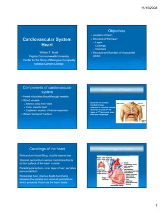
- 1. 11/15/2008 Objectives Cardiovascular System Heart William T. Budd Virginia Commonwealth University Center for the Study of Biological Complexity Medical Careers College Components of cardiovascular system Location of heart Structure of the heart Layers Coverings Chambers Structure and function of myocardial valves Location of Heart HeartHeart- circulates blood through vessels Blood vessels ArteriesArteries- away from heart VeinsVeins- towards heart CapillariesCapillaries- location of internal respiration BloodBlood- transport medium Coverings of the heart • posterior to sternum • medial to lungs • anterior to vertebral column • base lies beneath 2nd rib • apex at 5th intercostal space • lies upon diaphragm Coverings of Heart Pericardium-loose fitting, double layered sac Visceral pericardium-serous membrane that is on the surface of the heart muscle Parietal pericardium- inner layer of sac; secretes pericardial fluid Pericardial fluid- (Serous fluid)-fluid that is between the parietal and visceral pericardium which prevents friction as the heart beats. 1
- 2. 11/15/2008 Layers of heart tissue Epicardium Myocardium Endocardium Endocardium Inner lining Smooth surface that permits blood to move easily through the heart without agglutination. Continuous with lining of blood vessels Myocardium Middle layer made of cardiac muscle Forms the bulk of the heart wall Contains the septum - a thick muscular septumwall that completely separates the blood in the right side of the heart from the blood in the left side. Epicardium Protective, outer layer of the heart wall same as the visceral pericardium The coronary blood vessels that nourish the heart wall are located here Wall of the Heart Endocardium – lines the heart chambers Myocardium – Strong, muscular layer of heart Epicardium – Outer, serous layer of heart 2
- 3. 11/15/2008 Chambers Right Atrium Thinner wall than ventricles Receives deoxygenated blood from vena cava Passes blood through tricuspid valve into right ventricle Chambers Left Atrium Receives freshly oxygenated blood from pulmonary vein Passes blood to left ventricle through mitral valve Chambers Right Ventricle Thicker wall than atria Comprises most of anterior surface of heart Circulates deoxygenated blood to lungs through the pulmonic valve into pulmonary trunk Chambers Left Ventricle Receives blood from left atrium Thickest myocardial wall Forms apex of heart Sends blood to systemic circulation via aorta 3
- 4. 11/15/2008 Septa Interatrial septum Muscular division b/w atria Foramen ovale- opening in fetus ovaleFossa ovalis- shallow depression; remnants of ovalisforamen ovale Interventricular septum Thick muscular wall Seperates ventricles Heart Valves FunctionFunction- prevent blood from flowing backwards Responds to changes in pressure Two types of valves in heart Semilunar valves Located at exit of ventricles, originiate from endothelial lining of veins Heart contains two semilunar valves Pulmonic Aortic (Frequently damaged by Htn) Htn) Atrioventricular valves (AV) SemiSemi-lunar valves Atrioventricular Valves Valve cusps are connected to papillary muscles Chordae tendineaetendineaetiny collagen cords that anchor cusps of valve to papillary muscles Atrioventricular Valves Left AV valve (Mitral, bicuspid) Contains 2 cusps Subject to abuse Right AV valve (Tricuspid) Contains 3 cusps Not subjected to great abuses 4
- 5. 11/15/2008 Vessels of Heart Coronary Arteries Aorta Vena Cava Pulmonary Veins Pulmonary Trunk and pulmonary arteries •2 Main Coronary Arteries •Right CA- branches into some marginal arteries; supplies RV and posterior of heart •Left CA- branches into AIA (LAD) and circumflex; supplies LV Coronary Veins Transport deoxygenated blood to coronary sinus Coronary Sinus drains into RA Contractile Cells of Heart Remember myosin and actin Presence of calcium in cytoplasm leads to contraction Action potential is 30X longer than skeletal muscle Cardiac Myocyte Physiology Striated cells Adhere to sliding filament theory Shorter cells, branched, one nucleus per cell Connected tightly by intercalated disks Contain gap junctions De Polarization Re Na+ Na+ Na+ Na+ Na+ K+ Na+ K+ K+ Na+ K+ - 70mV Na+ K+ Na+ 30 mV 130 mV K+ Cardiac Cell Ca++ Ca++ Ca++ 5
- 6. 11/15/2008 Conduction System Cardiac cells are automaticitic They can depolarize spontaneously Autorhythmic cells NonNon-contractile cells, selfself-excitable, generate spontaneous action potentials, Trigger heart contractions Conduction System Located in SA node AV node AV bundle Bundle branches Purkinkie system Intrinsic Rates Three potential areas capable of beginning cardiac conduction SA Node- Located in right atria; 60-100 bpm Node60AV Node- Located at AV junction; 40-60 bpm Node40Ventricular System- Ventricles; < 40 System• Rate depends upon where in ventricles conduction originates Atrial Depolarization Visualized as P wave Normal duration is 0.120.12-0.16 seconds Atrial Delay Visualized as PR interval Normal duration is 0.120.12- 0.20 seconds 6
- 7. 11/15/2008 Ventricular Depolarization Visualized as QRS complex Normal duration is less than 0.12 seconds Ventricular Repolarization Visualized as T wave Normal duration is 0.160.16- 0.20 seconds Absolute and relative period of refraction Innervation of heart Heart rate can be influenced by autonomic nervous system Sympathetic Speeds up heart rate and increases force of contraction Parasympathetic Slows down heart rate 7