SlideShare ist ein Scribd-Unternehmen logo
1 von 69
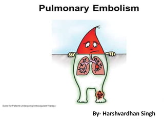
By- Harshvardhan Singh
 Pulmonary embolism (PE) is a blockage of the main
artery of the lung, or one of its branches by a
substance that has travelled from elsewhere in the
body through the bloodstream (embolism).
 Most commonly results from DVT that breaks off and
migrates to the lung, a process termed venous
thromboembolism.
 A small proportion of cases are caused by the
embolization of air, fat, or talc in drugs of
intravenous drug abusers.
• The obstruction of the blood flow through the
lungs and the resultant pressure on the right
ventricle of the heart leads to the symptoms and
signs of PE. The risk of PE is increased in various
situations, such as cancer or prolonged bed rest.
• This is an extremely common and highly lethal
condition that is a leading cause of death in all
age groups.
• One of the most prevalent disease processes
responsible for in-patient mortality (30%)
• Overlooked diagnosis
• 3rd most common cause of death.
• 2nd most commoncause of unexpected
death in most age groups.
• 60% of patients dying in the hospital have
had a PE.
• Diagnosis has been missed in about 70%
of the cases.
MOST COMMON SYMPTOMS
 Dyspnea (73%)
 Pleuritic chest pain (66%)
 Coughing
 Hemoptysis
RISK FACTORS
• Condition of stoppage or reduced blood flow
through the veins.
• Prolonged immobilization - fracture, paralysis,
bedridden due to illness or being elderly.
• Trauma and surgery
• Oral Contraception - combined with cigarette
smoking is well documented as a cause of
sudden death in healthy women.
• Pregnancy - cause is contributed to a
decrease in fibrinolytic function.
• Congenital( Hereditary antithrombin def, Leidin
V mutation)
ECG FINDINGS
• Sinus tachycardia: M/C abnormality
• Right heart strain pattern: seen as a RBBB
pattern or right axis deviation
• SIQIIITIII pattern: Deep S wave in lead I, Q
wave and T wave inversion in lead III;
This sign is seen as classical of PE
CHEST X-RAY FINDINGS
14% Normal
68% Atelectasis or parenchymal density
48% Pleural Effusion
35% Pleural based opacity
24% Elevated diaphragm
15% Prominent central pulmonary artery
7% Westermark’s sign
7% Cardiomegaly
5% Pulmonary edema
Initial CxR always NORMAL.
 May show – Collapse, consolidation, small pleural
effusion, elevated diaphragm.
 Pleural based opacities with convex medial margins are
also known as a Hampton's Hump
• Westermark sign – Dilatation of pulmonary
vessels proximal to embolism along with
collapse of distal vessels, often with a sharp
cut off.
Focal area of oligemia in the right middle zone and cutoff of the pulmonary artery
in the upper lobe of the right lung.
CTpulmonary angiography confirmed the presence of a thrombus in the right
pulmonary artery, with an occlusive thrombus in the pulmonary arteries of the
right upper and middle lobes
PALLA’S SIGN- enlarged right
descending pulmonary artery.
1.Diagnostic Criteriafor acute PE
 Complete arterial occlusion
 The artery may be enlarged compared with
adjacent patent vessels
ACUTE PE WITHIN THE POSTEROBASAL SEGMENTOF RLL.
2.Diagnostic Criteriafor acute PE
 A partial filling defect surrounded by
contrast material
 “polo mint” sign
 “railway track” sign
AcutePartial filling defect surrounded by contrast material (railway track sign) .
3.Diagnostic Criteriafor acute PE
 A peripheral intraluminal filling defect
 Acute angles with the arterial wall
AcuEccentrically positioned partial filling defect,which is surroundedby contrast material and forms
acute angleswith the arterial wall.
4.Diagnostic Criteriafor acute PE
 Peripheral wedge-shaped areas
of hyperattenuation
 Linear bands
Not specific for pulmonary embolism.
SPeripheral wedge-shaped area of hyperattenuation in the lung (arrow), a finding thatmay represent an
infarct,as well as a linear band (arrowhead).
5.Diagnostic Criteriafor acute PE
If Pulmonary arteries are indeterminate.
Lungs are clear.
To evaluate for pulmonary embolism do-
 Ventilation-perfusion scintigraphy
 Repeat CT pulmonary angiography
CHRONIC PE
1.Diagnostic Criteriafor chronic PE
 Complete occlusion
 smaller than adjacent patent vessels
Complete occlusion of vessels (arrowheads)that are smallerthan adjacentpatent vessels. Note the
collateral blood supply from a branch of the right hemidiaphragmatic artery (arrow).
2.Diagnostic Criteriafor chronic PE
 Peripheral, crescent-shaped, obtuse
angles with vessel wall
Chronic pulmonary embolism
3.Diagnostic Criteriafor chronic PE
 Smaller rencanalized arteries
4.Diagnostic Criteriafor chronic PE
 A web or flap within a contrast material–
filled artery
5.Diagnostic Criteriafor chronic PE
 Bronchial or other systemic collateral
vessels
Large chronic PE in the main and left main pulmonary arteries (arrowhead).
6.Diagnostic Criteriafor chronic PE
 Calcification within eccentric vessel
Pulmonary arterial wall calcificatio n (arrows), a secondary sign of chronic pulmonary
embolism.
7.Diagnostic Criteriafor chronic PE
 PA diameter > 30 mm
Pulmonary arterial HTN secondaryto chronic PE--PA41 mm in diameter
8.Diagnostic Criteriafor chronic PE
 Pericardial fluid
Acute Short axis of the right ventricle (dashed line)is wider than thatof the left ventricle (solid line)
Conclusion
Acute Chronic
Impacted artery large small
Angle acute obtuse
Others Polomint/railway track Recanalisation
Web/flap
Collateral arteries
Calcification
Mosaic
PHTN
Mosaic
right heart strain right heart strain
D-DIMER.
• For D-dimer <500ng/mL, negative predictive
value (NPV) 91-99%
• For D-dimer >500ng/mL, sens=93%, spec=25%,
and positive predictive value (PPV) = 30%
• Test is also useful for DVT rule out
VENTILATION-PERFUSION (V/Q) SCANS
• Identifies only ~50% of patients with PE.
• Abnormal (high + intermediate + low prob)
scans detect 98% of PE's but has low specificity
• About 60% of V/Q scans will be indeterminant
• Of intermediate probability scans, ~33% occur
with angiographically proven PE
LOWER EXTREMITY DOPPLER
USG.
• First-line if radiographic imaging
contraindicated or not readily available.
•Not likely required in patient with negative CT-PA
• Helpful to rule out DVT in patient with non-
diagnostic V/Q scan
• To evaluate for DVT as possible cause of PE or
to help rule in PE
• Up to 40% of patients with DVT without PE
symptoms will HAVE a PE by angiography.
• Serial US should be probably be performed in
patients with abnormal V/Q scans and positive
D-Dimers.
• These USG should be carried out on days 1, 3,
7, and 14.
PULMONARY ANGIOGRAPHY.
 GOLD STANDARD.
 Positive angiogram provides 100% certainty
that an obstruction exists in the pulmonary
artery.
 Negative angiogram provides > 90% certainty
in the exclusion of PE.
 Catherterisation of the subclavian vein
 Subclavian vein – Superior vena cava – right
atrium – right ventricle – main pulmonary artery
 Contrast
 DSA
Contrast Pulmonary Angiography
MAGNETIC RESONANCE
ANGIOGRAPHY (MRA)
• MRA for diagnosing PE are evolving rapidly
• Estimated sensitivity ~80% (~100% for larger
emboli), specificity 95%
• Non-invasive with little morbidity
• Dynamic gadolinium enhancement is used,
allowing high quality images
• Strongly consider prior to standard invasive
pulmonary angiography.
Thromboembolic material in both pulmonary arteries
(dotted arrows.
CT angiography clearly shows central embolic material (arrow).
(b) Coronal MR, showed similar information
(c) Axial image showing the thrombus
• Advantages
 Lack of ionizing
radiation
 Limitations
 Respiratory and cardiac motion artifact
 Suboptimal resolution for peripheral
pulmonary arteries
 Complicated blood flow patterns
CAUSESOF MISDIAGNOSIS OF
PULMONARY EMBOLISM
Causes of Misdiagnosis of PE:
Pathologic Factors
 Mucus Plug
Causes of Misdiagnosis of PE:
Pathologic Factors
 Perivascular Edema
Causes of Misdiagnosis of PE:
Pathologic Factors
 Localized Increase in Vascular Resistance
Causes of Misdiagnosis of PE:
Pathologic Factors
 Primary Pulmonary Artery Sarcoma(rare)
Causes of Misdiagnosis of PE:
Pathologic Factors
 Tumor Emboli
Causes of Misdiagnosis of PE:
Pathologic Factors
 Idiopathic Pulmonary Hypertension
Causes of Misdiagnosis of PE:
Pathologic Factors
 Proximal Interruption of the Pulmonary Artery
• Pulmonary embolus is a major health problem that is
highly treatable when diagnosed, but carries high risk for
morbidity and mortality is undiagnosed.
• Radiography is a major diagnostic tool in finding
pulmonary embolus.
• There are several diagnostic studies like CT, nuclear V/Q
scan and angiographic procedures that diagnose
pulmonary embolus. The main cause of PE is venous
thromboembolus that is diagnosed with ultrasound.
• Treatment can reduce the risk of recurring disease to
below 2%, which is medically acceptable.
Pulmonary embolism radiology imaging

Weitere ähnliche Inhalte

Was ist angesagt?

Presentation1.pptx, radiological signs in thoracic radiology.
Presentation1.pptx, radiological signs in thoracic radiology.Presentation1.pptx, radiological signs in thoracic radiology.
Presentation1.pptx, radiological signs in thoracic radiology.Abdellah Nazeer
 
Collapse consolidation
Collapse consolidationCollapse consolidation
Collapse consolidationairwave12
 
An approach to cardiac xray Dr. Muhammad Bin Zulfiqar
An approach to cardiac xray Dr. Muhammad Bin ZulfiqarAn approach to cardiac xray Dr. Muhammad Bin Zulfiqar
An approach to cardiac xray Dr. Muhammad Bin ZulfiqarDr. Muhammad Bin Zulfiqar
 
Solitary pulmonary nodules radiology pk ppt
Solitary pulmonary nodules  radiology pk pptSolitary pulmonary nodules  radiology pk ppt
Solitary pulmonary nodules radiology pk pptDr pradeep Kumar
 
Radiological imaging of mediastinal masses
Radiological imaging of mediastinal massesRadiological imaging of mediastinal masses
Radiological imaging of mediastinal massesPankaj Kaira
 
Presentation1.pptx radiological imaging of mediastinal masses .
Presentation1.pptx radiological imaging of mediastinal masses .Presentation1.pptx radiological imaging of mediastinal masses .
Presentation1.pptx radiological imaging of mediastinal masses .Abdellah Nazeer
 
Collapse & consolidation made simple - chest X-rayz
Collapse & consolidation made simple - chest X-rayzCollapse & consolidation made simple - chest X-rayz
Collapse & consolidation made simple - chest X-rayzDrNikrish Hegde
 
Signs in Chest Xray
Signs in Chest Xray Signs in Chest Xray
Signs in Chest Xray Archana Koshy
 
Pulmonary embolism imaging
Pulmonary embolism imagingPulmonary embolism imaging
Pulmonary embolism imagingDr. Soe Moe Htoo
 
Radiology Spotters mixed collection ppt
Radiology Spotters mixed collection pptRadiology Spotters mixed collection ppt
Radiology Spotters mixed collection pptDr pradeep Kumar
 
Presentation1.pptx, radiological imaging of copd.
Presentation1.pptx, radiological imaging of copd.Presentation1.pptx, radiological imaging of copd.
Presentation1.pptx, radiological imaging of copd.Abdellah Nazeer
 
Radiological features of Lung cancer Dr. Muhammad Bin Zulfiqar
Radiological features of Lung cancer Dr. Muhammad Bin ZulfiqarRadiological features of Lung cancer Dr. Muhammad Bin Zulfiqar
Radiological features of Lung cancer Dr. Muhammad Bin ZulfiqarDr. Muhammad Bin Zulfiqar
 
Imaging in mediastinal masses by Dr. Milan Silwal
Imaging in mediastinal masses by Dr. Milan SilwalImaging in mediastinal masses by Dr. Milan Silwal
Imaging in mediastinal masses by Dr. Milan SilwalMilan Silwal
 
Radiological imaging of pulmonary neoplasms
Radiological imaging of pulmonary neoplasmsRadiological imaging of pulmonary neoplasms
Radiological imaging of pulmonary neoplasmsPankaj Kaira
 
Diagnostic Imaging of Congenital Pulmonary Abnormalities
Diagnostic Imaging of Congenital Pulmonary AbnormalitiesDiagnostic Imaging of Congenital Pulmonary Abnormalities
Diagnostic Imaging of Congenital Pulmonary AbnormalitiesMohamed M.A. Zaitoun
 
Ascites and Pleural Effusion
 Ascites and Pleural Effusion Ascites and Pleural Effusion
Ascites and Pleural EffusionMedia Genie
 

Was ist angesagt? (20)

Presentation1.pptx, radiological signs in thoracic radiology.
Presentation1.pptx, radiological signs in thoracic radiology.Presentation1.pptx, radiological signs in thoracic radiology.
Presentation1.pptx, radiological signs in thoracic radiology.
 
Collapse consolidation
Collapse consolidationCollapse consolidation
Collapse consolidation
 
An approach to cardiac xray Dr. Muhammad Bin Zulfiqar
An approach to cardiac xray Dr. Muhammad Bin ZulfiqarAn approach to cardiac xray Dr. Muhammad Bin Zulfiqar
An approach to cardiac xray Dr. Muhammad Bin Zulfiqar
 
Solitary pulmonary nodules radiology pk ppt
Solitary pulmonary nodules  radiology pk pptSolitary pulmonary nodules  radiology pk ppt
Solitary pulmonary nodules radiology pk ppt
 
Radiological imaging of mediastinal masses
Radiological imaging of mediastinal massesRadiological imaging of mediastinal masses
Radiological imaging of mediastinal masses
 
Presentation1.pptx radiological imaging of mediastinal masses .
Presentation1.pptx radiological imaging of mediastinal masses .Presentation1.pptx radiological imaging of mediastinal masses .
Presentation1.pptx radiological imaging of mediastinal masses .
 
Collapse & consolidation made simple - chest X-rayz
Collapse & consolidation made simple - chest X-rayzCollapse & consolidation made simple - chest X-rayz
Collapse & consolidation made simple - chest X-rayz
 
Signs in Chest Xray
Signs in Chest Xray Signs in Chest Xray
Signs in Chest Xray
 
Pulmonary embolism imaging
Pulmonary embolism imagingPulmonary embolism imaging
Pulmonary embolism imaging
 
Radiology Spotters mixed collection ppt
Radiology Spotters mixed collection pptRadiology Spotters mixed collection ppt
Radiology Spotters mixed collection ppt
 
CXR: Lung Mass - Mediastinal Mass
CXR: Lung Mass - Mediastinal MassCXR: Lung Mass - Mediastinal Mass
CXR: Lung Mass - Mediastinal Mass
 
Presentation1.pptx, radiological imaging of copd.
Presentation1.pptx, radiological imaging of copd.Presentation1.pptx, radiological imaging of copd.
Presentation1.pptx, radiological imaging of copd.
 
CTA OF PULMONARY EMBOLISM
CTA OF PULMONARY EMBOLISMCTA OF PULMONARY EMBOLISM
CTA OF PULMONARY EMBOLISM
 
Radiological features of Lung cancer Dr. Muhammad Bin Zulfiqar
Radiological features of Lung cancer Dr. Muhammad Bin ZulfiqarRadiological features of Lung cancer Dr. Muhammad Bin Zulfiqar
Radiological features of Lung cancer Dr. Muhammad Bin Zulfiqar
 
Spots with keys (2)
Spots with keys (2)Spots with keys (2)
Spots with keys (2)
 
Imaging in mediastinal masses by Dr. Milan Silwal
Imaging in mediastinal masses by Dr. Milan SilwalImaging in mediastinal masses by Dr. Milan Silwal
Imaging in mediastinal masses by Dr. Milan Silwal
 
Radiological imaging of pulmonary neoplasms
Radiological imaging of pulmonary neoplasmsRadiological imaging of pulmonary neoplasms
Radiological imaging of pulmonary neoplasms
 
CT CHEST ANATOMY
CT CHEST ANATOMYCT CHEST ANATOMY
CT CHEST ANATOMY
 
Diagnostic Imaging of Congenital Pulmonary Abnormalities
Diagnostic Imaging of Congenital Pulmonary AbnormalitiesDiagnostic Imaging of Congenital Pulmonary Abnormalities
Diagnostic Imaging of Congenital Pulmonary Abnormalities
 
Ascites and Pleural Effusion
 Ascites and Pleural Effusion Ascites and Pleural Effusion
Ascites and Pleural Effusion
 

Ähnlich wie Pulmonary embolism radiology imaging

Pulmonary embolism,overview
Pulmonary embolism,overviewPulmonary embolism,overview
Pulmonary embolism,overviewEman Mahmoud
 
deep vein thrombosis and pe
deep vein thrombosis and pedeep vein thrombosis and pe
deep vein thrombosis and pePraveen Shukla
 
Pulmonary Embolism- Diagnosis by Dr.Tinku Joseph
Pulmonary Embolism- Diagnosis  by Dr.Tinku JosephPulmonary Embolism- Diagnosis  by Dr.Tinku Joseph
Pulmonary Embolism- Diagnosis by Dr.Tinku JosephDr.Tinku Joseph
 
Pulmonary embolism@ghanem@.2013
Pulmonary embolism@ghanem@.2013Pulmonary embolism@ghanem@.2013
Pulmonary embolism@ghanem@.2013Islam Ghanem
 
Acute Pulmonary Embolism: Introduction, Clinical presentation, Classification...
Acute Pulmonary Embolism: Introduction, Clinical presentation, Classification...Acute Pulmonary Embolism: Introduction, Clinical presentation, Classification...
Acute Pulmonary Embolism: Introduction, Clinical presentation, Classification...RichardKhoi
 
Diagnosis of Pulmonary Embolism - by Dr KD DELE
Diagnosis of Pulmonary Embolism - by Dr KD DELEDiagnosis of Pulmonary Embolism - by Dr KD DELE
Diagnosis of Pulmonary Embolism - by Dr KD DELEKemi Dele-Ijagbulu
 
Pulmonary embolisim
Pulmonary embolisimPulmonary embolisim
Pulmonary embolisimdratin75
 
pulomonary thromboembolism.pptx
pulomonary thromboembolism.pptxpulomonary thromboembolism.pptx
pulomonary thromboembolism.pptxSureshChevagoni1
 
VP pulmonary thmboembolism.pptx
VP pulmonary thmboembolism.pptxVP pulmonary thmboembolism.pptx
VP pulmonary thmboembolism.pptxvishwanath0908
 
Pulmonary embolism
Pulmonary embolismPulmonary embolism
Pulmonary embolismoday abdow
 
4.Pe(English终)
4.Pe(English终)4.Pe(English终)
4.Pe(English终)Deep Deep
 
Pulmonaryembolism ....tanmay new
Pulmonaryembolism ....tanmay newPulmonaryembolism ....tanmay new
Pulmonaryembolism ....tanmay newTanmay Jain
 
Pulmonary angiography
Pulmonary angiographyPulmonary angiography
Pulmonary angiographyPankaj Kaira
 
Update on Pulmonary Embolism
Update on Pulmonary EmbolismUpdate on Pulmonary Embolism
Update on Pulmonary EmbolismMuhanad Majeed
 

Ähnlich wie Pulmonary embolism radiology imaging (20)

Pulmonary embolism,overview
Pulmonary embolism,overviewPulmonary embolism,overview
Pulmonary embolism,overview
 
deep vein thrombosis and pe
deep vein thrombosis and pedeep vein thrombosis and pe
deep vein thrombosis and pe
 
Chronic PE
Chronic PEChronic PE
Chronic PE
 
Pulmonary embolism
Pulmonary embolismPulmonary embolism
Pulmonary embolism
 
Pulmonary Embolism- Diagnosis by Dr.Tinku Joseph
Pulmonary Embolism- Diagnosis  by Dr.Tinku JosephPulmonary Embolism- Diagnosis  by Dr.Tinku Joseph
Pulmonary Embolism- Diagnosis by Dr.Tinku Joseph
 
Pulmonary embolism@ghanem@.2013
Pulmonary embolism@ghanem@.2013Pulmonary embolism@ghanem@.2013
Pulmonary embolism@ghanem@.2013
 
Acute Pulmonary Embolism: Introduction, Clinical presentation, Classification...
Acute Pulmonary Embolism: Introduction, Clinical presentation, Classification...Acute Pulmonary Embolism: Introduction, Clinical presentation, Classification...
Acute Pulmonary Embolism: Introduction, Clinical presentation, Classification...
 
Diagnosis of Pulmonary Embolism - by Dr KD DELE
Diagnosis of Pulmonary Embolism - by Dr KD DELEDiagnosis of Pulmonary Embolism - by Dr KD DELE
Diagnosis of Pulmonary Embolism - by Dr KD DELE
 
Pulmonary Embolism
Pulmonary Embolism Pulmonary Embolism
Pulmonary Embolism
 
Pulmonary embolisim
Pulmonary embolisimPulmonary embolisim
Pulmonary embolisim
 
Pulmonary embolism
Pulmonary embolism Pulmonary embolism
Pulmonary embolism
 
Mac mahon rc 2016 pe
Mac mahon rc 2016 peMac mahon rc 2016 pe
Mac mahon rc 2016 pe
 
pulomonary thromboembolism.pptx
pulomonary thromboembolism.pptxpulomonary thromboembolism.pptx
pulomonary thromboembolism.pptx
 
VP pulmonary thmboembolism.pptx
VP pulmonary thmboembolism.pptxVP pulmonary thmboembolism.pptx
VP pulmonary thmboembolism.pptx
 
Pulmonaryembolism final
Pulmonaryembolism finalPulmonaryembolism final
Pulmonaryembolism final
 
Pulmonary embolism
Pulmonary embolismPulmonary embolism
Pulmonary embolism
 
4.Pe(English终)
4.Pe(English终)4.Pe(English终)
4.Pe(English终)
 
Pulmonaryembolism ....tanmay new
Pulmonaryembolism ....tanmay newPulmonaryembolism ....tanmay new
Pulmonaryembolism ....tanmay new
 
Pulmonary angiography
Pulmonary angiographyPulmonary angiography
Pulmonary angiography
 
Update on Pulmonary Embolism
Update on Pulmonary EmbolismUpdate on Pulmonary Embolism
Update on Pulmonary Embolism
 

Kürzlich hochgeladen

💎VVIP Kolkata Call Girls Parganas🩱7001035870🩱Independent Girl ( Ac Rooms Avai...
💎VVIP Kolkata Call Girls Parganas🩱7001035870🩱Independent Girl ( Ac Rooms Avai...💎VVIP Kolkata Call Girls Parganas🩱7001035870🩱Independent Girl ( Ac Rooms Avai...
💎VVIP Kolkata Call Girls Parganas🩱7001035870🩱Independent Girl ( Ac Rooms Avai...Taniya Sharma
 
Call Girl Coimbatore Prisha☎️ 8250192130 Independent Escort Service Coimbatore
Call Girl Coimbatore Prisha☎️  8250192130 Independent Escort Service CoimbatoreCall Girl Coimbatore Prisha☎️  8250192130 Independent Escort Service Coimbatore
Call Girl Coimbatore Prisha☎️ 8250192130 Independent Escort Service Coimbatorenarwatsonia7
 
VIP Call Girls Tirunelveli Aaradhya 8250192130 Independent Escort Service Tir...
VIP Call Girls Tirunelveli Aaradhya 8250192130 Independent Escort Service Tir...VIP Call Girls Tirunelveli Aaradhya 8250192130 Independent Escort Service Tir...
VIP Call Girls Tirunelveli Aaradhya 8250192130 Independent Escort Service Tir...narwatsonia7
 
Top Rated Bangalore Call Girls Richmond Circle ⟟ 8250192130 ⟟ Call Me For Gen...
Top Rated Bangalore Call Girls Richmond Circle ⟟ 8250192130 ⟟ Call Me For Gen...Top Rated Bangalore Call Girls Richmond Circle ⟟ 8250192130 ⟟ Call Me For Gen...
Top Rated Bangalore Call Girls Richmond Circle ⟟ 8250192130 ⟟ Call Me For Gen...narwatsonia7
 
Bangalore Call Girls Hebbal Kempapura Number 7001035870 Meetin With Bangalor...
Bangalore Call Girls Hebbal Kempapura Number 7001035870  Meetin With Bangalor...Bangalore Call Girls Hebbal Kempapura Number 7001035870  Meetin With Bangalor...
Bangalore Call Girls Hebbal Kempapura Number 7001035870 Meetin With Bangalor...narwatsonia7
 
Call Girls Service Surat Samaira ❤️🍑 8250192130 👄 Independent Escort Service ...
Call Girls Service Surat Samaira ❤️🍑 8250192130 👄 Independent Escort Service ...Call Girls Service Surat Samaira ❤️🍑 8250192130 👄 Independent Escort Service ...
Call Girls Service Surat Samaira ❤️🍑 8250192130 👄 Independent Escort Service ...CALL GIRLS
 
VIP Russian Call Girls in Varanasi Samaira 8250192130 Independent Escort Serv...
VIP Russian Call Girls in Varanasi Samaira 8250192130 Independent Escort Serv...VIP Russian Call Girls in Varanasi Samaira 8250192130 Independent Escort Serv...
VIP Russian Call Girls in Varanasi Samaira 8250192130 Independent Escort Serv...Neha Kaur
 
VIP Service Call Girls Sindhi Colony 📳 7877925207 For 18+ VIP Call Girl At Th...
VIP Service Call Girls Sindhi Colony 📳 7877925207 For 18+ VIP Call Girl At Th...VIP Service Call Girls Sindhi Colony 📳 7877925207 For 18+ VIP Call Girl At Th...
VIP Service Call Girls Sindhi Colony 📳 7877925207 For 18+ VIP Call Girl At Th...jageshsingh5554
 
Call Girls Varanasi Just Call 9907093804 Top Class Call Girl Service Available
Call Girls Varanasi Just Call 9907093804 Top Class Call Girl Service AvailableCall Girls Varanasi Just Call 9907093804 Top Class Call Girl Service Available
Call Girls Varanasi Just Call 9907093804 Top Class Call Girl Service AvailableDipal Arora
 
Call Girls Coimbatore Just Call 9907093804 Top Class Call Girl Service Available
Call Girls Coimbatore Just Call 9907093804 Top Class Call Girl Service AvailableCall Girls Coimbatore Just Call 9907093804 Top Class Call Girl Service Available
Call Girls Coimbatore Just Call 9907093804 Top Class Call Girl Service AvailableDipal Arora
 
Top Rated Bangalore Call Girls Mg Road ⟟ 8250192130 ⟟ Call Me For Genuine Sex...
Top Rated Bangalore Call Girls Mg Road ⟟ 8250192130 ⟟ Call Me For Genuine Sex...Top Rated Bangalore Call Girls Mg Road ⟟ 8250192130 ⟟ Call Me For Genuine Sex...
Top Rated Bangalore Call Girls Mg Road ⟟ 8250192130 ⟟ Call Me For Genuine Sex...narwatsonia7
 
Call Girl Number in Vashi Mumbai📲 9833363713 💞 Full Night Enjoy
Call Girl Number in Vashi Mumbai📲 9833363713 💞 Full Night EnjoyCall Girl Number in Vashi Mumbai📲 9833363713 💞 Full Night Enjoy
Call Girl Number in Vashi Mumbai📲 9833363713 💞 Full Night Enjoybabeytanya
 
Call Girls Service Jaipur Grishma WhatsApp ❤8445551418 VIP Call Girls Jaipur
Call Girls Service Jaipur Grishma WhatsApp ❤8445551418 VIP Call Girls JaipurCall Girls Service Jaipur Grishma WhatsApp ❤8445551418 VIP Call Girls Jaipur
Call Girls Service Jaipur Grishma WhatsApp ❤8445551418 VIP Call Girls Jaipurparulsinha
 
Kesar Bagh Call Girl Price 9548273370 , Lucknow Call Girls Service
Kesar Bagh Call Girl Price 9548273370 , Lucknow Call Girls ServiceKesar Bagh Call Girl Price 9548273370 , Lucknow Call Girls Service
Kesar Bagh Call Girl Price 9548273370 , Lucknow Call Girls Servicemakika9823
 
Low Rate Call Girls Kochi Anika 8250192130 Independent Escort Service Kochi
Low Rate Call Girls Kochi Anika 8250192130 Independent Escort Service KochiLow Rate Call Girls Kochi Anika 8250192130 Independent Escort Service Kochi
Low Rate Call Girls Kochi Anika 8250192130 Independent Escort Service KochiSuhani Kapoor
 
Book Paid Powai Call Girls Mumbai 𖠋 9930245274 𖠋Low Budget Full Independent H...
Book Paid Powai Call Girls Mumbai 𖠋 9930245274 𖠋Low Budget Full Independent H...Book Paid Powai Call Girls Mumbai 𖠋 9930245274 𖠋Low Budget Full Independent H...
Book Paid Powai Call Girls Mumbai 𖠋 9930245274 𖠋Low Budget Full Independent H...Call Girls in Nagpur High Profile
 
Russian Escorts Girls Nehru Place ZINATHI 🔝9711199012 ☪ 24/7 Call Girls Delhi
Russian Escorts Girls  Nehru Place ZINATHI 🔝9711199012 ☪ 24/7 Call Girls DelhiRussian Escorts Girls  Nehru Place ZINATHI 🔝9711199012 ☪ 24/7 Call Girls Delhi
Russian Escorts Girls Nehru Place ZINATHI 🔝9711199012 ☪ 24/7 Call Girls DelhiAlinaDevecerski
 
♛VVIP Hyderabad Call Girls Chintalkunta🖕7001035870🖕Riya Kappor Top Call Girl ...
♛VVIP Hyderabad Call Girls Chintalkunta🖕7001035870🖕Riya Kappor Top Call Girl ...♛VVIP Hyderabad Call Girls Chintalkunta🖕7001035870🖕Riya Kappor Top Call Girl ...
♛VVIP Hyderabad Call Girls Chintalkunta🖕7001035870🖕Riya Kappor Top Call Girl ...astropune
 
Call Girls Horamavu WhatsApp Number 7001035870 Meeting With Bangalore Escorts
Call Girls Horamavu WhatsApp Number 7001035870 Meeting With Bangalore EscortsCall Girls Horamavu WhatsApp Number 7001035870 Meeting With Bangalore Escorts
Call Girls Horamavu WhatsApp Number 7001035870 Meeting With Bangalore Escortsvidya singh
 

Kürzlich hochgeladen (20)

💎VVIP Kolkata Call Girls Parganas🩱7001035870🩱Independent Girl ( Ac Rooms Avai...
💎VVIP Kolkata Call Girls Parganas🩱7001035870🩱Independent Girl ( Ac Rooms Avai...💎VVIP Kolkata Call Girls Parganas🩱7001035870🩱Independent Girl ( Ac Rooms Avai...
💎VVIP Kolkata Call Girls Parganas🩱7001035870🩱Independent Girl ( Ac Rooms Avai...
 
Escort Service Call Girls In Sarita Vihar,, 99530°56974 Delhi NCR
Escort Service Call Girls In Sarita Vihar,, 99530°56974 Delhi NCREscort Service Call Girls In Sarita Vihar,, 99530°56974 Delhi NCR
Escort Service Call Girls In Sarita Vihar,, 99530°56974 Delhi NCR
 
Call Girl Coimbatore Prisha☎️ 8250192130 Independent Escort Service Coimbatore
Call Girl Coimbatore Prisha☎️  8250192130 Independent Escort Service CoimbatoreCall Girl Coimbatore Prisha☎️  8250192130 Independent Escort Service Coimbatore
Call Girl Coimbatore Prisha☎️ 8250192130 Independent Escort Service Coimbatore
 
VIP Call Girls Tirunelveli Aaradhya 8250192130 Independent Escort Service Tir...
VIP Call Girls Tirunelveli Aaradhya 8250192130 Independent Escort Service Tir...VIP Call Girls Tirunelveli Aaradhya 8250192130 Independent Escort Service Tir...
VIP Call Girls Tirunelveli Aaradhya 8250192130 Independent Escort Service Tir...
 
Top Rated Bangalore Call Girls Richmond Circle ⟟ 8250192130 ⟟ Call Me For Gen...
Top Rated Bangalore Call Girls Richmond Circle ⟟ 8250192130 ⟟ Call Me For Gen...Top Rated Bangalore Call Girls Richmond Circle ⟟ 8250192130 ⟟ Call Me For Gen...
Top Rated Bangalore Call Girls Richmond Circle ⟟ 8250192130 ⟟ Call Me For Gen...
 
Bangalore Call Girls Hebbal Kempapura Number 7001035870 Meetin With Bangalor...
Bangalore Call Girls Hebbal Kempapura Number 7001035870  Meetin With Bangalor...Bangalore Call Girls Hebbal Kempapura Number 7001035870  Meetin With Bangalor...
Bangalore Call Girls Hebbal Kempapura Number 7001035870 Meetin With Bangalor...
 
Call Girls Service Surat Samaira ❤️🍑 8250192130 👄 Independent Escort Service ...
Call Girls Service Surat Samaira ❤️🍑 8250192130 👄 Independent Escort Service ...Call Girls Service Surat Samaira ❤️🍑 8250192130 👄 Independent Escort Service ...
Call Girls Service Surat Samaira ❤️🍑 8250192130 👄 Independent Escort Service ...
 
VIP Russian Call Girls in Varanasi Samaira 8250192130 Independent Escort Serv...
VIP Russian Call Girls in Varanasi Samaira 8250192130 Independent Escort Serv...VIP Russian Call Girls in Varanasi Samaira 8250192130 Independent Escort Serv...
VIP Russian Call Girls in Varanasi Samaira 8250192130 Independent Escort Serv...
 
VIP Service Call Girls Sindhi Colony 📳 7877925207 For 18+ VIP Call Girl At Th...
VIP Service Call Girls Sindhi Colony 📳 7877925207 For 18+ VIP Call Girl At Th...VIP Service Call Girls Sindhi Colony 📳 7877925207 For 18+ VIP Call Girl At Th...
VIP Service Call Girls Sindhi Colony 📳 7877925207 For 18+ VIP Call Girl At Th...
 
Call Girls Varanasi Just Call 9907093804 Top Class Call Girl Service Available
Call Girls Varanasi Just Call 9907093804 Top Class Call Girl Service AvailableCall Girls Varanasi Just Call 9907093804 Top Class Call Girl Service Available
Call Girls Varanasi Just Call 9907093804 Top Class Call Girl Service Available
 
Call Girls Coimbatore Just Call 9907093804 Top Class Call Girl Service Available
Call Girls Coimbatore Just Call 9907093804 Top Class Call Girl Service AvailableCall Girls Coimbatore Just Call 9907093804 Top Class Call Girl Service Available
Call Girls Coimbatore Just Call 9907093804 Top Class Call Girl Service Available
 
Top Rated Bangalore Call Girls Mg Road ⟟ 8250192130 ⟟ Call Me For Genuine Sex...
Top Rated Bangalore Call Girls Mg Road ⟟ 8250192130 ⟟ Call Me For Genuine Sex...Top Rated Bangalore Call Girls Mg Road ⟟ 8250192130 ⟟ Call Me For Genuine Sex...
Top Rated Bangalore Call Girls Mg Road ⟟ 8250192130 ⟟ Call Me For Genuine Sex...
 
Call Girl Number in Vashi Mumbai📲 9833363713 💞 Full Night Enjoy
Call Girl Number in Vashi Mumbai📲 9833363713 💞 Full Night EnjoyCall Girl Number in Vashi Mumbai📲 9833363713 💞 Full Night Enjoy
Call Girl Number in Vashi Mumbai📲 9833363713 💞 Full Night Enjoy
 
Call Girls Service Jaipur Grishma WhatsApp ❤8445551418 VIP Call Girls Jaipur
Call Girls Service Jaipur Grishma WhatsApp ❤8445551418 VIP Call Girls JaipurCall Girls Service Jaipur Grishma WhatsApp ❤8445551418 VIP Call Girls Jaipur
Call Girls Service Jaipur Grishma WhatsApp ❤8445551418 VIP Call Girls Jaipur
 
Kesar Bagh Call Girl Price 9548273370 , Lucknow Call Girls Service
Kesar Bagh Call Girl Price 9548273370 , Lucknow Call Girls ServiceKesar Bagh Call Girl Price 9548273370 , Lucknow Call Girls Service
Kesar Bagh Call Girl Price 9548273370 , Lucknow Call Girls Service
 
Low Rate Call Girls Kochi Anika 8250192130 Independent Escort Service Kochi
Low Rate Call Girls Kochi Anika 8250192130 Independent Escort Service KochiLow Rate Call Girls Kochi Anika 8250192130 Independent Escort Service Kochi
Low Rate Call Girls Kochi Anika 8250192130 Independent Escort Service Kochi
 
Book Paid Powai Call Girls Mumbai 𖠋 9930245274 𖠋Low Budget Full Independent H...
Book Paid Powai Call Girls Mumbai 𖠋 9930245274 𖠋Low Budget Full Independent H...Book Paid Powai Call Girls Mumbai 𖠋 9930245274 𖠋Low Budget Full Independent H...
Book Paid Powai Call Girls Mumbai 𖠋 9930245274 𖠋Low Budget Full Independent H...
 
Russian Escorts Girls Nehru Place ZINATHI 🔝9711199012 ☪ 24/7 Call Girls Delhi
Russian Escorts Girls  Nehru Place ZINATHI 🔝9711199012 ☪ 24/7 Call Girls DelhiRussian Escorts Girls  Nehru Place ZINATHI 🔝9711199012 ☪ 24/7 Call Girls Delhi
Russian Escorts Girls Nehru Place ZINATHI 🔝9711199012 ☪ 24/7 Call Girls Delhi
 
♛VVIP Hyderabad Call Girls Chintalkunta🖕7001035870🖕Riya Kappor Top Call Girl ...
♛VVIP Hyderabad Call Girls Chintalkunta🖕7001035870🖕Riya Kappor Top Call Girl ...♛VVIP Hyderabad Call Girls Chintalkunta🖕7001035870🖕Riya Kappor Top Call Girl ...
♛VVIP Hyderabad Call Girls Chintalkunta🖕7001035870🖕Riya Kappor Top Call Girl ...
 
Call Girls Horamavu WhatsApp Number 7001035870 Meeting With Bangalore Escorts
Call Girls Horamavu WhatsApp Number 7001035870 Meeting With Bangalore EscortsCall Girls Horamavu WhatsApp Number 7001035870 Meeting With Bangalore Escorts
Call Girls Horamavu WhatsApp Number 7001035870 Meeting With Bangalore Escorts
 

Pulmonary embolism radiology imaging

  • 2.  Pulmonary embolism (PE) is a blockage of the main artery of the lung, or one of its branches by a substance that has travelled from elsewhere in the body through the bloodstream (embolism).  Most commonly results from DVT that breaks off and migrates to the lung, a process termed venous thromboembolism.  A small proportion of cases are caused by the embolization of air, fat, or talc in drugs of intravenous drug abusers.
  • 3. • The obstruction of the blood flow through the lungs and the resultant pressure on the right ventricle of the heart leads to the symptoms and signs of PE. The risk of PE is increased in various situations, such as cancer or prolonged bed rest. • This is an extremely common and highly lethal condition that is a leading cause of death in all age groups. • One of the most prevalent disease processes responsible for in-patient mortality (30%) • Overlooked diagnosis
  • 4. • 3rd most common cause of death. • 2nd most commoncause of unexpected death in most age groups. • 60% of patients dying in the hospital have had a PE. • Diagnosis has been missed in about 70% of the cases.
  • 5. MOST COMMON SYMPTOMS  Dyspnea (73%)  Pleuritic chest pain (66%)  Coughing  Hemoptysis
  • 6. RISK FACTORS • Condition of stoppage or reduced blood flow through the veins. • Prolonged immobilization - fracture, paralysis, bedridden due to illness or being elderly. • Trauma and surgery • Oral Contraception - combined with cigarette smoking is well documented as a cause of sudden death in healthy women. • Pregnancy - cause is contributed to a decrease in fibrinolytic function. • Congenital( Hereditary antithrombin def, Leidin V mutation)
  • 7. ECG FINDINGS • Sinus tachycardia: M/C abnormality • Right heart strain pattern: seen as a RBBB pattern or right axis deviation • SIQIIITIII pattern: Deep S wave in lead I, Q wave and T wave inversion in lead III; This sign is seen as classical of PE
  • 8.
  • 9.
  • 10.
  • 11. CHEST X-RAY FINDINGS 14% Normal 68% Atelectasis or parenchymal density 48% Pleural Effusion 35% Pleural based opacity 24% Elevated diaphragm 15% Prominent central pulmonary artery 7% Westermark’s sign 7% Cardiomegaly 5% Pulmonary edema
  • 13.  May show – Collapse, consolidation, small pleural effusion, elevated diaphragm.  Pleural based opacities with convex medial margins are also known as a Hampton's Hump
  • 14.
  • 15. • Westermark sign – Dilatation of pulmonary vessels proximal to embolism along with collapse of distal vessels, often with a sharp cut off.
  • 16. Focal area of oligemia in the right middle zone and cutoff of the pulmonary artery in the upper lobe of the right lung. CTpulmonary angiography confirmed the presence of a thrombus in the right pulmonary artery, with an occlusive thrombus in the pulmonary arteries of the right upper and middle lobes
  • 17. PALLA’S SIGN- enlarged right descending pulmonary artery.
  • 18. 1.Diagnostic Criteriafor acute PE  Complete arterial occlusion  The artery may be enlarged compared with adjacent patent vessels
  • 19.
  • 20. ACUTE PE WITHIN THE POSTEROBASAL SEGMENTOF RLL.
  • 21. 2.Diagnostic Criteriafor acute PE  A partial filling defect surrounded by contrast material  “polo mint” sign  “railway track” sign
  • 22.
  • 23. AcutePartial filling defect surrounded by contrast material (railway track sign) .
  • 24. 3.Diagnostic Criteriafor acute PE  A peripheral intraluminal filling defect  Acute angles with the arterial wall
  • 25. AcuEccentrically positioned partial filling defect,which is surroundedby contrast material and forms acute angleswith the arterial wall.
  • 26. 4.Diagnostic Criteriafor acute PE  Peripheral wedge-shaped areas of hyperattenuation  Linear bands Not specific for pulmonary embolism.
  • 27. SPeripheral wedge-shaped area of hyperattenuation in the lung (arrow), a finding thatmay represent an infarct,as well as a linear band (arrowhead).
  • 28. 5.Diagnostic Criteriafor acute PE If Pulmonary arteries are indeterminate. Lungs are clear. To evaluate for pulmonary embolism do-  Ventilation-perfusion scintigraphy  Repeat CT pulmonary angiography
  • 30. 1.Diagnostic Criteriafor chronic PE  Complete occlusion  smaller than adjacent patent vessels
  • 31. Complete occlusion of vessels (arrowheads)that are smallerthan adjacentpatent vessels. Note the collateral blood supply from a branch of the right hemidiaphragmatic artery (arrow).
  • 32. 2.Diagnostic Criteriafor chronic PE  Peripheral, crescent-shaped, obtuse angles with vessel wall
  • 34. 3.Diagnostic Criteriafor chronic PE  Smaller rencanalized arteries
  • 35.
  • 36. 4.Diagnostic Criteriafor chronic PE  A web or flap within a contrast material– filled artery
  • 37.
  • 38. 5.Diagnostic Criteriafor chronic PE  Bronchial or other systemic collateral vessels
  • 39. Large chronic PE in the main and left main pulmonary arteries (arrowhead).
  • 40. 6.Diagnostic Criteriafor chronic PE  Calcification within eccentric vessel
  • 41. Pulmonary arterial wall calcificatio n (arrows), a secondary sign of chronic pulmonary embolism.
  • 42. 7.Diagnostic Criteriafor chronic PE  PA diameter > 30 mm
  • 43. Pulmonary arterial HTN secondaryto chronic PE--PA41 mm in diameter
  • 44. 8.Diagnostic Criteriafor chronic PE  Pericardial fluid
  • 45. Acute Short axis of the right ventricle (dashed line)is wider than thatof the left ventricle (solid line)
  • 46. Conclusion Acute Chronic Impacted artery large small Angle acute obtuse Others Polomint/railway track Recanalisation Web/flap Collateral arteries Calcification Mosaic PHTN Mosaic right heart strain right heart strain
  • 47. D-DIMER. • For D-dimer <500ng/mL, negative predictive value (NPV) 91-99% • For D-dimer >500ng/mL, sens=93%, spec=25%, and positive predictive value (PPV) = 30% • Test is also useful for DVT rule out
  • 48. VENTILATION-PERFUSION (V/Q) SCANS • Identifies only ~50% of patients with PE. • Abnormal (high + intermediate + low prob) scans detect 98% of PE's but has low specificity • About 60% of V/Q scans will be indeterminant • Of intermediate probability scans, ~33% occur with angiographically proven PE
  • 49.
  • 50.
  • 51. LOWER EXTREMITY DOPPLER USG. • First-line if radiographic imaging contraindicated or not readily available. •Not likely required in patient with negative CT-PA • Helpful to rule out DVT in patient with non- diagnostic V/Q scan
  • 52. • To evaluate for DVT as possible cause of PE or to help rule in PE • Up to 40% of patients with DVT without PE symptoms will HAVE a PE by angiography. • Serial US should be probably be performed in patients with abnormal V/Q scans and positive D-Dimers. • These USG should be carried out on days 1, 3, 7, and 14.
  • 53. PULMONARY ANGIOGRAPHY.  GOLD STANDARD.  Positive angiogram provides 100% certainty that an obstruction exists in the pulmonary artery.  Negative angiogram provides > 90% certainty in the exclusion of PE.
  • 54.  Catherterisation of the subclavian vein  Subclavian vein – Superior vena cava – right atrium – right ventricle – main pulmonary artery  Contrast  DSA
  • 56. MAGNETIC RESONANCE ANGIOGRAPHY (MRA) • MRA for diagnosing PE are evolving rapidly • Estimated sensitivity ~80% (~100% for larger emboli), specificity 95% • Non-invasive with little morbidity • Dynamic gadolinium enhancement is used, allowing high quality images • Strongly consider prior to standard invasive pulmonary angiography.
  • 57. Thromboembolic material in both pulmonary arteries (dotted arrows.
  • 58. CT angiography clearly shows central embolic material (arrow). (b) Coronal MR, showed similar information (c) Axial image showing the thrombus
  • 59. • Advantages  Lack of ionizing radiation  Limitations  Respiratory and cardiac motion artifact  Suboptimal resolution for peripheral pulmonary arteries  Complicated blood flow patterns
  • 61. Causes of Misdiagnosis of PE: Pathologic Factors  Mucus Plug
  • 62. Causes of Misdiagnosis of PE: Pathologic Factors  Perivascular Edema
  • 63. Causes of Misdiagnosis of PE: Pathologic Factors  Localized Increase in Vascular Resistance
  • 64. Causes of Misdiagnosis of PE: Pathologic Factors  Primary Pulmonary Artery Sarcoma(rare)
  • 65. Causes of Misdiagnosis of PE: Pathologic Factors  Tumor Emboli
  • 66. Causes of Misdiagnosis of PE: Pathologic Factors  Idiopathic Pulmonary Hypertension
  • 67. Causes of Misdiagnosis of PE: Pathologic Factors  Proximal Interruption of the Pulmonary Artery
  • 68. • Pulmonary embolus is a major health problem that is highly treatable when diagnosed, but carries high risk for morbidity and mortality is undiagnosed. • Radiography is a major diagnostic tool in finding pulmonary embolus. • There are several diagnostic studies like CT, nuclear V/Q scan and angiographic procedures that diagnose pulmonary embolus. The main cause of PE is venous thromboembolus that is diagnosed with ultrasound. • Treatment can reduce the risk of recurring disease to below 2%, which is medically acceptable.