Call Girls Jabalpur Just Call 8250077686 Top Class Call Girl Service Available
Respiratory System
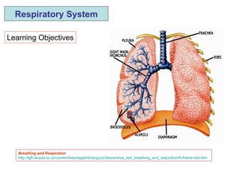
1. Learning Objectives Respiratory System Breathing and Respiration http://lgfl.skoool.co.uk/content/keystage4/biology/pc/lessons/uk_ks4_breathing_and_respiration/h-frame-ns6.htm
2. Cells require a continuous supply of energy in order to survive - achieved by cell respiration, which occurs in the cytoplasm and mitochondria of the cell. Aerobic respiration requires O 2 from air (via the respiratory system) and energy rich molecules (principally glucose) from food (via the digestive system). Results in the complete breakdown of glucose to CO 2 and H 2 O , with the production of a large amount of energy ( ATP ) CO 2 , a waste product of respiration is excreted via the respiratory system Cell Respiration Anaerobic respiration occurs in the absence of oxygen (e.g. in muscles during strenuous exercise). Glucose is partially broken down to lactic acid , with the production of a lower amount of energy
3. Energy - needed for movement (muscle contraction), growth , keeping warm , , etc ctive transport Aerobic Respiration Anaerobic respiration Uses oxygen In absence of oxygen CO 2 produced No CO 2 produced Energy (large amount) Energy (small amount) Rapid release of limited energy Glucose + Oxygen During vigorous exercise (no O 2 available) Produces lactic acid Muscle fatigue (and cramp) Carbon dioxide + Water Glucose + ENERGY (large amount) Lactic acid + ENERGY (limited amount)
6. Tests for CO 2 and O2 CO2 test – bubble through lime water (calcium hydroxide)) – clear to cloudy (milky) – forms calcium carbonate (chalk) O2 – glowing splint – glows brighter – supports combustion Differences between inspired & expired air http://lgfl.skoool.co.uk/content/keystage4/biology/pc/modules/breathing_respiration/inhaled_exhaled_air/index.html
7. Oxygenated blood to pulmonary vein Deoxygenated blood from pulmonary artery O 2 CO 2 CO 2 from plasma O 2 into red cells http://lgfl.skoool.co.uk/content/keystage4/biology/pc/modules/breathing_respiration/gas_exchange/index.html
10. Nose/Nasal cavity Filters and warms incoming air Pharynx Common passageway for respiratory and digestive system Larynx (Voice box) Opening to trachea – contains vocal cords – epiglottis prevents food entering the trachea Trachea (windpipe) C-shaped rings of cartilage – prevent collapse; lined with mucus secreting, and, ciliated cells; lining covered with mucus – mucus traps pathogens and dust and cilia propel mucus with foreign material (pathogens + dust) towards throat (swallowed or expectorated)
11. 2 Bronchii (R + L) – Right more vertical and wider than left - safety Bronchioles – narrower branches – no cartilage – smooth muscle Alveoli (~ 600 million; SA ~ 100 m2) – Site of gas exchange; O2 from alveoli to blood; CO2 from blood to alveoli Spherical and many – large surface area for diffusion Thin wall – short pathway for diffusion Moist lining - allows gases to dissolve Rich blood supply – capillaries surrounding the alveoli – maintain concentration gradient
12. Inhalation (Inspiration) External intercostal muscles contract Pulls rib cage upwards and outwards At the same time diaphragm muscle contracts – becomes flat Cause an increase in the volume of the chest cavity Results in a decrease in pressure Air drawn into lungs from atmosphere
13. Exhalation (Expiration) External intercostal muscles relax Rib cage moves down Diaphragm relaxes – becomes dome shape Volume of cavity decreases Increase in pressure Air forced out into atmosphere 1 inspiration + 1 expiration = 1 breath Breathing rate = 12-15 breaths per minute Internal intercostals and abdominal muscles contract in forced expiration – e.g. blowing a balloon
14.
15. Breathing is coordinated in the respiratory centre – located in the medulla oblongata in the brain Vagus nerve – impulses from stretch receptors in lungs to expiratory centre which cuts off the inspiratory centre – causing the diaphragm and intercostal muscles to relax Recoil of elastic lungs and decrease in thoracic volume due to lowering of rib cage causes an increase in pressure and expiration Phrenic nerve – from medulla to diaphragm and lungs Intercostal nerve – from medulla to intercostal muscles
16. Breathing movements can be recorded as a trace (graph) on a spirometer Tidal volume (TV) Volume of air inspired or expired In one breath = 500 ml Vital capacity (VC) Volume of air moved out from lungs after forced inspiration Residual volume (RV) Volume of air remaining in lungs after forced expiration
17. Haemoglobin (respiratory pigment) Hb is present in RBCs – transports O2 from lungs to tissues - needs Fe for its formation – combines reversibly with O2 Combines irreversibly with carbon monoxide (poisonous) – prevents carriage of O2 – reduced respiration - fatal Transports some CO2 (tissues to lungs) - picks up CO2 at in tissues and releases at in the (lungs). Most CO2 carried dissolved in plasma Hb has a high affinity for O2 at high concentrations of O2 (lungs) and low affinity at low O2 concentrations (tissues).This can be demonstrated experimentally by an oxygen dissociation curve
18. Hb in the red cells of deoxygenated blood picks up O2 in the lungs (alveoli) where the concentration (partial pressure; tension) of oxygen is high and CO2 low – Hb + O2 Oxyhaemoglobin Hb unloads the O2 in tissues where the O2 concentration is low and CO2 concentration is high – due to respiration. The CO2 causes oxyhaemoglobin to unload its oxygen – Oxyhaemoglobin Hb + O2
19. Lungs High O2; Low CO2 Tissues Low O2; High CO2 % Saturation of Hb Partial pressure of oxygen / mm mercury
20. Effects of Exercise During exercise the CO 2 level in blood rises This is detected by chemoreceptors in medulla of the brain and receptors in the aortic arch and carotid arteries. Other receptors detect changes in O2, pH, and temperature Stimulates respiratory centre. The cardiovascular centre is also stimulated Rate (up to 30 breaths/min) and depth of breathing increases Rate (up to 130 bpm) and force of contraction of the heart increases Objective is to deliver more O 2 and glucose to cells and remove CO 2 , lactic acid and heat
21. Artificial Ventilation (mouth to mouth resucitation) Expired air is rich in CO2 (and also has some O2)– stimulates breathing by stimulating respiratory centre Similarly in treatment of carbon dioxide poisoning – a mixture of CO2 and O2 is administered in order to stimulate the respiratory centre
22. Exercise and Oxygen Debt During vigorous exercise the body needs a lot more energy and therefore more oxygen and glucose for respiration. Breathing is deeper and faster - rushing the extra oxygen to the muscles in dilated blood vessels. More energy is released - to meet the higher level of demand. Soon a point is reached when the body cannot breathe any faster or harder, and aerobic respiration cannot continue due to the resulting shortage of oxygen – cannot meet the energy demand Muscles start to respire anaerobically - producing lactic acid, which accumulates in the muscles and causes muscle fatigue and cramps. Lactic acid has to be broken down to carbon dioxide and water immediately the exercise has finished. This is an oxidisation reaction, and requires oxygen.
23. This extra oxygen needed to neutralise the harmful effects of anaerobic respiration is called an oxygen debt . To get the extra oxygen to 'pay back' the debt, the body continues to breathe deeply for some time after vigorous activity has ceased. When all the lactic acid in the muscles is broken down the oxygen debt is repaid and normal aerobic respiration resumes. In a fit person the breathing and pulse rate return to normal after exercise much more quickly. In a fit person aerobic respiration is more efficient, and so build up less of an oxygen debt while exercising, and need less extra oxygen to breakdown any lactic acid in their muscles resulting from anaerobic respiration. A fit person has a more muscular (larger) heart – beating with a greater force - pumping more blood per contraction than the heart of an unfit person – faster recovery due to faster supply of glucose and oxygen and faster removal of waste and heat
24.
25. In emphysema, the walls of the air sacs in the lungs break down and over-inflate. This affects the lungs' ability to use oxygen. Symptoms include chronic cough, shortness of breath, and recurrent lung infections. The breathing difficulties can decrease the person's ability to perform daily chores. Advanced cases of emphysema can result in severe breathing difficulty. Although smoking is the major cause of emphysema, other factors, such as exposure to air pollution, dust, fumes, and irritants, may contribute to emphysema. In addition, there is a hereditary form of emphysema that is caused by a deficiency of a protein known as alpha1-antitrypsin. Asthma is chronic airway inflammation characterized by recurrent episodes of coughing, chest tightness, breathlessness, and wheezing resulting from obstructed and narrowed air passages. Asthma symptoms can be intermittent, mild persistent, moderate persistent, or severe persistent. Chronic bronchitis is an inflammation of the large air passages in the lungs called bronchi. It is caused by exposure to irritants such as smoking, pollution, allergens, or recurrent infections. Over time, this irritation causes the production of excessive mucous, airflow obstruction, and cough commonly known as "smoker's cough." A person with chronic bronchitis has a cough that produces large amounts of mucous most days of the month, 3 months of the year for 2 consecutive years. Pneumonia is an infection of the lungs that causes fluid to fill the lung's tiny air sacs. As a result, less oxygen is delivered to the blood, putting an enormous strain on the heart and lungs. The outlook is usually good for people with normal lungs and good body defenses. However, pneumonia often strikes people who are already ill or elderly. It is, in fact, the sixth leading cause of death in the United States.
26.
27.
28.
29.
30.
31.
32.
33.
34.
35.
36.
37.
38.
39.
40.
41.
42.
43.
44.
45.
46.
47.
48.
49.
50.
51.
52. Muscles and Joints Immediate effects when first exercising: Muscles contract more often Blood flow to muscles increases Muscle temperature rises Little effect on bones and joints Effects of regular training : Muscles increase in size (hypertrophy) Muscular endurance improves Muscles, tendons and ligaments around joints get stronger Joints become more stable and flexibility at joints increases Bone width and density increases The cardiovascular system Immediate effects when first exercising: Increased heart rate Heart contracts more powerfully – increased stroke volume. Blood diverted to muscles, eg it is diverted from the digestive system to the muscles. Blood temperature rises. Blood vessels near skin open to allow heat to be lost. Effects of regular training: Heart muscle increases in size and strength. Cardiac output increases. Lower resting heart rate, quicker recovery from exercise. Reduced risk of heart disease. Increased number of capillaries in muscles. Increased volume of blood and red blood cells.
53. The respiratory system Immediate effects when first exercising: Increased rate and depth of breathing – rise in tidal volume Effects of regular training: Increased strength of diaphragm and intercostal muscles: Greater number of alveoli ]. Increased ability of the lungs to extract oxygen from the air. Increased vital capacity Increased amount of oxygen delivered to, and carbon dioxide removed from, the body.