2. F.Nicol et al.
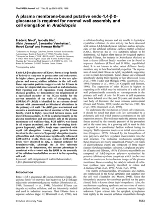
Fig. 1. kor mutant phenotype. (A) Seven-day-old light-grown wild-type (left) and korrigan (right) seedlings. (B) Six week-old greenhouse-grown
plants: wild-type (left) and korrigan (right). (C) Seven-day-old dark-grown wild-type (left), korrigan (middle) and kor transformed with the 8.5 kb
complementing genomic fragment (right) seedlings. (D) Fifteen-day-old light-grown wild-type (left) and korrigan (right) seedlings. (E) Seven-day-
old light-grown wild-type roots. (F) Seven-day-old light-grown korrigan roots. Calcofluor-stained transverse sections through hypocotyls of 7-day-old
dark-grown (G) wild-type and (H) korrigan seedlings. Arrows indicate wall separations often observed in the kor mutant. Scale bar represents 65 µm.
5564
3. Dwarf mutant deficient for endo-1,4-β-D-glucanase
family in plant cell expansion. The gene was cloned from
an A.thaliana dwarf mutant, korrigan (kor), showing a
defect in the elongation of all non-tip-growing cells
examined. KOR encodes an evolutionarily conserved integ-
ral membrane protein which is primarily located in the
plasma membrane. KOR-transcript levels in the hypocotyl
correlated positively with cell elongation and were not
affected in mutants overproducing free auxin or ethylene,
or mutants deficient in gibberellins. In contrast, a mutant
deficient in brassinosteroids showed reduced KOR-mRNA
levels. The observed structural alterations in the mutant
cell wall suggest a role for KOR in the correct assembly
of the cellulose–hemicellulose network in the expanding
cell wall.
Results
kor, a novel cell elongation mutant
kor was found as a single allele in a screen for short
hypocotyl mutants among dark-grown T-DNA or chemic-
ally mutagenized seedlings. Despite the relatively large
scale of the screen, no other alleles for KOR were identified
although several alleles were found for other loci like
PROCUSTE (eight alleles, H.Hofte, unpublished data and
¨
Desnos et al., 1996) and BOTERO (six alleles, H.Hofte, ¨
unpublished data).
The segregation in the progeny of kor heterozygous
plants showed a 3:1 ratio (1136 wild-type and 386 kor
seedlings among 1522 seeds sown; χ2 0.031; P 0.05)
which demonstrates that the mutant phenotype segregates
as a single, recessive, nuclear locus.
The genetic map position of the locus was deduced
from the localization of the cloned KOR gene (see below)
on two yeast artificial chromosomes (CIC4G5 and
Fig. 2. Reduced hypocotyl and root growth in kor mutants. CIC8D5) containing the microsatellite marker nga129,
(A) Hypocotyl and (B) root lengths were measured as a function of which maps to the bottom of chromosome V (position
the number of days after transfer to the growth chamber.
81.7).
The hypocotyl of 7-day-old dark-grown kor seedlings
hydrogen bonds (Hayashi and Maclachlan, 1984; Hayashi, was four times shorter (Figures 1C and 2A) than that of
1989). Electron microscopic evidence indicates that xylo- the wild-type, but elongation kinetics were unchanged.
glucan molecules form cross-bridges between cellulose For both the mutant and wild-type, the hypocotyl elongated
microfibrils, thereby establishing a load-bearing cellulose– between day 2 and 7, after which a plateau was reached
xyloglucan network (McCann et al., 1990). Studies on (Figure 2A). kor hypocotyls grown in white light were
tomato cell cultures adapted to the inhibitor of cellulose only slightly shorter than those of the wild-type (Figures
deposition, 2,6-dichlorobenzonitrile (DCB), indicate that 1A and 2A). Root growth was also reduced in the mutant
cellulose–hemicellulose on the one hand, and pectins on compared with wild-type (Figures 1C and 2B) and the
the other hand, form two independent networks in the difference was most pronounced in the light. In light
primary wall. DCB-adapted cell walls have greatly reduced conditions the root growth rate of the wild-type was
cellulose contents. As a result, xyloglucans cannot bind significantly higher (0.19 cm/day) than in dark-grown
to cellulose and are secreted into the medium. Surprisingly, plants (0.12 cm/day). The size of all other organs investi-
in the absence of a cellulose–hemicellulose network, the gated was also reduced in kor, including stems, rosette
wall integrity and strength is maintained by a Ca2 -linked leaves, flowers and siliques (Figure 1B and D). Despite
network of acidic pectins, as shown by the extreme the reduced size, mutant plants were fully fertile. No
susceptibility of these walls to Ca2 -chelators. differences in size between the mutant and wild-type were
Wall stress relaxation may involve slippage or hydro- observed for tip-growing cells such as trichomes and root
lysis of the load-bearing xyloglucan cross-bridges hairs (data not shown). Furthermore, the normal Mendelian
(Cleland, 1971; Albersheim, 1975; Hayashi and segregation ratio for the mutant phenotype suggested that
Maclachlan, 1984) and it has frequently been suggested pollen tube elongation was unaffected.
that EGases, together with other cell-wall proteins, such Scanning electron microscopy (SEM) of mutant dark-
as expansins and xyloglucan-endo-transglycosylases, may grown hypocotyls revealed an irregular surface (Figure
play a role in this process. 3J) consisting of epidermal cells with irregular shapes.
To our knowledge, this work represents the first demon- Wide cells alternated with narrow cells and some cells
stration of an essential role for a member of the EGase had collapsed entirely or had failed to expand. Although
5565
4. F.Nicol et al.
Fig. 3. A cell elongation defect in kor mutants. Scanning electron micrographs (SEM) of 7-day-old (A, D) light-grown seedlings and (E, H) 7-day-
old dark-grown seedlings. (B, C) Transverse sections of 7-day-old light-grown wild-type (B) and kor (C) and (F, G) dark-grown wild-type (F) and
kor (G) hypocotyls. (I, J) SEM of 7-day-old light-grown wild-type (I) and kor (J) cotyledons. Arrows indicate the unexpanded epidermal cells. Scale
bar represents 100 µm.
7-day-old dark-grown kor hypocotyls showed a length from the wild-type (Figure 3A), but differences were less
comparable with that of wild-type light-grown hypocotyls pronounced than in the dark (Figure 3E and H).
(Figure 2A), the apical hook, the closed cotyledons and In kor cotyledons, cell shape was highly variable.
the epidermal differentiation pattern (Gendreau et al., Areas with almost fully expanded, jigsaw puzzle-like cells
1997) indicated that the mutation did not affect other alternated with islands of poorly expanded cells with a
aspects of skotomorphogenic development. Besides the smooth surface (Figure 3I and J), suggesting a stochastic
epidermis, all other hypocotyl cells were also affected in expression of the mutant phenotype.
the mutant as shown in transverse sections (Figure 3C
KOR encodes a member of the endo-1,4-β-D-
and G) compared with the wild-type (Figure 3B and F).
glucanase family
No changes in radial organization could be observed.
However, all cells, including those in the central cylinder, The T-DNA segregated as a single locus and could not
showed increased radial expansion. Similar observations be separated genetically from the kor mutation, indicating
were made for the root (Figure 1E and F). In light-grown a close linkage ( 3 cM). Southern analysis of DNA
kor hypocotyls (Figure 3D), cell shape was also different extracted from kor and wild-type plants, digested with
5566
5. Dwarf mutant deficient for endo-1,4-β-D-glucanase
Fig. 4. KOR, a putative membrane-bound endo-1,4-β-D-glucanase. (A) Domain structure: unique restriction sites in the gene sequence are shown,
introns are represented by black arrows, the T-DNA is inserted 200 bp upstream of the ATG-initiation codon. The predicted N-terminal cytosolic tail,
the membrane anchor and the extracellular EGase domain and the proline-rich C-terminal domain are indicated. (B) Hydrophobicity plot according
to Kyte and Doolittle (1982). (C) Alignment of KOR with plant and bacterial EGases: AvoCel1, Tomcel3 and celD are the amino acids sequences of
a soluble EGase from avocado without the signal peptide, a membrane-bound EGase from tomato, an EGase from Clostridium thermocellum,
respectively. The predicted amino acid sequence of a rice EST is included. urepresents the residues essential for catalytic activity identified in celD
which are also conserved in KOR (Asp198, Asp201, H516, Glu555); *indicates the eight predicted glycosylation sites. The thick black line overlines
the predicted transmembrane domain in KOR (Von Heijne, 1986). The accession numbers are as in Figure 5.
several enzymes and probed with DNA fragments corres- right border was isolated and used to obtain genomic
ponding to either the left or right T-DNA border revealed clones. Complementation tests were carried out with an
a single, simple T-DNA insertion. Plant DNA flanking the 8.5 kb DNA fragment, presumably containing the entire
5567
6. F.Nicol et al.
gene, cloned into a binary T-DNA vector carrying a
hygromycin-resistance gene. Plants heterozygous for kor
were transformed by in planta infiltration (Bechtold et al.,
1993). All primary transformants had a wild-type pheno-
type in the light. The T2 progeny obtained after selfing of
one of these transformants segregated for kor with a 1:15
ratio (65 kor and 1167 wild-type seedlings of 1232 seeds
sown; χ2 0.12; P 0.05) which was expected for an
unlinked insertion complementing the mutant phenotype.
In addition, all phenotypically mutant T2 seedlings were
hygromycin-sensitive, whereas all hygromycin-resistant
seedlings had a wild-type phenotype in the light and in
the dark. The transformation efficiency of plants homo-
zygous for kor was much less efficient and yielded only
a single transformant. This transformant showed a wild-
type phenotype. The T2 progeny from this transformant
was 100% kanamycin-resistant, confirming the homo-
zygous state of the T-DNA tagged kor mutation, and
segregated the wild-type phenotype in a 3:1 ratio (315
wild-type and 95 kor seedlings of 410 seeds sown; χ2
0.75; P 0.05). All wild-type plants were also hygromycin
resistant, indicating the presence of the transgene. Figure
1C shows that the presence of one copy of the transgene
completely restores the phenotype of dark-grown seedlings
homozygous for the kor mutation to wild-type. The Fig. 5. KOR represents a distinct, evolutionarily conserved subclass of
adult phenotype was also entirely complemented in the plant EGases. Similarity tree (Genetics Computer Group- version 9.0)
transformant (data not shown). All these data indicate that of type E EGases after removal of the predicted signal sequences. This
family consists of two subgroups whereby subgroup E2 possesses both
the 8.5 kb fragment had complemented the mutation and
microbial and plant members. Sequences and DDJB/EMBL/GenBank
therefore should contain the KOR gene. accession numbers are: korrigan (U37702), tomcel3 (X97189), rice
A 2.3 kb cDNA clone was isolated using the right (EST; D46633), tomcel1 (U13054), pepper cel1 (X87323), BAC
border T-DNA flanking fragment, and the sequence was (M57400), Atglucanase (U17888), EGL1 (L41046), tomcel2 (U13055),
peppercel3 (X83711), peppercel2 (X97190), poplarcel1 (D32166),
compared with 3 kb of genomic sequence flanking the
avocel1 (M17634), tomcel4 (U20590), avocel2 (X55790), CstcelZ
right border (DDJB/EMBL/GenBank accession No.
(X55299) and CtcelD (X04584).
AF073875). The genomic sequence contained five Open
Reading Frames (ORFs) corresponding to the cDNA
sequence interrupted by four introns (Figure 4A) with Heijne, 1986; Figure 4A and B). The N-terminal stretch
consensus splice-donor and acceptor sites. The first ATG of 100 amino acids distinguishes KOR and Cel3 from
codon is most likely to be the initiator codon, since it is other plant EGases. Interestingly, this sequence is highly
preceded by an in-frame stop codon at conserved between dicots and monocots The sequence
–46 bp. The T-DNA is inserted 200 bp upstream of the shows 94% identity between KOR and Cel3, and one rice
initiator ATG, presumably in the promoter. The cDNA EST encodes a polypeptide showing 82% identity to this
sequence is 100% identical to an A.thaliana sequence segment in KOR (Figure 4C). KOR, Cel3 and the rice
present in the public databases (DDJB/EMBL/GenBank EST are more similar to each other than to other plant
accession No. U37702). The predicted amino acid EGases suggesting a functional specialization preceding
sequence of 622 residues contains eight potential N- the divergence between monocots and dicots in evolution
glycosylation sites and is similar over almost its entire (Figure 5). KOR and Cel3 also can be distinguished from
length to EGases from plants and bacteria. For instance, other EGases by the presence of a C-terminal extension
the amino acid sequence is 43.3% identical to avocado of 30 amino acids rich in prolines (Figure 4A).
Cel1 sequence and 44.4% identical to pea EGL1. In the
KOR is an integral membrane protein
bacterial enzyme CelD, site-directed mutagenesis and
chemical modification techniques have identified four To determine the intracellular localization of KOR, a
amino acids (D198/201, H516 and E555) as being poten- polyclonal rabbit antiserum was raised against a chimeric
tially important in catalysis (Chauvaux et al., 1992). As protein produced in Escherichia coli, composed of bac-
in other plant EGases, these four residues are also con- terial glutathione-S-transferase fused to the 65 N-terminal
served in the KOR sequence (Figure 4C). KOR is highly amino acids of KOR.
similar (81.8% identity) to Cel3, a recently identified Immunoblotting with the immuno-purified antiserum
membrane-bound EGase family member in tomato demonstrated the presence of a 70 kDa protein in wild-
(Brummel et al., 1997). In contrast to all other plant type leaf microsomes but not in soluble fractions (Figure
EGases, the sequences of KOR and Cel3 do not contain 6A, lanes 1 and 2). No cross-reacting band was detected in
a predicted cleavable signal peptide. Instead, the mature the mutant, demonstrating the specificity of the antiserum
protein is predicted to be a type II integral membrane (Figure 6A; lanes 3 and 4). After treatment of the
protein anchored in the membrane by a stretch of highly microsomes with Na2CO3 (pH 11), a procedure known to
hydrophobic amino acids located in the N-terminus (Von strip peripheral proteins from membranes, the cross-
5568
7. Dwarf mutant deficient for endo-1,4-β-D-glucanase
external epidermal wall (0.9 0.38 µm [n 156] in kor
0.31 µm [n
versus 0.56 162] in the wild-type) and
0.19 µm [n
cortical walls (0.37 10] in kor versus
0.14 0.05 µm [n 10] in the wild-type). The distribution
of polysaccharides was visualized by periodic acid-thiocar-
bohydrazyde-silver proteinate (PATAg) staining (Thiery, ´
1967; Roland and Vian, 1991). In epidermal walls three
domains could be distinguished including an intensely
stained cuticle, a poorly stained outer layer and a strongly
stained inner layer. Wild-type walls had a regular surface
and visible layers of material could be distinguished in
the innermost layer (Figure 8A). In the external epidermal
wall of kor, the staining was much more irregular, i.e.
highly electron-dense deposits were visible at the cyto-
plasmic side of the wall (see white arrows on Figure 8B),
whereas in other areas staining was practically absent. In
mutant cortical cell walls irregular fibrillar material could
be observed (Figure 8D) compared with the wild-type
(Figure 8C).
Fig. 6. KOR is an integral membrane protein. (A) Immunoblot of
To visualize cellulosic material, sections were stained
wild-type (lanes 1 and 2) and kor (lanes 3 and 4) leaf microsomes
(lanes 1 and 3) and supernatant (lanes 2 and 4). (B) Immunoblot of with calcofluor (Figure 1G and H). In the wild-type a
wild-type leaf microsomes before and after Na2CO3 (pH 11)
regular distribution of fluorescent material could be
extraction. A polyclonal rabbit antiserum raised against the N-terminus
observed in epidermal and cortical cell walls. Separation
of KOR (anti-NKOR) was used in (A) and (B) as primary antibody.
of cortical walls could be observed occasionally, but
(C) Immunoblot of wild-type leaf microsomes before and after
Na2CO3 (pH 11) extraction, with a polyclonal rabbit antiserum against exclusively at cell corners. In the mutant, walls of adjacent
¨
the soluble ER-localized protein BIP as primary antiserum (Hofte cortical cells were separated at numerous places distributed
et al., 1992).
over the entire cell surface (Figure 1H, arrows), and the
fluorescent staining was more irregular.
reacting protein remained in the membrane fraction (Figure Xyloglucan (XG) epitopes were visualized in external
6B). As a control, the soluble binding protein (BIP), epidermal walls with a polyclonal anti-XG antiserum.
present in the reticulum lumen (Denecke et al., 1993), Gold particles could be observed throughout the wall
was recovered in the soluble fraction (Figure 6C). These section for both wild-type and mutant (Figure 8E and F).
results show that KOR is an integral membrane protein. To assess the contribution of the cellulose–xyloglucan
network to the integrity of the wall, an extraction was
carried out with the Ca2 -chelator CDTA, a procedure
KOR is preferentially located in the plasma
known to selectively extract Ca2 -bridged pectates from
membrane
Microsomal membrane vesicles prepared from an A.thali- the wall (Jauneau et al., 1992). In the wild-type, CDTA
ana suspension culture were separated using two consecut- extraction caused the wall to swell, but with the preserva-
ive rounds of free-flow electrophoresis (Canut et al., tion of the overall wall structure (Figure 8G). Fibrillar
1988). A first separation yielded three peaks (Figure 7A), material decorated with antibodies could be seen in the
respectively enriched for (1) plasma membranes, (2) innermost half of the wall. CDTA-treated kor mutant walls
endoplasmic reticulum (ER) membranes and (3) tonoplast contained XG in amounts comparable with that observed
markers. Fractions 1 and 3 were pooled and subjected to in the wild-type as indicated by the gold-labeling; however,
a second separation, yielding fractions 4 and 6 (Figure CDTA not only caused mutant walls to swell, but invariably
7B) that were highly enriched for plasma membrane and resulted in the separation of the most recently deposited
tonoplast, respectively (Table I). Fraction 2 was also material from the rest of the wall. Disordered fibrillar
subjected to a further round of purification giving fraction material could be observed in the ruptured areas
5 (Figure 7C), which was further enriched for the ER (Figure 8H).
marker. Essentially immunoblotting localized KOR to the
Regulation of KOR-mRNA levels
plasma membrane-enriched fraction (Figure 7D), no cross-
reacting protein was detected in the ER fraction. A faint Northern blot analysis using a probe specific for KOR
70 kDa band could be detected in the tonoplast fraction detected a single transcript of 2 kb in wild-type plants.
together with some smaller molecular weight bands. These The mRNA was found in all organs, with the highest
bands were reproducibly present in different experiments levels in stems and roots, and the lowest in siliques (5.5
and might be the result of the proteolytic cleavage of the times less than in stems; Figure 9A and B). Although not
protein in the vacuole. visible on Figure 9A, a faint band was also detected in
kor seedlings and adult plants (Figure 9B).
Cell wall alterations in kor To investigate a potential role for KOR in the control
Using microscopy, the walls of mutant and wild-type were of cell elongation, KOR-mRNA levels were monitored
investigated on transverse hypocotyl sections of 7-day-old during hypocotyl development in the dark (Figure 10).
dark-grown seedlings. Transmission electron microscopy No KOR mRNA could be detected in seeds imbibed for 3 h
(TEM) showed that fixed mutant cell walls were invariably or in 1-day-old seedlings. Hypocotyls started elongating
thicker than wild-type walls. This was true both for the between day 1 and 2 after transfer to the culture room.
5569
8. F.Nicol et al.
Fig. 7. KOR is primarily located in the plasma membrane. Separation of microsomal membranes by free-flow electrophoresis. (A) Separation profiles
of microsomal membranes. (B) Profile obtained after a second separation round of pooled fractions 1 and 3 and (C) after a second separation round
of fraction 2. Profiles were monitored at 280 nm. Fractions 4 and 6 are respectively enriched for plasma membrane and tonoplast markers as
summarized in Table I. (D) Immunoblot analysis of membrane fractions (50 µg protein/lane) with the same rabbit anti-NKOR primary antiserum as
in Figure 8.
auxin. KOR-mRNA levels were ~50% higher in sur2
Table I. Enrichment of free-flow electrophoresis fractions for plasma
compared with the wild-type (Figure 11B); however, this
membrane- and tonoplast-associated ATPase activities
difference was not reproducible in independent experi-
ments (data not shown). Mutant eto seedlings overproduce
Marker enzymes (fractions)
ethylene (Vogel et al., 1998) and show the characteristic
4 6
triple response of reduced length and increased width of
hypocotyl and root, and an exagerated apical hook. Despite
Total ATPase activity 1297 231
the dramatic differences in hypocotyl length, no significant
(nmoles Pi/ min.mg protein)
Inhibition (%) by KNO3 (tonoplast) –1 89 differences in KOR-mRNA levels were observed (Figure
Inhibition (%) by NaN3 1 12 11B). GA treatment (see Materials and methods) of wild-
(mitochondria)
type plants did not affect hypocotyl length and caused a
Inhibition (%) by V2O5 86 15
small increase in KOR-mRNA levels, which was not
(plasma membrane)
reproducible in different experiments. The mutant ga1
Fractions are as in Figure 7. (abc33) is deficient for the first committed step in GA
biosynthesis (Dubreucq et al., 1996) and failed to elongate
The growth rate increased between day 2 and 4 after normally in the presence of low concentrations of GA4 7.
which it dropped (Figure 10A). At day 2, concomitant Despite the dramatic difference in hypocotyl length, KOR-
with the onset of hypocotyl elongation, the mRNA mRNA levels did not differ significantly between ga1
appeared. KOR-mRNA levels were highest during the (abc33) and wild-type plants (Figure 11C).
linear growth phase of the hypocotyl (day 4 to 6) and
decreased at day 7, at least 1 day, and possibly 2 days,
Discussion
after the drop in growth rate (Figure 10B and C).
Using a set of mutants, we investigated the effect of Our screen yielded a single KOR allele whereas multiple
cell elongation-controlling hormones on the hypocotyl alleles were found for other dwarf loci. An explanation
length and KOR-mRNA levels of 4-day-old dark-grown for the low allele frequency could be that kor is a rare
seedlings (Figure 11). The mutant det2 is deficient for an leaky mutation caused by a T-DNA insertion in the
early step of the brassinosteroid biosynthetic pathway promoter, and that the null allele has a more drastic
(Fujioka et al., 1997). Hypocotyls of det2 were three times phenotype, not recovered in our screen. In accordance
shorter than those of the wild-type. Interestingly, det2 with this, Northern blots of RNA from kor revealed a
seedlings showed 3-fold lower KOR-mRNA levels than faint band hybridizing to the KOR probe, suggesting that
the wild-type. The addition of 10–7 M brassinolide (BR) residual KOR activity may be present in the mutant.
to the medium partially restored hypocotyl growth of Indeed, it is likely that this faint band does not correspond
det2 and also caused an increase in KOR-mRNA levels. to the mRNA from a related gene, since the same probe
Treatment of wild-type plants with BR caused a slight did not reveal the presence of a second cross-reacting
decrease in length and in KOR-mRNA levels (Figure sequence on low-stringency Southern blots (data not
11A). Seedlings homozygous for the sur2 mutation contain shown).
increased levels of free indolacetic acid (IAA) and reduced Despite the usual precautions that need to be taken
levels of conjugated IAA (Delarue et al., 1998). Figure 1 when deducing a gene function from the phenotype of a
shows that sur2 hypocotyls were shorter than those of the single mutant allele, the following observations together
wild-type. This has been observed previously and reflects indicate that the reduced expression of KOR is the primary
the inhibitory effect on elongation of high levels of free cause of the growth defect in the mutant. First, the dwarf
5570
9. Dwarf mutant deficient for endo-1,4-β-D-glucanase
Fig. 9. Ubiquitous expression of KOR mRNA. (A) Northern blot with
8 µg total RNA from wild-type and kor tissues: Se, 7-day-old light-
grown seedlings; L, leaves; St, stems; Fl, flowers; Si, siliques; R,
roots; and Ap, the entire aerial portion of an adult plant, were
hybridized with the 765 bp PstI–PstI fragment (between positions 184
and 879 in the KOR genomic sequence, see Figure 4). A faint 2 kb
transcript was detected with this probe, in kor mRNA. (B) Relative
quantities of KOR mRNA after normalization with the hybridization
signal obtained with a 25S RNA gene probe.
levels were strongly reduced, which is consistent with the
recessive nature of the mutant phenotype. Fourthly, all
aspects of the mutant phenotype were complemented by
an ectopically inserted 8.5 kb DNA fragment containing
the wild-type KOR gene.
Although the enzymatic activity remains to be demon-
strated, the following observations suggest that KOR
encodes a bona fide EGase. The predicted KOR sequence
is similar to secreted plant EGases over almost its entire
length, and the residues essential for catalytic activity in
bacterial CelD are conserved. In addition, Brummel et al.
(1997) observed EGase activity using the artificial sub-
strate CMC in sucrose density/gradient fractions enriched
for the tomato ortholog of KOR, Cel3. We have also
Fig. 8. Cell wall defects in kor mutants. Transmission electron observed membrane-associated CMC-ase activity in
microscopy of transverse hypocotyl sections. (A, B) Outer epidermal
A.thaliana microsomes (F.Nicol, unpublished data), but it
and (C, D) cortical cell walls sections of 7-day-old dark-grown (A, C)
remains to be determined whether this indeed corresponds
wild-type and (B, D) korrigan hypocotyls. Sections were stained for
to the activity of KOR.
polysaccharides with PATAg. Note the irregular surface and the
Whereas almost all other characterized EGase family
absence of stratified microfibrils in the inner part of the mutant wall.
White arrows indicate aggregates of highly electron-dense material members in plants and bacteria are soluble secreted
frequently observed at the cytoplasmic side of mutant walls. (E, F, G,
enzymes (Brummell et al., 1994), KOR and its tomato
H) Outer epidermal sections of 7-day-old dark-grown (E, G) wild-type
ortholog Cel3 are unique in that they are both integral
and (F, H) korrigan hypocotyls submitted (G, H) or not (E, F) to a
membrane proteins. The membrane location was suggested
CDTA extraction. Xyloglucans were visualized with polyclonal anti-
XG-antibodies and polysaccharides stained by the PATAg method. C, by the presence of a predicted N-terminal transmembrane
cytoplasm; Cu, cuticle. Scale bar represents 0.6 µm.
anchor in the sequence, and was confirmed experimentally
for both proteins. Based upon the charge distribution
mutant phenotype cosegregated with a T-DNA insertion around the transmembrane anchor, the protein is predicted
200 bp upstream of the initiation codon of KOR. Secondly, to be a type II membrane protein, with the N-terminus
no major DNA rearrangements surrounding the T-DNA facing the cytosol. This orientation was experimentally
were observed, suggesting that no other genes were confirmed for Cel3, and therefore, in view of the strong
inactivated as a result of the T-DNA insertion (data not sequence conservation (82% amino acid identity), is also
shown). Thirdly, in the mutant, KOR-mRNA and protein likely to be true for KOR. Essentially, as for Cel3, KOR
5571
10. F.Nicol et al.
Fig. 11. Effect of cell elongation-controlling hormones on the
hypocotyl length of 4-day-old seedlings and on KOR-mRNA levels.
(A) Effect of brassinosteroids (BR). Wild-type seedlings (Col0
ecotype) non-treated (lane 1) or treated (lane 2) with 10–7 M BR. det2
seedlings non-treated (lane 3) or treated (lane 4) with BR. (B) Wild-
type seedlings (Col0 ecotype; lane 5), auxin-overproducing mutant
sur2 (lane 6) and ethylene-overproducing mutant eto (lane 7).
(C) Effect of GA4 7. Wild-type seedlings (WS ecotype) untreated
(lane 8) or treated (lane 9) with 10–7 M GA4 7, and ga1 (abc33)
mutant treated with 10–7 M GA4 7 (lane 10).
The N-terminal part of KOR, containing the cytosolic
domain and the membrane anchor sequence, is highly
conserved between rice, tomato and A.thaliana. The mem-
brane location therefore appears to be under a strong
Fig. 10. KOR mRNA appears after germination together with the selective pressure and predates the divergence between
initiation of hypocotyl elongation in the dark. (A) Growth kinetics of
monocots and dicots. The membrane location of a cell
wild-type dark-grown hypocotyls and growth rate (inset) in cm/day.
wall-modifying enzyme may have the following advant-
(B) Relative quantities of KOR mRNA after normalization with the
ages. First, in contrast to soluble secreted proteins, mem-
signal from a 25S RNA probe. (C) Northern blot with RNA isolated
from seeds imbibed for 3 h and from 1- to 7-day-old dark-grown wild- brane proteins may be more easily targeted to a particular
type seedlings. The same probe as in Figure 9 was used.
subdomain of the cell wall; for instance, it might be more
favorable to express an elongation-promoting enzyme in
was found in the plasma membrane fraction. We also the longitudinal and not in the transverse wall. Secondly,
observed a minor portion of KOR in the internal membrane it may be advantageous to confine the protein to the
fraction enriched for the tonoplast and different from the innermost area of the wall, where it may contribute to the
ER. Cel3 was also found in internal membranes but in construction of the expanding wall. Thirdly, the regulation
the fraction enriched for the Golgi apparatus. It is not of the abundance of the protein in plasma membrane
known whether this discrepancy is a result of the different through endo- or exocytosis may be a way to control the
separation procedures used in the two studies (linear amount of enzyme in the wall. In this respect, the
sucrose gradient versus free-flow electrophoresis) or observation that a 70 kDa and some smaller cross-reacting
reflects differences specific to the two species or the bands were present in the tonoplast-enriched fraction
physiology of the tissue investigated (tomato root versus might point to a plasma membrane retrieval and vacuolar
A.thaliana suspension cultures). In any case, these data degradation mechanism. Fourthly, KOR may form part of
suggest a complex regulation of the intracellular targeting a complex with other membrane and/or cytosolic proteins.
of KOR/Cel3. A detailed localization, together with pulse– We are currently investigating these different possibilities.
chase experiments, are needed to determine exactly in
A role for KOR in cell elongation
which intracellular compartments the KOR protein resides
during the different stages of cellular growth and differen- This study demonstrates, for the first time to our know-
tiation. ledge, an essential role for a member of a family of cell
5572
11. Dwarf mutant deficient for endo-1,4-β-D-glucanase
wall hydrolytic enzymes in normal cell elongation. In the properties of kor walls. Secondly, polysaccharides were
mutant, cell divisions during embryogenesis and in the deposited in an irregular fashion in both external epidermal
meristems take place normally (data not shown), sug- and cortical cell walls. Thirdly, the extraction of the wall
with CDTA, a treatment that selectively removes Ca2 -
gesting no role for KOR in cell plate formation during
cytokinesis, although in the absence of a true null allele, linked acidic pectins, caused a separation of the innermost
this possibility cannot be ruled out entirely. In contrast, layers from the rest of the wall, which was never observed
the mutant phenotype indicates a role for KOR in the in wild-type walls. This interesting observation suggests
elongation of all cells investigated, except for tip-growing that, in kor, the cellulose–xyloglucan network that remains
following removal of the Ca2 -pectate network, is unable
cells. The increased diameter of mutant cells, clearly
observed in dark-grown hypocotyls, suggests a role for to maintain the integrity of the innermost layers of the
KOR in the control of the direction of cell expansion. wall. A similar situation has been observed previously in
Increased radial expansion was also observed for the DCB-adapted tomato cell cultures. In these cells a cellu-
lose–xyloglucan network was practically absent, and Ca2 -
cellulose-deficient mutant rsw1 (Arioli et al., 1998) or
wild-type plants cultured in the presence of cellulose linked acidic pectins were required for maintaining the
biosynthesis inhibitors, indicating a central role for cellu- integrity and strength of the wall.
lose in determining the cell shape. As discussed below, Assuming that KOR is an EGase, how could the
the observed cellular phenotype in kor would be compat- absence, or the reduced levels of KOR cause such changes
ible with a defect in the cellulose–hemicellulose network. in the cellulose–XG network? EGases hydrolyse 1,4-β
Consistent with the mutant phenotype, KOR mRNA was linkages behind unsubstituted glucose residues. Cellulose,
found in all growing organs examined. In the developing xylans and xyloglucans contain 1,4-β linkages. KOR, like
hypocotyl, KOR-mRNA levels were correlated with cell all known plant EGases, lacks a cellulose-binding domain
elongation. The mRNA appeared at the onset of cell and would be unable to hydrolyse crystalline cellulose.
elongation in the hypocotyl and reached a maximum Xylans mainly accumulate during secondary wall forma-
during the linear growth phase. Interestingly, the drop in tion, and the absence of a xylanase would not be expected
growth rate after the fourth day of culture was not preceded to cause a primary cell wall phenotype. In the current
but followed by a drop in KOR-mRNA levels, indicating model for the primary wall in dicots, the reduction of
that the growth arrest is not caused by a reduction in xyloglucanase activity would be compatible with reduced
KOR-mRNA levels. wall loosening and cell elongation. In addition, it has been
We did not observe pronounced variations in KOR- proposed that xyloglucans need to be cleaved in order to
mRNA levels between the different hormone mutants, permit the incorporation of new microfibrils into the
except for the brassinosteroid-deficient mutant det2 in existing cellulose–xyloglucan network (Hayashi, 1989).
which the reduced hypocotyl length was associated with In this context, the observed unordered accumulation of
a modest but reproducible reduction in KOR mRNA. It wall material at the cytoplasmic side of mutant walls and
remains to be seen whether KOR-mRNA levels are under the reduced coherence of the CDTA-extracted walls would
direct brassinosteroid control, or whether the reduced be compatible with a reduced ability to cleave load-
expression level is an indirect result of reduced cell bearing xyloglucans in the mutant. In this view though, it
elongation. Interestingly, the hypocotyl lengths of auxin remains to be explained why a highly conserved KOR
and ethylene overproducers as well as the GA-deficient ortholog is present in rice, since in grasses XGs do
mutant were more or less affected without a significant not play a predominant role in primary wall structure
reduction in KOR mRNA, suggesting a specific action of (Carpita, 1996).
BR on KOR expression. BR has been shown to regulate In conclusion, a mutation causing a reduction in the
the mRNA levels of TCH4 encoding a xyloglucan-endo- amount of KOR, a putative EGase primarily located in
transglycosylase (XET)-homolog (Kauschmann et al., the plasma membrane, caused a cell elongation defect
1996). XETs are thought to be involved in the molecular in all non-tip-growing cells investigated. In dark-grown
grafting reactions required for the insertion of new poly- hypocotyls, the growth defect is associated with pro-
saccharides into the cell wall. The mutant phenotype also nounced changes in the cellulose–xyloglucan network
suggests a similar role for KOR. This raises the interesting within the primary wall. We are currently investigating
possibility that growth control by brassinosteroids is the substrate specificity of KOR, the architecture of mutant
mediated through the expression of a set of enzymes walls, and the regulation of the expression and localization
involved in the construction of the cell wall. More detailed of the protein. These studies will further clarify the
studies are needed to determine the relationship between biochemical, cellular and developmental function of KOR,
hormone action, cell elongation and the expression of and provide new insights into the molecular mechanisms
KOR at the transcriptional but also at different post- involved in the construction of the wall and the control
transcription levels. of cell elongation in plants.
A role for KOR in the assembly of the cellulose–
Materials and methods
hemicellulose network in the primary wall
A number of changes in the architecture of primary walls Plant strains and growth conditions
were observed in kor. First, fixed mutant walls were Seeds of A.thaliana Heyhn, ecotypes Wassilewskija (WS) and Columbia
(Col0) were provided by K.Feldman (University of Arizona, Tucson,
thicker than those of the wild-type. This could be an
USA). Mutants used were the GA-deficient mutant abc33, allelic to
indirect result of the fixation procedure rather than a true ga1 (Dubreucq et al., 1996; provided by B.Dubreucq), the ethylene-
increase in thickness of the unfixed wall. In either case, overproducing mutant eto (Guzman and Ecker, 1990; provided by ABRC,
the results point to an alteration of the physicochemical Columbus, Ohio, USA), the auxin-overproducing mutant sur2 (Delarue
5573
12. F.Nicol et al.
et al., 1998; provided by C.Bellini) and the brassinosteroid-deficient Northern blots were performed using Hybond N membranes and hybrid-
mutant cop7, allelic to det2 (X.-W.Deng, personnal communication; ized to random primed probes following the manufacturer’s instructions
provided by X.-W.Deng). Seeds were sterilized as described by Santoni (Amersham, Buckinghamshire, UK). Northern blots were carried out
et al. (1994). Seeds were allowed to germinate on nutrient medium as with a 765 bp PstI–PstI fragment from the genomic clone as a probe.
described by Estelle and Somerville (1987) with 1% sucrose, with or For the hormone experiment, the Northern blot was hybridized with the
without GA4 7 (Sigma) or brassinosteroids (provided by S.Fujioka, 273 bp BglII–BglII cDNA fragment as a probe.
RIKEN, Wako-shi, Japan) in a growth chamber. For the GA experiment,
seeds were imbibed in a GA4 7 solution (10–5 M) at 4°C for 48 h and Molecular cloning of the KORRIGAN gene
washed five times with sterilized water before being sown on the Standard molecular cloning techniques were performed as described by
appropriate medium. Plants were grown in a 16h light (200 µmol/m2/s), Sambrook et al. (1989). Southern blot analysis revealed the presence of
8 h dark cycle. Temperatures were 20 and 15°C, respectively, during a single T-DNA insertion. Genomic DNA of the korrigan mutant was
day and night. Seeds were imbibed for 48 h at 4°C and exposed to white digested by MspI and ligated with T4-DNA ligase. A right border
light (200 µmol/m2/s) before transfer to final growth conditions. For flanking sequence was obtained through Inverse Polymerase Chain
dark conditions, plates were wrapped in two layers of aluminium foil. Reaction (IPCR) using the method described by Lindsey and Topping
Days of growth were counted after transfer of Petri dishes from 4°C to (1996). The PCR reaction was performed using the T-DNA oligonucleo-
the growth chamber. A.thaliana suspension cultures were obtained and tides TAG7 (5 -GGACTGACCACCCCAAGTGC-3 ) and TAG8 (5 -
maintained as described by Axelos et al. (1992). ACTCGACGGCCTGTGGGCAT-3 ). The 600 bp flanking fragment was
used to probe an A.thaliana genomic phage library made by John
Hypocotyl length measurements Mulligan and distributed via the EEC-BRIDGE Arabidopsis DNA Stock
Seedlings (30–40) were spread out on agar plates and magnified using Center. A positive phage clone was digested with PstI and subcloned
a photographic enlarger. The projected image was traced with a pencil into the pZero vector (Invitrogen, Leek, The Netherlands). DNA sequen-
on a sheet of paper. Lengths were measured using image analysis cing was performed using an ABI 373A automated sequencer (Applied
software (Optimas 4.1) as described by Gendreau et al. (1997). Biosystems). A 8.5 kb XhoI DNA fragment presumed to contain the
entire KORRIGAN gene was introduced into the XhoI site of the binary
Microscopy vector pBIB-HYG (Becker, 1990) containing a hygromycin resistance
SEM and light microscopy were done as described by Desnos et al. marker. The recombinant binary vector was introduced into Agrobacter-
(1996). Arabidopsis thaliana seedlings were fixed, dehydrated and ium strain C58C1 (pMP90; Koncz and Schell, 1986) through electropor-
embedded in LR White or Spurr’s epoxy medium as described previously ation. The T-DNA was transformed into plants heterozygous and
(His et al., 1997). Transverse sections (90 nm in thickness) were mounted homozygous for korrigan using the infiltration protocol described by
on gold grids (Ø: 3.05 mm, Oxford Instruments, Orsay, France) and Bechtold et al. (1993). Transformants were selected in vitro, on a medium
containing hygromycin (100 µg/ml). The F2 progeny of individual
polysaccharides were visualized through PATAg staining (Roland and
Vian, 1991). For visualization of XGs in the wall, 7-day-old dark-grown hygromycin resistant plants were analyzed for the segregation of korrigan
seedlings were fixed with 4% of glutaraldehyde in 0.1 M cacodylate mutants. A cDNA library (D’Alessio, 1992) was also screened with the
buffer (pH 7.2) at room temperature for 90 min. This treatment was 765 bp PstI–PstI genomic fragment. The KORRIGAN predicted amino
followed by a CDTA-Na2 extraction at 4°C for 24 h. Seedlings were acid sequence was used to search the protein sequence databases using
then submitted to a post-fixation in a solution of 2% (w/v) osmium the programs BLASTX and TBLASTN (Altschul et al., 1990). Endo-
tetroxide for 1 h. Specimens were then briefly rinsed in deionized water 1,4-β-D-glucanases were aligned using the Pileup program within the
and dehydrated by bathing in graded mixtures of ethanol and water (10, GCG program package, version 9.0 (Genetics Computer Group). Identical
20, 40, 60, 80, 90, 95 and 100%, v/v). They were bathed in a solution amino acids were visualized using the program SeqVu, version 1.1
of graded London Resin White (LRW, Oxford Agar)/ethanol (v/2v, v/v, (Garvan Institute, Sydney, Australia).
2v/v) for 30 min each and embedded in LRW overnight at 60°C.
Mapping
Transverse sections (90 nm in thickness) were mounted on gold grids.
Samples were then treated in a 3% milk [Tris saline buffer 0.1% PCR screening of the CIC library was performed as described by Creusot
Tween (TTBS)] solution for 30 min. Sections were washed in TTBS et al. (1995) using ordered YACs pooled in three dimensions. Two
buffer and incubated at 4°C for 12 h with a 1:100 dilution of the XG oligonucleotides, B (5 -GAGACGCAGCAGAGTTGGTT-3 ) and C (5 -
antiserum (Moore et al., 1986). Grids were washed with TTBS buffer AGATCATCAATGGAATCAGCAG-3 ), the 5 ends of which respect-
and incubated for 1 h with a 1:40 dilution of the secondary goat anti- ively correspond to nucleotides 194 and 744 of the KOR cDNA sequence,
rabbit antiserum linked to 10 nm gold particles. Grids were then washed were used to amplify by PCR a unique 550 bp fragment from genomic
for 30 min in TTBS and milliQ water. Polysacharides were visualized A.thaliana Columbia DNA. The PCR primers identified two YAC clones
on those sections through PATAg staining. (CIC 4G5 and CIC 8D5) located at the bottom of chromosome V and
containing the microsatellite marker nga129 (Lister and Dean, 1994)
Isolation of the korrigan mutant and genetic analysis which mapped the gene to chromosome V, position 81.7 cM.
Approximately 12 000 individual T-DNA-mutagenized T2 lines (Bechtold
Protein expression and immunological techniques
et al., 1993) and about 800 M2 families from single ethylmethane
sulfonate (EMS) mutagenized M1 plants (Desnos et al., 1996) were A cDNA fragment encoding the 65 N-terminal amino acids (NKOR;
screened for short hypocotyl mutants in the dark. Progeny of putative NcoI/86–BglII/279) was cloned into pGEX2T6 (Pharmacia) in frame
kor heterozygotes (kanamycin resistant seedlings with a wild-type with the glutathion S-transferase (GST) protein. The expression and the
phenotype) were allowed to germinate on non-selective medium and purification of the fusion GST–NKOR protein was perfomed using the
scored for the appearance of the mutant phenotype. The mutant phenotype recommendations of the manufacturer. The 37 kDa soluble purified
segregated in a 1:3 ratio in all progeny of all plants tested. Seedlings recombinant protein was used to produce a rabbit antiserum following
showing a mutant phenotype were transferred to a selective medium. the standard immunization protocols of Eurogentec (Seraing, Belgium).
All the phenotypically mutant seedlings (800) showed resistance to The antiserum was purified according to the procedure described by Lin
kanamycin, indicating a tight linkage between the inserted T-DNA and et al. (1989). Immunoblots were carried out with a 1:500 dilution of the
the mutant phenotype ( 3 cM). NKOR rabbit antiserum or a 1:10 000 dilution of the anti-BIP rabbit
antiserum (Hofte et al., 1992) and a secondary anti-rabbit IgG F(ab )2
¨
Southern and Northern blot analysis fragment linked to horseradish peroxidase, according to the specification
Arabidopsis thaliana genomic DNA was isolated as previously described of the manufacturer (Amersham) on Hybond C membranes (Amersham).
(Bouchez et al., 1996). For RNA extraction, tissues from different Signals were revealed using the Amersham ECL System.
parts of 4- to 5-week-old wild-type (WS) plants were harvested and
Microsome preparation
immediately frozen in liquid nitrogen. For root-RNA preparations, plants
were grown in liquid culture medium (described in Estelle and Somerville, Microsomes were prepared from freshly harvested leaves of 6-week-old
1987; but without agarose) for 15 days then harvested and frozen in plants. Tissues (500 mg fresh weight) were ground in 2.5 ml extraction
liquid nitrogen. For expression kinetics, seedlings were grown in the buffer (12% w/w sucrose, 50 mM Tris–HCl pH 7.8, 1 mM EDTA,
dark in Petri dishes containing medium covered by filter paper, then 1 mM DTT, 0.04 mM leupeptine, 0.02 mM lepstatine, 1 mg/ml
harvested at the appropriate day under a green safe light and frozen in polyvinylpyrolidone) and centrifuged at 3000 g for 10 min to remove
liquid nitrogen. Seeds were imbibed in water for 3 h and frozen. RNA cell walls and debris. The supernatant was layered over 1 ml of 16%
was extracted as described by Sambrook et al. (1989). Southern and (w/w) sucrose and microsomes were pelleted from the supernatant
5574
13. Dwarf mutant deficient for endo-1,4-β-D-glucanase
through centrifugation for 60 min at 35 000 r.p.m. in a TST 55 rotor Carpita,N.C. (1996) Structure and biogenesis of the cells walls of grasses.
(Kontron instruments) at 4°C. The supernatant was TCA-precipitated Annu. Rev. Plant Physiol., 47, 445–476.
and resuspended in the buffer described above. To strip peripheral Carpita,N.C. and Gibeaut,D.M. (1993) Structural models of primary cell
proteins from microsomes, the pellet was resuspended in 50 mM Tris walls in flowering plants: consistency of molecular structure with the
with protease inhibitors and adjusted to 100 mM Na2CO3 (pH 11.5). physical properties of the walls during growth. Plant J., 3, 1–30.
Membranes were pelleted from the supernatant through centrifugation Cass,L.G., Kirven,K.A. and Christoffersen,R.E. (1990) Isolation and
for 60 min at 40 000 r.p.m. in a TST 55 rotor (Kontron instrument) at characterization of a cellulase gene family member expressed during
4°C. The pellet was resuspended in 100 mM Na2CO3 (pH 11.5) and avocado fruit ripening. Mol. Gen. Genet., 223, 76–86.
centrifuged again. The pellet was resuspended in extraction buffer and ´
Chauvaux,S., Beguin,P. and Aubert,J.-P. (1992) Site-directed mutagenesis
the supernatant combined and TCA precipitated. Proteins were quantified of essential carboxylic residues in Clostridium thermocellum
using the protein assay kit according to the specification of the manufac- endoglucanase CelD. J. Biol. Chem., 267, 4472–4478.
turer (Sigma Diagnostics, St Louis, USA) using bovine serum albumin Cleland,R. (1971) Cell wall extension. Annu. Rev. Plant Physiol., 22,
as standard. 197–222.
Cosgrove,D.J. (1997) Relaxation in a high-stress environment: The
Free-flow electrophoresis molecular bases of extensible cell walls and cell enlargement. Plant
Membrane preparation and free-flow electrophoresis were performed Cell, 9, 1031–1041.
exactly as described previously by Canut et al. (1988). The specific Creusot,F. et al. (1995) The CIC library: a large insert YAC library for
activities of marker enzymes were determined as described previously genome mapping in Arabidopsis thaliana. Plant J., 8, 763–770.
(Canut et al., 1990). D’Alessio,J.M. (1992) Automatic subcloning of cDNA. Focus, 14, 76–79.
Delarue,M., Prinsen,E., Van Onckelen,H., Caboche,M. and Bellini,C.
(1998) Sur2 mutations of Arabidopsis thaliana define a new locus
Acknowledgements involved in the control of auxin homeostasis. Plant J., 14, 603–611.
Del Campillo,E. and Bennett,A.B. (1996) Pedicel breakstrength and
The authors thank Thierry Desprez for assistance with sequence compar-
cellulase gene expression during tomato flower abscission. Plant
isons; Christine Camilleri and Sophie Desloire for the mapping of the
Physiol., 111, 813–820.
KOR locus; Madeleine Lemain, Anne-Marie Jaunet, Brigitte Gelie for
Denecke,J., Ek,B., Caspers,M., Sinjorgo,K.M.C. and Palva,E.T. (1993)
SEM; Claude Pethe for assistance with the biochemical techniques;
Analysis of sorting signals responsible for the accumulation of soluble
Catherine Bellini, Nicole Bechtold and Georges Pelletier for generating
reticuloplasmins in the plant endoplasmic reticulum. J. Exp. Bot., 44,
the T-DNA insertion lines and providing assistance in the mutant screen;
213–221.
Jacques Goujeaud and Joel Talbotec for the assistance in the greenhouse;
¨
Desnos,T., Orbovic,V., Bellini,C., Kronenberger,J., Caboche,M., Traas,J.
Xing-Wang Deng, Catherine Bellini and Bertrand Dubreucq for kindly
and Hofte,H. (1996) Procuste1 mutants identify two distinct genetic
¨
providing the mutants cop7, sur2 and ga1 (abc33), respectively; the
pathways controlling hypocotyl cell elongation, respectively in dark-
Arabidopsis Biological Resource Center for providing us eto seeds and
and light-grown Arabidopsis seedlings. Development, 122, 683–693.
genomic and cDNA libraries; Andrew Staehelin for the anti-XG antibody;
Dubreucq,B., Grappin,P. and Caboche,M. (1996) A new method for the
Michel Caboche, Jan Traas, Gwenaelle Joliff, Bernard Henrissat, Pierre
identification and isolation of genes essential for Arabidopsis thaliana
Beguin for stimulating discussions and Heather McKhann, Michel
seed germination. Mol. Gen. Genet., 252, 42–50.
Caboche and Ageeth van Tuinen for the critical reading of the manuscript.
Estelle,M.A. and Somerville,C. (1987) Auxin-resistant mutants of
`
This work was supported by a grant from the Ministere de la Recherche
Arabidopsis thaliana with an altered morphology. Mol. Gen. Genet.,
et de la Technologie to F.N., and grant ACC-SV No. 9501006 from the
206, 200–206.
`
Ministere de la Recherche et de la Technologie to H.H.
Ferrarese,L., Trainotti,L., Moretto,P., Polverino de Laureto,P., Rascio,N.
and Casadoro,G. (1995) Differential ethylene-inducible expression of
References cellulase in pepper plants. Plant Mol. Biol., 29, 735–747.
Fujioka,S. et al. (1997) The Arabidopsis deetiolated2 mutant is blocked
Albersheim,P. (1975) The walls of growing plant cells. Sci. Am., 232, early in brassinosteroid biosynthesis. Plant Cell, 9, 1951–1962.
80–95. Gendreau,E., Traas,J., Desnos,T., Grandjean,O., Caboche,M. and
Altschul,S.F., Gish,W., Miller,W., Myers,E.W. and Lipman,D.J. (1990) ¨
Hofte,H. (1997) Cellular basis of hypocotyl growth in Arabidopsis
Basic local alignment search tool. J. Mol. Biol., 215, 403–410. thaliana. Plant Physiol., 114, 295–305.
Arioli,T. et al. (1998) Molecular analysis of cellulose biosynthesis in Guzman,P. and Ecker,J.R. (1990) Exploiting the triple response of
Arabidopsis. Science, 279, 717–720. Arabidopsis to identify ethylene-related mutants. Plant Cell, 2, 513–
Axelos,M., Curie,C., Mazzolini,L., Bardet,C. and Lescure,B. (1992) A 523.
protocol for transient gene expression in Arabidopsis thaliana Hayashi,T. (1989) Xyloglucans in the primary cell wall. Annu. Rev.
protoplasts isolated from cell suspension cultures. Plant Physiol. Plant Physiol. Plant Mol. Biol., 40, 139–168.
Biochem., 30, 123–128. Hayashi,T. and Maclachlan,G. (1984) Pea xyloglucan and cellulose. I.
Bechtold,N., Ellis,J. and Pelletier,G. (1993) In planta Agrobacterium Macromolecular organization. Plant Physiol., 75, 596–604.
mediated gene transfer by infiltration of adult Arabidopsis thaliana Henrissat,B., Claeyssens,M., Tomme,P., Lemesle,L. and Mornon,J.-P.
plants. C.R. Acad. Sci. Paris, 316, 1194–1199.
(1989) Cellulase families revealed by hydrophobic cluster analysis.
Becker,D. (1990) Binary vector which allow the exchange of plant
Gene, 81, 83–95.
selectable markers and reporter genes. Nucleic Acids Res., 18, 203.
His,I., Driowich,A. and Jauneau,A. (1997) Distribution of cell wall
Bouchez,D., Vittorioso,P., Courtial,B. and Camilleri,C. (1996)
polysaccharides in the epidermis of flax hypocotyl seedlings: calcium
Kanamycin rescue: a simple technique for the recovery of T-DNA
induced-acidification of pectins. Plant Physiol. Biochem., 35, 631–644.
flanking sequence. Plant Mol. Biol. Rep., 14, 115–123.
Hofte,H. and Chrispeels,M.J. (1992) Protein sorting to the vacuolar
¨
Brummell,D.A., Lashbrook,C.C. and Bennett,A.B. (1994) Plant endo-
membrane. Plant Cell, 4, 995–1004.
1,4-β-D-glucanases. Structure, properties and physiological function.
Hoson,T. and Nevins,D.J. (1989) β-D-glucan antibodies inhibit auxin-
In Himmel,M.E., Baker,J.O. and Overend,R.P. (eds), Enzymatic
induced elongation and changes in the cell wall of Zea coleoptile
Conversion of Biomass for Fuels Production. American Chemical
segments. Plant Physiol., 90, 1353–1358.
Society, Washington DC, pp. 100–129.
Inouhe,M. and Nevins,D.J. (1991) Inhibition of auxin-induced cell
Brummell,D.A., Catala,C., Lashbrook,C.C. and Bennett,A.B. (1997) A
elongation of maize coleoptiles by antibodies specific for cell wall
membrane-anchored E-type endo-1,4-β-glucanase is localized on Golgi
glucanases. Plant Physiol., 96, 426–431.
and plasma membranes of higher plants. Proc. Natl Acad. Sci. USA,
Jauneau,A., Morvan,C., Lefebvre,F., Demarty,M., Ripoll,C. and
94, 4794–4799.
Thellier,M. (1992) Differential extractability of calcium and pectin
´
Canut,H., Brightman,A., Boudet,A.M. and Morre,D.J. (1988) Plasma
subtances in different wall regions of epicotyl cells in young flax
membrane vesicles of opposite sideness from soybean hypocotyls by
plants. J. Histochem. Cytochem., 40, 1183–1189.
preparative free-flow electrophoresis. Plant Physiol., 86, 631–637.
Kauschmann,A., Jessop,A., Koncz,C., Szekeres,M., Willmitzer,L. and
´
Canut,H., Brightman,A., Boudet,A.M. and Morre,D.J. (1990) Tonoplast
Altmann,T. (1996) Genetic evidence for an essential role of
vesicles of opposite sideness from soybean hypocotyls by preparative
brassinosteroids in plant development. Plant J., 9, 701–713.
free-flow electrophoresis. Plant Physiol., 94, 1149–1156.
5575
14. F.Nicol et al.
Koncz,C. and Schell,J. (1986) The promoter of TL-DNA gene controls
the tissue-specific expression of chimaeric genes carried by a novel
type of Agrobacterium tumefaciens. Mol. Gen. Genet., 204, 383–396.
Kyte,J. and Doolittle,R.F. (1982) A simple method for displaying the
hydropathic character of a protein. J. Mol. Biol., 157, 105–132.
Lashbrook,C.C., Gonzalez-Bosch,C. and Benett,A.B. (1994) Two
divergent endo-β-1,4-glucanase genes exhibit overlapping expression
in ripening fruit and abscissing flowers. Plant Cell, 6, 1485–1493.
Lin,M., Turpin,D.H. and Plaxton,W.C. (1989) Pyruvate kinase isozymes
from the green alga, Selenastrum minutum. Arch. Biochem. Biophys.,
269, 219–227.
Lindsey,K. and Topping,J.F. (1996) T-DNA-mediated insertional
mutagenesis. In Foster,G.D. and Twell,D. (eds), Plant Gene Isolation.
Principles and Practice. Wiley, Chichester, UK, pp. 275–300.
Lister,C. and Dean,C. (1994) Recombinant inbred lines for mapping
RFLP and phenotypic markers in Arabidopsis thaliana. In 4th
International Congress of Plant Molecular Biology. The International
Society for Molecular Biology.
McCann,M.C., Wells,B. and Roberts,K. (1990) Direct visualization of
cross-links in the primary plant cell wall. J. Cell Sci., 96, 3223–3334.
Moore,P.J., Darvill,A.G., Albersheim,P. and Staehelin,L.A. (1986)
Immunogold localization of xyloglucan and rhamnogalacturonan 1 in
the cell wall of suspension-cultured sycamore cells. Plant Physiol.,
82, 787–794.
Pear,J.R., Kawagoe,Y., Schreckengost,W.E., Delmer,D.P. and
Stalker,D.M. (1996) Higher plants contain homologs of the bacterial
celA genes encoding the catalytic subunit of cellulose synthase. Proc.
Natl Acad. Sci. USA, 93, 12637–12642.
Roland,J.C. and Vian,B. (1991) General preparation and staining of thin
sections. In Hall,J.L. and Hawes,C. (eds) Electron Microscopy of
Plant Cells. Academic Press, London, UK, pp. 1–66.
Sambrook,J., Fritsch,E.F. and Maniatis,T. (1989) Molecular Cloning: a
Laboratory Manual. Cold Spring Harbor Laboratory Press, Cold
Spring Harbour, NY.
Santoni,V., Bellini,C. and Caboche,M. (1994) Use of two-dimensional
protein-pattern analysis for the characterization of Arabidopsis thaliana
mutants. Planta, 192, 557–566.
Staehelin,L.A. and Moore,I. (1995) The plant golgi apparatus: structure,
functional organization and trafficking mechanisms. Annu. Rev. Plant.
Physiol. Plant Mol. Biol., 46, 261–288.
´ ´
Thiery,J.-P. (1967) Mise en evidence des polysaccharides sur coupes
´
fines en microscopie electronique. J. Microscopy, 6, 987–1018.
Tucker,M.L. and Milligan,S.B. (1991) Sequence analysis and comparison
of avocado fruit and bean abscission cellulases. Plant Physiol., 95,
928–933.
Vogel,J.P., Woeste,K.E., Theologis,A. and Kieber,J.J. (1998) Recessive
and dominant mutations in the ethylene biosynthetic gene ACS5 of
Arabidopsis confer cytokinin insensitivity and ethylene
overproduction, respectively. Proc. Natl Acad. Sci. USA, 95, 4766–
4771.
Von Heijne,G. (1986) A new method for predicting signal sequence
cleavage sites. Nucleic Acids Res., 14, 4683–4690.
Wu,S.C., Blumer,J.M., Darvill,A.G. and Albersheim,P. (1996)
Characterization of an endo-β-1,4-glucanase gene induced by auxin
in elongating pea epicotyls. Plant Physiol., 110, 163–170.
Received September 12, 1997; revised July 2, 1998;
accepted August 11, 1998
5576