Empfohlen

Empfohlen
Weitere ähnliche Inhalte
Ähnlich wie Carbon Scaffolds For Critical Tendon Defects
Ähnlich wie Carbon Scaffolds For Critical Tendon Defects (20)
Kürzlich hochgeladen
Kürzlich hochgeladen (20)
Carbon Scaffolds For Critical Tendon Defects
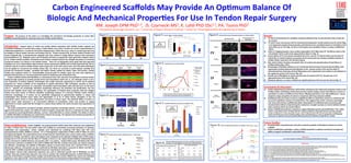
- 1. Carbon Engineered Scaffolds May Provide An Op9mum Balance Of Perspec(ve Biologic And Mechanical Proper9es For Use In Tendon Repair Surgery Advantage Solu(ons, LLC RM. Joseph DPM PhD1,3, JS. Czarnecki MS2, K. Lafdi PhD DSc2,3, PA. Tsonis PhD3 1 Perspec@ve Advantage Solu@ons, LLC, 2 University of Dayton Research Ins@tute, 3 Center for Tissue Regenera@on & Engineering at Dayton Purpose: The purpose of this study is to inves9gate the mechanical and biologic proper9es of carbon fiber Figure 1: Reconstructed Cri9cal Size Defect of Achilles Tendon Figure 5: Load to Failure of Carbon Scaffolds Figure 9: Confocal Microscopy of Fibroblast Adhesion on Carbon Scaffolds Results: scaffolds as poten9al graSs for repairing cri9cal-‐size Achilles tendon defects. Light Microscopy Immunoflourescence 1) CF and CV demonstrate less variability in physical alributes (Fig. 2) and load failure (Fig. 5) than the Microscopy GJ controls. 2) CF and CV were more porous than GJ and demonstrated greater tensile modulus than GJ control (Figs. * 2, 4). Regression analysis showed porosity and thickness to be a predic9ve factors in load failure of GJ Introduc9on: Surgical repair of cri9cal size tendon defects associated with Achilles tendon ruptures are Denotes Sta9s9cal Significance Between Groups Sample 1 control but not CF of CV (Fig . 8). Stress and modulus were predic9ve factors in carbon scaffold load formidable challenges in reconstruc9ve surgery. Cri9cal defects occur when a tendon can not be re-‐approximated or failure. 1c sufficiently lengthened to reconstruct the tendon following injury. When this occurs, there is a tendon gap that must 3) Micro-‐CT imaging showed a more random organiza9on of CV fibers than CF. The fibrous nature of GJ be bridged to restore tendon structure and func9on (Fig 1a). Tendon transfers (Fig. 1b Flexor Hallucis transfer) and CV extracellular matrix could not be appreciated on Micro-‐CT (Fig. 3). tendon allograSs (Fig. 1c Semitendonosis allograS) have been u9lized to bridge deficits however both techniques 4) Load to failure of CF most closely resembled that of GJ controls. CV load to failure was less than CF have limita9ons (1, 2). AllograSs pose a risk of infec9on and have no capacity for repair given their acellular nature 1a 1b 1d 1e and GJ (Fig. 5). Carbon modulus was greater the GJ control and more closely resembles modulus of (3, 4). Tendon transfers provide a func9onal muscle tendon complex however the strength and power of commonly Sample 2 Achilles tendon reported in the literature (Fig.6). transferred tendons are inferior to the Achilles tendon. There are no biologically ac9ve graSs with FDA approved 5) Max. load to failure of carbon was greater than GJ control and reported values of load failure of indica9ons for use in cri9cal defect repair however several commercial scaffolds such as GraS Jacket (GJ) (Fig. 1d) are Figure 6: Tensile Behavior of Carbon Scaffolds rou9nely used to re-‐enforce Achilles tendon repair surgery (5). In the most severe cases, all of the above techniques Figure 2: Physical Proper9es of Carbon Scaffolds and GraS Jacket Controls Achilles tendon (Figs. 5 &6 ). 6) Morphology of fibroblast adhesion on CF consistently demonstrated cell clustering and diffuse ac9n may be applied to reconstruct the Achilles tendon (Fig. 1e). There are currently no graS implants available that organiza9on similarly observed with adhesion to the vascular surface of GJ controls while CV provide the mechanical strength , durability and cellular ac9vity needed for tendon repair and func9on. Limited Sample 1 demonstrated less clustering and focal filamentous ac9n organiza9on more similar to cell adhesion to studies have explored carbon as a biocompa9ble structural graS for Achilles repair yet none have explored its the epidermal surface of GJ control. (Fig. 10). poten9al dual func9ons as a structural graS and vehicle for delivering cells and biofactors (6). 7) Growth rate of fibroblasts on carbon was less than GJ control GJ>CF>CV. Growth rate on CF Carbon scaffolds seeded with fibroblasts or mesenchymal stem cells may have the poten9al to enhance tendon healing through synthesis of anabolic growth factors and extracellular matrix (10, 11, 12). Although carbon is not a CF 8) approached that of GJ control (Fig. 10). Fibroblast adhesion on carbon scaffolds was significantly lower than GJ controls cultures (Fig. 7). new component of medical implants, technologies that enable nanoscale engineering of carbon and its physical proper9es are. Carbon scaffolds can be engineered to specific dimensions, fiber orienta9on, porosity as well as Sample 2 mechanical strength and texture at a nanoscale. Extracellular matrix interac9ons with cells also occur on this same scale to regulate cell morphology. Fibroblast morphology influences cell anabolism and prolifera9on, two key Conclusions & Discussion: 1) Carbon engineering has poten9al to yield tendon subs9tutes with mechanical proper9es similar to the process that regulate 9ssue repair and healing. The organiza9on of Poly(D,L-‐lac9c-‐co-‐glycolic acid) and collagen Achilles tendon and biologic ac9vity that promotes tendon healing. Factors that influence variance in meshes have been shown to influence fibroblast morphology however it is unknown whether carbon similarly load failure in carbon are different from factors affec9ng load failure in GJ. These differences may be influences morphology or if carbon can be specifically manipulated to geometrically stabilize or induce cell Figure 3: Micro-‐CT Imaging of Scaffold Organiza9on *Achilles modulus data extracted from: Abrahams, M. Mechanical Behavior of Tendon IN VITRO: A Preliminary Report. Med. & Biol. Eng 1967;5(5):433-‐443 Sample 1 related to unique proper9es of carbon in comparison with 9ssue derived scaffolds. morphologies that promote 9ssue repair and regenera9on (7, 8, 9). In na9ve 9ssues, disrup9on or loss of Dermal Surface 2) Scaffold porosity was shown to have an insignificant affect on load failure variance in carbon but did extracellular matrix results in 9ssue degenera9on and failure of 9ssue repair. The poten9al ability of carbon to account for significant variance in GJ load failure. This suggests that carbon may have advantages support 9ssue repair processes in an environment deficient of extracellular matrix may provide an unique opportunity to create a graS with the strength of a mature 9ssue without the matrix structure and organiza9on of Figure 7: Fibroblasts Adhesion and Survival to Scaffolds In Vitro GJ over some natural 9ssue scaffolds that allow maximiza9on of scaffold porosity to promote graS vasculariza9on without compromising the mechanical strength necessary to restore Achilles func9on mature 9ssue. Sample 2 * when repairing cri9cal size tendon defects. This study compared the mechanical strength, physical proper9es and biologic capacity of two carbon scaffolds CV CV Epidermal Surface Denotes Sta9s9cal 3) Carbon scaffolds demonstrate a capacity to support fibroblast growth and prolifera9on. This makes against GraS Jacket test controls. The ability of carbon to support cell growth and act as a substrate for 9ssue repair (Surface) (Deep) Significance Between Groups carbon a poten9al carrier of living cells for sustained delivery of growth factors and extracellular was assessed by examining fibroblast adhesion, survival and prolifera9on on carbon scaffolds. The ability of carbon matrix synthesis to a site of tendon repair. Some refinement in CF and CV proper9es will likely be to stabilize fibroblast morphology was examined by confocal microscopy. The physical and mechanical proper9es of (Red = Ac9n Filaments, Blue = Nuclei) necessary to op9mize and prolong fibroblast growth and adhesion proper9es to carbon. carbon were examined by Micro-‐CT and MTS tes9ng. GJ CV CF GJ CF CF Future Studies: Materials&Methods: Carbon Scaffolds: Two polyacrylonitrile (PAN) carbon fiber materials were engineered (Surface) (Deep) Figure 10: Rates of Fibroblast Prolifera9on on Scaffolds In Vitro (A 450nm-‐A690nm) 1) Characteriza9on of growth factor and matrix molecule synthesis of fibroblasts cultured on carbon to make a Carbon Veil ( CV) and Carbon Fabric (CF) scaffold by conven9onal processing techniques of spinning, scaffolds stabiliza9on and carboniza9on. Carbon scaffolds were fabricated by combining PAN fibers with 10% w/w 2) Further modifica9on and design a carbon scaffolds proper9es to op9mize mechanical strength and polycaprolactone (14kDa, SIGMA) in acetone . CV is a heterogeneous organiza9on of fibers while CF fibers are more ability to support cell adhesion and prolifera9on homogenously aligned. The sequen9al processes of spinning and carboniza9on were used to make carbon scaffolds. Figure 4: Rela9onship of Carbon Scaffold Porosity to Tensile Stress Mechanical Tes9ng: Load failure, stress and modulus were compared across hydrated scaffolds with an MTS using a Disclosure loading rate of 2.54 mm /min, (N=10). Micro-‐CT Tes9ng: A micro-‐ct x-‐ray system (Scanco Medical, Switzerland) was *Test Control Samples of GraSJacket were donated by Wright Medical Technology* used to characterize scaffold thickness and porosity with 7 um sec9ons. Confocal Microscopy: Fibroblasts (CRL 2703 Stepwise Mul9variate Regression of Carbon Scaffolds’ Load Failure as cell line) were cultured on scaffolds in vitro with in DMEM with 10% FBS. Confocal microscopy was used to evaluate Figure 8: a Func9on of Tensile Modulus, Porosity, Stress, Scaffold Thickness References cell adhesion, morphology and viability of fibroblast cultures at several 9me points over a period of 96 hrs using Carbon Scaffolds (Combined Scaffolds N=40 ) GraS Jacket Control (N=20) 1.Suenark, P. and P. Suebpongsiri, Clinical outcomes of flexor hallucis longus transfer for the treatment of Achilles tendinosis rupture. J Med Assoc Thai, 2009. 92 Suppl 6: p. S226-‐31. Metamorph soSware. Cell growth was also confirmed by fibroblast absorbance measured at 450nm and 690 nm 2.Nellas, Z.J., B.G. Loder, and S.J. Wertheimer, Reconstruc9on of an Achilles tendon defect u9lizing an Achilles tendon allogra;. J Foot Ankle Surg, 1996. 35(2): p. 144-‐8; discussion 190. 3.Kainer, M.A., et al., Clostridium infec9ons associated with musculoskeletal-‐9ssue allogra;s. N Engl J Med, 2004. 350(25): p. 2564-‐71. using the WST-‐1 viability assay (Roche Applied Science) per manufacturer instruc9ons. 4.Barbour, S.A. and W. King, The safe and effec9ve use of allogra; 9ssue-‐-‐an update. Am J Sports Med, 2003. 31(5): p. 791-‐7. 5.Barber, F.A., et al., A biomechanical study of Achilles tendon repair augmenta9on using Gra;Jacket matrix. Foot Ankle Int, 2008. 29(3): p. 329-‐33. Data Analysis: Second order polynomial regression was used to determine growth rate of fibroblasts on each 6.Parsons, J.R., et al., Long-‐term follow-‐up of achilles tendon repair with an absorbable polymer carbon fiber composite. Foot Ankle, 1989. 9(4): p. 179-‐84. 7.Ricci, J.L., et al., Morphological characteris9cs of tendon cells cultured on synthe9c fibers. J Biomed Mater Res, 1984. 18(9): p. 1073-‐87. scaffold. Step-‐wise mul9variate regression was used to examine the rela9onship between scaffold thickness and 8.Ricci, J.L., A.G. Gona, and H. Alexander, In vitro tendon cell growth rates on a synthe9c fiber scaffold material and on standard culture plates. J Biomed Mater Res, 1991. 25(5): p. 651-‐66. 9.Bashur, C.A., L.A. Dahlgren, and A.S. Goldstein, Effect of fiber diameter and orienta9on on fibroblast morphology and prolifera9on on electrospun poly(D,L-‐lac9c-‐co-‐glycolic acid) meshes. Biomaterials, porosity on load failure in pooled carbon samples and GraS Jacket (GJ) controls. ANOVA was used to test sta9s9cal 2006. 27(33): p. 5681-‐8. 10.Chen, J.L., et al., Efficacy of hESC-‐MSCs in kniQed silk-‐collagen scaffold for tendon 9ssue engineering and their roles. Biomaterials. 31(36): p. 9438-‐51. significance with alpha of 0.05. GraS Jacket (GJ) control samples were donated by Wright Medical Technologies. 11.Chen, X., et al., Stepwise differen9a9on of human embryonic stem cells promotes tendon regenera9on by secre9ng fetal tendon matrix and differen9a9on factors. Stem Cells, 2009. 27(6): p. 1276-‐87. 12.Liu, W., et al., Repair of tendon defect with dermal fibroblast engineered tendon in a porcine model. Tissue Eng, 2006. 12(4): p. 775-‐88