SlideShare ist ein Scribd-Unternehmen logo
1 von 121
The  lower  portion of the appendicular skeleton consists of the  pelvic girdle  and the  lower extremities .
According to  your text , the term  “pelvic girdle”  refers only to the left and right ossa coxae (each of which is composed of the ilium, ischium, and pubis).  However, some texts also include the sacrum. Ilium Iliac crest Acetabulum
The  pelvis  consists of the two ossa coxae (singular = os coxae), the sacrum, and the coccyx.
The pelvic girdle  protects  and supports  several abdominal organs
Each  os coxae  consist of three bones fused together Acetabulum
Each  os coxae  (hip bone) consists of the ilium, ischium, and pubis.  These three bones fuse between 13-15 years of age. Acetabulum
The  ilium  is the largest and uppermost of the pelvic bones
The  auricular surface  is the portion of the ilium that articulates with the sacrum
The  iliac crest  terminates anteriorly at the  anterior superior iliac spine .
 
This person is actually pressing on the  anterior superior iliac spine .
The  iliac crest  terminates posteriorly at the  posterior superior iliac spine
 
 
Bone marrow biopsy  in posterior portion of the iliac crest
The  ischium  is the posterior-inferior component of the os coxae.  It is the portion of the pelvis we sit on.
Note the  ischial tuberosity  of the ischium on which we sit.
 
The  pubis  is the anterior component of the os coxae.
 
 
WHAT PORTION OF YOUR PELVIS IS  MOST PROXIMAL  TO YOUR BELT? A COCCYX B SACRUM C ILIUM D ISCHIUM E PUBIS
The  false pelvis  (shown in green) is the upper portion of the pelvic basin and is enclosed by the wing-like sides of the flared iliac bones.
The  true pelvis  (shown in pink) is the lower portion of the pelvic basin and it encloses the pelvic cavity and forms a deep narrower region that contains the pelvic organs.
During birth (parturition) the fetus passes through the  true pelvis  during a normal vaginal delivery
Pelvimetry   is the measurement of the size/shape of the  pelvic outlet Inferior view
Fetus’ head  ready to pass through the true pelvis and pelvic outlet during birth
 
Are you comfortable and relaxed?
The old ways are sometimes the best
The  squatting position  can increase the diameter of the true pelvis
[object Object],[object Object]
Birth (parturition) in the squatting position with support from partner.
Supported squatting position for childbirth
During pregnancy there is a special hormone called  relaxin  that is released by the  corpus luteum  of the ovary
Relaxin  loosens the  symphysis pubis  and the  sacroiliac joint  to increase the diameter of the true pelvis and pelvic outlet to facilitate parturition.
Male Female Note the female pelvis has wider flair of iliac crests, has a more spherical true pelvis, and the pubic arch is wider than the male
Articular surface of  pubic bones  at symphysis pubis
The  femur  is the only bone of the thigh.  However, we will also discuss the patella.  The femur is the strongest bone in the body. It is a useful study technique to  compare and contrast  the bones of the lower extremity with their counterparts in the upper extremity.
The proximal rounded  head  of the femur articulates with the acetabulum of the os coxae.  Below the head is a constricted  neck  that is a common site for fractures of the femur in elderly persons.
A tiny ligament connects the acetabulum to the  fovea  in the proximal head of the femur
shaft The constricted  neck  joins the  shaft  of the femur..
The  shaft of the femur has a slight medial bow  to bring the knee joints more in line with the body’s plane of gravity.  This degree of convergence is more pronounced in females.
Because of a female’s wider hips the medial bow of the femur is more noticeable and the knees are in closer proximity to each other.
It is technically  incorrect  to say that an elderly person, usually a female, has broken her “hip”.  It is more accurate to state that they broke the  neck of their femur .
The  greater trochanter , which develops because of pulling stress on the periosteum, is found on the proximal lateral portion of the femur.
Features of the  femur
The  patella  is a sesamoid bone positioned on the anterior surface of the femur.  It does NOT articulate with the tibia.
Patella  on femur
Note largest facet is lateral when apex is inferior.
WHICH OF THE FOLLOWING DOES  NOT  ARTICULATE WITH THE FEMUR? A TIBIAL TUBEROSITY B PUBIS C SESAMOID BONE D ILIUM E ISCHIUM
The  lower leg , or crural region, contains two bones: the  tibia , which is a weight-bearing bone, and the  fibula , which not a weight-bearing bone but is important for muscle attachment. posterior anterior
Interosseous membrane
The  tibia  is the larger and more medial of the two bones of the crural region.  At its  proximal end  the tibia articulates with the femur.  At its  distal end  the tibia articulates with the talus.
The  intercondylar eminence  of the tibia is just a small stabilizing bump in the center of the tibial plateau. The  medial and lateral condyles of the tibia  articulate with the medial and lateral condyles of the distal femur. Anterior Posterior
Anterior view The fibula and the tibia at their proximal ends form the  superior tibiofibular joint
The  tibial tuberosity  of the tibia is the distal attachment site of the patellar ligament.
About one finger’s width below the tibial tuberosity, and just medial, is a flat surface on the anterior proximal portion of the tibia  where interosseous infusions of life-saving fluids can be administered  by punching a large needle through the bone into the medullary cavity.  This is easiest in children under five years of age.
An  interosseous infusion needle  used for punching through bone
Interosseous infusion
Interosseous infusion of life-saving fluid  works best in children under five years of age .  Unlike veins, which may collapse when blood pressure falls, the medullary cavity of the tibia ALWAYS stays open.
The  anterior crest of the tibia  can be easily palpated.  The  medial malleolus  is at the distal end of the tibia and forms a medial knob that is also easily palpated.  It is erroneously referred to as an ankle bone. Anterior Posterior
A child receiving life-saving fluid via interosseous infusion  into the medial malleolus  of the tibia.  As was stated previously, such I.O. infusions work best in children under five years of age.
for interosseous (I.O.) infusion of life-saving fluids So both the proximal AND distal ends of the tibia can be used for I.O. infusions.
The inferior portions of the tibia and fibula articulate to form the  inferior tibiofibular joint
The  fibula  does  not  articulate with the femur but it does with the talus. The distal end of the fibula forms the  lateral malleolus,  which is easily palpated on the lateral side of the ankle.  It is erroneously referred to as an ankle bone.
 
There are seven  tarsal bones  that contribute to the ankle.  We will only discuss two of these.  The  talus  is the most superior of the tarsal bones and it articulates with the malleoli of the tibia and fibula.  The  calcaneus  forms the prominence of the heel.
The  calcaneus  forms the prominence of the heel.  It also serves as an attachment site for large calf muscles.
The foot contains  five metatarsal bones,  with #1 leading to the great toe.  All of the toes (digits) contain  three bones , except for the great toe (digit #1), that only contains  two.  These bones in the toes are called  phalanges .
There are three arches that support the weight of the body and provide leverage when walking:  transverse arch, medial longitudinal arch, and lateral longitudinal arch .
The ligamentous  medial and lateral   longitudinal arches  and   the  transverse arch  of the foot are maintained primarily by the foot bones themselves.
Talipes or clubfoot  is a congenital malformation in which the sole of the foot is twisted medially.  Read about other foot pathologies in the clinical view in the text.
ACCORDING TO THE CLINICAL VIEW IN YOUR TEXT ON PATHOLOGIES OF THE FOOT,  WHICH OF THE FOLLOWING CONDITIONS  IS OFTEN SEEN IN PERSONS WITH  POLIOMYELITIS ? A PES PLANUS B TALIPES EQUINOVARUS C PES CAVUS D METATARSAL STRESS FRACTURE E CLUBFOOT
Fracture of the pelvis in a child  caused by blunt trauma.  Open or closed fractures of the pelvis are associated with the risk of life-threatening blood loss.
Indirect forces can fracture the pelvis
Closed fractures of the pelvis may occur in elderly persons who suffer from osteoporosis.
Since it is a complete circle, stress to the pelvis usually results in fractures in  two places  when stress is applied.
Fractures of the pelvis can be associated with  life-threatening blood loss. Umbilicus Bleeding into tissues
Rapidly restoration of normal anatomy to control pelvic fracture bleeding is essential.
What many persons refer to as a “hip fracture” is  actually a fracture of the neck of the femur .
Fractures of the neck of the femur most commonly occur in elderly post-menopausal females with osteoporosis.
Risk-taking young adults can also fracture the neck of the femur in traumatic accidents.
Teenage male slipped on ice and suffered break in neck of femur
Repair of the teenage male’s femoral neck fracture with stabilizing screws.
Risk-taking  male  snowboarder’s  chest X-ray  after fracture of his femoral neck. Diaphragm Heart ?
Fracture of the neck of the femur in risk-taking male snowboarder.
This has to be painful!
His repair with a screwed on plate to the femoral shaft with an attached screw into the femoral neck.
Close-up of screw into femoral neck and head
Severe  trauma is needed to  fracture the femoral shaft  in young adults.
Since severe trauma is needed to fracture the massive femur in young adults, such fractures are often open.
Open or closed femoral fractures can be associated with  life-threatening blood loss .
Excessive internal bleeding  in femoral shaft fracture knee Thigh
Femoral shaft fractures are of special concern because the large elastic muscles which span the femur can pull jagged ends of the fractured bone through the muscles, blood vessels, and nerves causing  on-going damage  after the original fracture!
A  Hare Traction Splint  can stop this on-going trauma by pulling on the leg.
The Hare Traction Splint can be removed in the Operating Room  after  the femoral fracture is repaired with a metal plate and screws.  These plates and screws can be left in place for the patient’s lifetime, or removed after about a year.
 
X-ray of  comminuted patellar fracture
Repair of comminuted patellar  fracture with pins and wire.
 
Note runner (see his helmet) coming in leading with his knee The baseman is experiencing a tibia/fibula fracture and doesn’t know it yet.  He is still trying to make the play.
Close-up of collusion of knee with baseman’s tibia/fibula.
A former student of mine, who was playing as a baseman in a  coed softball game behind the HYPER building while wearing cleats, suffered a similar tibia/ fibula fracture.
The tibial nail or rod was inserted into his medullary cavity just below his tibial tuberosity so it extended through the fracture site and was screwed in place.
The nail or rod was inserted into the tibial medullary cavity through a hole drilled just below the tibial tuberosity.
Tibial rod or nail in place, inserted past the site of fracture, and screwed in place.  Note that the fibula is left as is as it is not a weight-bearing bone.
Fixation of the tibial rod or nail in place with screws.
View of the healed tibia and fibula of the male softball player mentioned earlier who sustained a tibia/fibula fracture behind the HYPER building.  This is  one year after the injury.   He opted to have the rod (nail) removed the next week.  The rod is removed by screwing a device into the proximal end of the rod and they using a slap hammer to pull it out.  It is essential that  all  the screws are removed before the hammering begins!
Long jumper experiencing  right tibial fracture  on takeoff.
Close-up of right tibial fracture that occurred on takeoff
Open fracture of tibia upon landing
WHICH OF THE FOLLOWING  IS TYPICALLY ASSOCIATED WITH POTENTIALLY FATAL  HYPOVOLEMIC SHOCK ? A ILIAC FRACTURE B POTTS FRACTURE C CRURAL FRACTURE D COLLES FRACTURE E CRUCIFIXION
Fractures in the ankle region were common before better boots were designed.  Now skiers are more likely to suffer boot-top fractures of the tibia/fibula  or  dislocate the knee.
A  Pott’s fracture  occurs when a side-ways force causes both malleoli to fracture.
Pott’s fracture
Calcaneal fracture
Repair of calcaneal fracture
All gangrenous (dead) tissue must be surgically amputated.  However, a surgeon can do it with style.
Note that at the end of the surgery the foot has retained its symmetry and aesthetics.

Weitere ähnliche Inhalte

Was ist angesagt?

Upper limb bones-joints-muscles-Dr.B.B.Gosai
Upper limb bones-joints-muscles-Dr.B.B.GosaiUpper limb bones-joints-muscles-Dr.B.B.Gosai
Upper limb bones-joints-muscles-Dr.B.B.GosaiDr.B.B. Gosai
 
Joints of the limbs
Joints of the limbsJoints of the limbs
Joints of the limbsTerri Leng
 
Knee joint anatomy final ppt
Knee joint anatomy final pptKnee joint anatomy final ppt
Knee joint anatomy final pptJemi Kansagara
 
Joints upper limb 2nd lecture10122010
Joints upper limb 2nd lecture10122010Joints upper limb 2nd lecture10122010
Joints upper limb 2nd lecture10122010Lawrence James
 
Kin191 A.Ch.6.Knee.Patellofemoral.Anatomy
Kin191 A.Ch.6.Knee.Patellofemoral.AnatomyKin191 A.Ch.6.Knee.Patellofemoral.Anatomy
Kin191 A.Ch.6.Knee.Patellofemoral.AnatomyJLS10
 
07 Appendicular Skeleton Pelvic Girdle And Lower Limbs
07 Appendicular Skeleton   Pelvic Girdle And Lower Limbs07 Appendicular Skeleton   Pelvic Girdle And Lower Limbs
07 Appendicular Skeleton Pelvic Girdle And Lower Limbsguest334add
 
Ankle joint & bones of foot
Ankle joint & bones of footAnkle joint & bones of foot
Ankle joint & bones of footM S
 
Anatomy of Lower leg, ankle, and foot
Anatomy of Lower leg, ankle, and footAnatomy of Lower leg, ankle, and foot
Anatomy of Lower leg, ankle, and footDr Vicky Kasundra
 
Lower limb complete
Lower limb completeLower limb complete
Lower limb completeLucidante1
 
Skeleton system- bones and their number with detailed description.
Skeleton system- bones and their number with detailed description.Skeleton system- bones and their number with detailed description.
Skeleton system- bones and their number with detailed description.bhartisharma175
 
Joints of the upper limb by Dr. MemoOna Huzaifa
Joints of the upper limb by Dr. MemoOna HuzaifaJoints of the upper limb by Dr. MemoOna Huzaifa
Joints of the upper limb by Dr. MemoOna HuzaifaHuzaifa Zahoor
 
knee joint
knee jointknee joint
knee jointhanan777
 
06 Appendicular Skeleton Pectoral Girdle And Upper Limbs
06 Appendicular Skeleton   Pectoral Girdle And Upper Limbs06 Appendicular Skeleton   Pectoral Girdle And Upper Limbs
06 Appendicular Skeleton Pectoral Girdle And Upper Limbsguest334add
 
Lower limb bones-joints-muscles-Dr.B.B.Gosai
Lower limb bones-joints-muscles-Dr.B.B.GosaiLower limb bones-joints-muscles-Dr.B.B.Gosai
Lower limb bones-joints-muscles-Dr.B.B.GosaiDr.B.B. Gosai
 

Was ist angesagt? (20)

Upper limb bones-joints-muscles-Dr.B.B.Gosai
Upper limb bones-joints-muscles-Dr.B.B.GosaiUpper limb bones-joints-muscles-Dr.B.B.Gosai
Upper limb bones-joints-muscles-Dr.B.B.Gosai
 
Joints of the limbs
Joints of the limbsJoints of the limbs
Joints of the limbs
 
Knee joint anatomy final ppt
Knee joint anatomy final pptKnee joint anatomy final ppt
Knee joint anatomy final ppt
 
Joints upper limb 2nd lecture10122010
Joints upper limb 2nd lecture10122010Joints upper limb 2nd lecture10122010
Joints upper limb 2nd lecture10122010
 
Kin191 A.Ch.6.Knee.Patellofemoral.Anatomy
Kin191 A.Ch.6.Knee.Patellofemoral.AnatomyKin191 A.Ch.6.Knee.Patellofemoral.Anatomy
Kin191 A.Ch.6.Knee.Patellofemoral.Anatomy
 
Ankle joint Anatomy
Ankle joint AnatomyAnkle joint Anatomy
Ankle joint Anatomy
 
07 Appendicular Skeleton Pelvic Girdle And Lower Limbs
07 Appendicular Skeleton   Pelvic Girdle And Lower Limbs07 Appendicular Skeleton   Pelvic Girdle And Lower Limbs
07 Appendicular Skeleton Pelvic Girdle And Lower Limbs
 
Ankle joint & bones of foot
Ankle joint & bones of footAnkle joint & bones of foot
Ankle joint & bones of foot
 
Upper limb bone
Upper limb bone Upper limb bone
Upper limb bone
 
Anatomy of Lower leg, ankle, and foot
Anatomy of Lower leg, ankle, and footAnatomy of Lower leg, ankle, and foot
Anatomy of Lower leg, ankle, and foot
 
Lower limb bones
Lower limb bonesLower limb bones
Lower limb bones
 
Lower limb complete
Lower limb completeLower limb complete
Lower limb complete
 
Anatomy of the Knee Joint
Anatomy of the Knee JointAnatomy of the Knee Joint
Anatomy of the Knee Joint
 
Skeleton system- bones and their number with detailed description.
Skeleton system- bones and their number with detailed description.Skeleton system- bones and their number with detailed description.
Skeleton system- bones and their number with detailed description.
 
Joints of the upper limb by Dr. MemoOna Huzaifa
Joints of the upper limb by Dr. MemoOna HuzaifaJoints of the upper limb by Dr. MemoOna Huzaifa
Joints of the upper limb by Dr. MemoOna Huzaifa
 
Knee Joint Anatomy
Knee Joint AnatomyKnee Joint Anatomy
Knee Joint Anatomy
 
Knee joint
Knee jointKnee joint
Knee joint
 
knee joint
knee jointknee joint
knee joint
 
06 Appendicular Skeleton Pectoral Girdle And Upper Limbs
06 Appendicular Skeleton   Pectoral Girdle And Upper Limbs06 Appendicular Skeleton   Pectoral Girdle And Upper Limbs
06 Appendicular Skeleton Pectoral Girdle And Upper Limbs
 
Lower limb bones-joints-muscles-Dr.B.B.Gosai
Lower limb bones-joints-muscles-Dr.B.B.GosaiLower limb bones-joints-muscles-Dr.B.B.Gosai
Lower limb bones-joints-muscles-Dr.B.B.Gosai
 

Andere mochten auch

Bones and joints 1 1
Bones and joints 1 1Bones and joints 1 1
Bones and joints 1 1David West
 
Classification of joints
Classification of jointsClassification of joints
Classification of jointsFarhan Ali
 
Structural classification of muscle by Dr. S. N. Singh
Structural classification of muscle by Dr. S. N. SinghStructural classification of muscle by Dr. S. N. Singh
Structural classification of muscle by Dr. S. N. SinghDr. Shyam Narayan Singh
 
Muscular System Ppt
Muscular System PptMuscular System Ppt
Muscular System Pptshas595
 
Macroscopic & Microscopic Structure of Muscular System
Macroscopic & Microscopic Structure of Muscular SystemMacroscopic & Microscopic Structure of Muscular System
Macroscopic & Microscopic Structure of Muscular SystemSado Anatomist
 
Classification of Joints
Classification of JointsClassification of Joints
Classification of JointsMaylord Demol
 
Chap10 powerpoint Muscular System
Chap10 powerpoint Muscular SystemChap10 powerpoint Muscular System
Chap10 powerpoint Muscular Systemkevperrino
 
Bryan English - classification of muscle injuries in sport
Bryan English - classification of muscle injuries in sportBryan English - classification of muscle injuries in sport
Bryan English - classification of muscle injuries in sportMuscleTech Network
 
Physical therapy management of pain,shimaa essa
Physical therapy management of pain,shimaa essaPhysical therapy management of pain,shimaa essa
Physical therapy management of pain,shimaa essaShimaa Essa
 
Types of movements at synovial joints
Types of movements at synovial jointsTypes of movements at synovial joints
Types of movements at synovial jointsDAWN V TOMY
 
Kinesiology & biomechanics
Kinesiology & biomechanics Kinesiology & biomechanics
Kinesiology & biomechanics Huda Alsafadi
 
classification of joints and characteristics of each type joint
classification of joints and characteristics of each type jointclassification of joints and characteristics of each type joint
classification of joints and characteristics of each type jointmuti ullah
 
Joints structure and classification
Joints structure and classification Joints structure and classification
Joints structure and classification trieducation
 
Skeletal and Muscular System
Skeletal and Muscular System Skeletal and Muscular System
Skeletal and Muscular System Jenny Dixon
 

Andere mochten auch (20)

Bones and joints 1 1
Bones and joints 1 1Bones and joints 1 1
Bones and joints 1 1
 
Classification of joints
Classification of jointsClassification of joints
Classification of joints
 
Structural classification of muscle by Dr. S. N. Singh
Structural classification of muscle by Dr. S. N. SinghStructural classification of muscle by Dr. S. N. Singh
Structural classification of muscle by Dr. S. N. Singh
 
Muscular System Ppt
Muscular System PptMuscular System Ppt
Muscular System Ppt
 
Macroscopic & Microscopic Structure of Muscular System
Macroscopic & Microscopic Structure of Muscular SystemMacroscopic & Microscopic Structure of Muscular System
Macroscopic & Microscopic Structure of Muscular System
 
Classification of Joints
Classification of JointsClassification of Joints
Classification of Joints
 
Muscle system
Muscle systemMuscle system
Muscle system
 
Chap10 powerpoint Muscular System
Chap10 powerpoint Muscular SystemChap10 powerpoint Muscular System
Chap10 powerpoint Muscular System
 
Bryan English - classification of muscle injuries in sport
Bryan English - classification of muscle injuries in sportBryan English - classification of muscle injuries in sport
Bryan English - classification of muscle injuries in sport
 
Physical therapy management of pain,shimaa essa
Physical therapy management of pain,shimaa essaPhysical therapy management of pain,shimaa essa
Physical therapy management of pain,shimaa essa
 
Types of movements at synovial joints
Types of movements at synovial jointsTypes of movements at synovial joints
Types of movements at synovial joints
 
Joints
JointsJoints
Joints
 
Anatomy of Joints & its classification
Anatomy of Joints & its classificationAnatomy of Joints & its classification
Anatomy of Joints & its classification
 
joints
 joints joints
joints
 
Kinesiology & biomechanics
Kinesiology & biomechanics Kinesiology & biomechanics
Kinesiology & biomechanics
 
classification of joints and characteristics of each type joint
classification of joints and characteristics of each type jointclassification of joints and characteristics of each type joint
classification of joints and characteristics of each type joint
 
Joints structure and classification
Joints structure and classification Joints structure and classification
Joints structure and classification
 
Skeletal and Muscular System
Skeletal and Muscular System Skeletal and Muscular System
Skeletal and Muscular System
 
Structure of skeletal muscle
Structure of skeletal muscleStructure of skeletal muscle
Structure of skeletal muscle
 
Muscular System
Muscular System Muscular System
Muscular System
 

Ähnlich wie 07 Appendicular Skeleton Pelvic Girdle And Lower Limbs

05 Axial Skeleton Vertebral Column And Thoracic Cage
05 Axial Skeleton   Vertebral Column And Thoracic Cage05 Axial Skeleton   Vertebral Column And Thoracic Cage
05 Axial Skeleton Vertebral Column And Thoracic CageKevin Young
 
05 Axial Skeleton Vertebral Column And Thoracic Cage
05 Axial Skeleton   Vertebral Column And Thoracic Cage05 Axial Skeleton   Vertebral Column And Thoracic Cage
05 Axial Skeleton Vertebral Column And Thoracic Cageguest295165
 
Appendicular Skeleton.ppt
Appendicular Skeleton.pptAppendicular Skeleton.ppt
Appendicular Skeleton.pptlovekeshSingh12
 
05 Axial Skeleton Vertebral Column And Thoracic Cage
05 Axial Skeleton   Vertebral Column And Thoracic Cage05 Axial Skeleton   Vertebral Column And Thoracic Cage
05 Axial Skeleton Vertebral Column And Thoracic Cageguest334add
 
female pelvis.pptx
female pelvis.pptxfemale pelvis.pptx
female pelvis.pptxmadhuB74
 
Anatomy of the Lower Limb.pdf
Anatomy of the Lower Limb.pdfAnatomy of the Lower Limb.pdf
Anatomy of the Lower Limb.pdfWalubitaWalubita1
 
Anatomy of hip and lower limb bones
Anatomy of hip and lower limb bonesAnatomy of hip and lower limb bones
Anatomy of hip and lower limb bonesAlio Hersi
 
Radiology of the lower extremity dr asif ali khan
Radiology of the lower extremity   dr asif ali khanRadiology of the lower extremity   dr asif ali khan
Radiology of the lower extremity dr asif ali khanYangtze university
 
femalepelvisppt-131030100023-phpapp01 (1).pptx
femalepelvisppt-131030100023-phpapp01 (1).pptxfemalepelvisppt-131030100023-phpapp01 (1).pptx
femalepelvisppt-131030100023-phpapp01 (1).pptxsainiboyRicky
 
OSTEOLOGY OF THE HIP JOINT presentattion.pptx
OSTEOLOGY OF THE HIP JOINT presentattion.pptxOSTEOLOGY OF THE HIP JOINT presentattion.pptx
OSTEOLOGY OF THE HIP JOINT presentattion.pptxOluseyi7
 
MSK L001 Back Vertebral column-pelvic girdle.pdf
MSK L001 Back Vertebral column-pelvic girdle.pdfMSK L001 Back Vertebral column-pelvic girdle.pdf
MSK L001 Back Vertebral column-pelvic girdle.pdfAHMED ASHOUR
 
Appendicular Skeleton- Lower limb
Appendicular Skeleton- Lower limbAppendicular Skeleton- Lower limb
Appendicular Skeleton- Lower limbZENITH PARMAR
 

Ähnlich wie 07 Appendicular Skeleton Pelvic Girdle And Lower Limbs (20)

Hip joint
Hip jointHip joint
Hip joint
 
05 Axial Skeleton Vertebral Column And Thoracic Cage
05 Axial Skeleton   Vertebral Column And Thoracic Cage05 Axial Skeleton   Vertebral Column And Thoracic Cage
05 Axial Skeleton Vertebral Column And Thoracic Cage
 
05 Axial Skeleton Vertebral Column And Thoracic Cage
05 Axial Skeleton   Vertebral Column And Thoracic Cage05 Axial Skeleton   Vertebral Column And Thoracic Cage
05 Axial Skeleton Vertebral Column And Thoracic Cage
 
Appendicular Skeleton.ppt
Appendicular Skeleton.pptAppendicular Skeleton.ppt
Appendicular Skeleton.ppt
 
Pelvis.pptx
Pelvis.pptxPelvis.pptx
Pelvis.pptx
 
05 Axial Skeleton Vertebral Column And Thoracic Cage
05 Axial Skeleton   Vertebral Column And Thoracic Cage05 Axial Skeleton   Vertebral Column And Thoracic Cage
05 Axial Skeleton Vertebral Column And Thoracic Cage
 
Pelvis
PelvisPelvis
Pelvis
 
The Pelvis.pptx
The Pelvis.pptxThe Pelvis.pptx
The Pelvis.pptx
 
Female pelvis ppt
Female pelvis pptFemale pelvis ppt
Female pelvis ppt
 
female pelvis.pptx
female pelvis.pptxfemale pelvis.pptx
female pelvis.pptx
 
Female pelvis
Female pelvisFemale pelvis
Female pelvis
 
anatomy of pelvic hip
anatomy of pelvic hipanatomy of pelvic hip
anatomy of pelvic hip
 
Anatomy of the Lower Limb.pdf
Anatomy of the Lower Limb.pdfAnatomy of the Lower Limb.pdf
Anatomy of the Lower Limb.pdf
 
Anatomy of hip and lower limb bones
Anatomy of hip and lower limb bonesAnatomy of hip and lower limb bones
Anatomy of hip and lower limb bones
 
Radiology of the lower extremity dr asif ali khan
Radiology of the lower extremity   dr asif ali khanRadiology of the lower extremity   dr asif ali khan
Radiology of the lower extremity dr asif ali khan
 
mutinta ndeleki.pptx
mutinta ndeleki.pptxmutinta ndeleki.pptx
mutinta ndeleki.pptx
 
femalepelvisppt-131030100023-phpapp01 (1).pptx
femalepelvisppt-131030100023-phpapp01 (1).pptxfemalepelvisppt-131030100023-phpapp01 (1).pptx
femalepelvisppt-131030100023-phpapp01 (1).pptx
 
OSTEOLOGY OF THE HIP JOINT presentattion.pptx
OSTEOLOGY OF THE HIP JOINT presentattion.pptxOSTEOLOGY OF THE HIP JOINT presentattion.pptx
OSTEOLOGY OF THE HIP JOINT presentattion.pptx
 
MSK L001 Back Vertebral column-pelvic girdle.pdf
MSK L001 Back Vertebral column-pelvic girdle.pdfMSK L001 Back Vertebral column-pelvic girdle.pdf
MSK L001 Back Vertebral column-pelvic girdle.pdf
 
Appendicular Skeleton- Lower limb
Appendicular Skeleton- Lower limbAppendicular Skeleton- Lower limb
Appendicular Skeleton- Lower limb
 

Mehr von Kevin Young

Environmental attitudes survey 2010
Environmental attitudes survey 2010Environmental attitudes survey 2010
Environmental attitudes survey 2010Kevin Young
 
Respiratory system slide show
Respiratory system slide showRespiratory system slide show
Respiratory system slide showKevin Young
 
Classification of articulations
Classification of articulationsClassification of articulations
Classification of articulationsKevin Young
 
Anatomical terminology
Anatomical terminologyAnatomical terminology
Anatomical terminologyKevin Young
 
Arteries & veins of the upper body
Arteries & veins of the upper bodyArteries & veins of the upper body
Arteries & veins of the upper bodyKevin Young
 
Autonomic Nervous System
Autonomic Nervous SystemAutonomic Nervous System
Autonomic Nervous SystemKevin Young
 
Peripheral Nervous System
Peripheral Nervous SystemPeripheral Nervous System
Peripheral Nervous SystemKevin Young
 
Central Nervous System
Central Nervous SystemCentral Nervous System
Central Nervous SystemKevin Young
 
10 Muscle Tissue And Organization
10 Muscle Tissue And Organization10 Muscle Tissue And Organization
10 Muscle Tissue And OrganizationKevin Young
 
03 Cartilage And Bone Connective Tissue
03 Cartilage And Bone Connective Tissue03 Cartilage And Bone Connective Tissue
03 Cartilage And Bone Connective TissueKevin Young
 
02 Integumentary System
02 Integumentary System02 Integumentary System
02 Integumentary SystemKevin Young
 

Mehr von Kevin Young (20)

Assignment1
Assignment1Assignment1
Assignment1
 
Environmental attitudes survey 2010
Environmental attitudes survey 2010Environmental attitudes survey 2010
Environmental attitudes survey 2010
 
Respiratory system slide show
Respiratory system slide showRespiratory system slide show
Respiratory system slide show
 
Articulations
ArticulationsArticulations
Articulations
 
Classification of articulations
Classification of articulationsClassification of articulations
Classification of articulations
 
Anatomical terminology
Anatomical terminologyAnatomical terminology
Anatomical terminology
 
The heart
The heartThe heart
The heart
 
Arteries & veins of the upper body
Arteries & veins of the upper bodyArteries & veins of the upper body
Arteries & veins of the upper body
 
ANS Essentials
ANS EssentialsANS Essentials
ANS Essentials
 
Autonomic Nervous System
Autonomic Nervous SystemAutonomic Nervous System
Autonomic Nervous System
 
Peripheral Nervous System
Peripheral Nervous SystemPeripheral Nervous System
Peripheral Nervous System
 
Central Nervous System
Central Nervous SystemCentral Nervous System
Central Nervous System
 
Muscles 3
Muscles 3Muscles 3
Muscles 3
 
Muscles 2
Muscles 2Muscles 2
Muscles 2
 
Axial Muscles
Axial MusclesAxial Muscles
Axial Muscles
 
10 Muscle Tissue And Organization
10 Muscle Tissue And Organization10 Muscle Tissue And Organization
10 Muscle Tissue And Organization
 
Muscles 1
Muscles 1Muscles 1
Muscles 1
 
03 Cartilage And Bone Connective Tissue
03 Cartilage And Bone Connective Tissue03 Cartilage And Bone Connective Tissue
03 Cartilage And Bone Connective Tissue
 
02 Integumentary System
02 Integumentary System02 Integumentary System
02 Integumentary System
 
09 Tissues
09 Tissues09 Tissues
09 Tissues
 

Kürzlich hochgeladen

Food Chain and Food Web (Ecosystem) EVS, B. Pharmacy 1st Year, Sem-II
Food Chain and Food Web (Ecosystem) EVS, B. Pharmacy 1st Year, Sem-IIFood Chain and Food Web (Ecosystem) EVS, B. Pharmacy 1st Year, Sem-II
Food Chain and Food Web (Ecosystem) EVS, B. Pharmacy 1st Year, Sem-IIShubhangi Sonawane
 
Class 11th Physics NEET formula sheet pdf
Class 11th Physics NEET formula sheet pdfClass 11th Physics NEET formula sheet pdf
Class 11th Physics NEET formula sheet pdfAyushMahapatra5
 
Energy Resources. ( B. Pharmacy, 1st Year, Sem-II) Natural Resources
Energy Resources. ( B. Pharmacy, 1st Year, Sem-II) Natural ResourcesEnergy Resources. ( B. Pharmacy, 1st Year, Sem-II) Natural Resources
Energy Resources. ( B. Pharmacy, 1st Year, Sem-II) Natural ResourcesShubhangi Sonawane
 
Activity 01 - Artificial Culture (1).pdf
Activity 01 - Artificial Culture (1).pdfActivity 01 - Artificial Culture (1).pdf
Activity 01 - Artificial Culture (1).pdfciinovamais
 
1029 - Danh muc Sach Giao Khoa 10 . pdf
1029 -  Danh muc Sach Giao Khoa 10 . pdf1029 -  Danh muc Sach Giao Khoa 10 . pdf
1029 - Danh muc Sach Giao Khoa 10 . pdfQucHHunhnh
 
Advanced Views - Calendar View in Odoo 17
Advanced Views - Calendar View in Odoo 17Advanced Views - Calendar View in Odoo 17
Advanced Views - Calendar View in Odoo 17Celine George
 
How to Give a Domain for a Field in Odoo 17
How to Give a Domain for a Field in Odoo 17How to Give a Domain for a Field in Odoo 17
How to Give a Domain for a Field in Odoo 17Celine George
 
PROCESS RECORDING FORMAT.docx
PROCESS      RECORDING        FORMAT.docxPROCESS      RECORDING        FORMAT.docx
PROCESS RECORDING FORMAT.docxPoojaSen20
 
The basics of sentences session 3pptx.pptx
The basics of sentences session 3pptx.pptxThe basics of sentences session 3pptx.pptx
The basics of sentences session 3pptx.pptxheathfieldcps1
 
Unit-IV- Pharma. Marketing Channels.pptx
Unit-IV- Pharma. Marketing Channels.pptxUnit-IV- Pharma. Marketing Channels.pptx
Unit-IV- Pharma. Marketing Channels.pptxVishalSingh1417
 
2024-NATIONAL-LEARNING-CAMP-AND-OTHER.pptx
2024-NATIONAL-LEARNING-CAMP-AND-OTHER.pptx2024-NATIONAL-LEARNING-CAMP-AND-OTHER.pptx
2024-NATIONAL-LEARNING-CAMP-AND-OTHER.pptxMaritesTamaniVerdade
 
TỔNG ÔN TẬP THI VÀO LỚP 10 MÔN TIẾNG ANH NĂM HỌC 2023 - 2024 CÓ ĐÁP ÁN (NGỮ Â...
TỔNG ÔN TẬP THI VÀO LỚP 10 MÔN TIẾNG ANH NĂM HỌC 2023 - 2024 CÓ ĐÁP ÁN (NGỮ Â...TỔNG ÔN TẬP THI VÀO LỚP 10 MÔN TIẾNG ANH NĂM HỌC 2023 - 2024 CÓ ĐÁP ÁN (NGỮ Â...
TỔNG ÔN TẬP THI VÀO LỚP 10 MÔN TIẾNG ANH NĂM HỌC 2023 - 2024 CÓ ĐÁP ÁN (NGỮ Â...Nguyen Thanh Tu Collection
 
Explore beautiful and ugly buildings. Mathematics helps us create beautiful d...
Explore beautiful and ugly buildings. Mathematics helps us create beautiful d...Explore beautiful and ugly buildings. Mathematics helps us create beautiful d...
Explore beautiful and ugly buildings. Mathematics helps us create beautiful d...christianmathematics
 
microwave assisted reaction. General introduction
microwave assisted reaction. General introductionmicrowave assisted reaction. General introduction
microwave assisted reaction. General introductionMaksud Ahmed
 
Application orientated numerical on hev.ppt
Application orientated numerical on hev.pptApplication orientated numerical on hev.ppt
Application orientated numerical on hev.pptRamjanShidvankar
 
Russian Escort Service in Delhi 11k Hotel Foreigner Russian Call Girls in Delhi
Russian Escort Service in Delhi 11k Hotel Foreigner Russian Call Girls in DelhiRussian Escort Service in Delhi 11k Hotel Foreigner Russian Call Girls in Delhi
Russian Escort Service in Delhi 11k Hotel Foreigner Russian Call Girls in Delhikauryashika82
 
Making and Justifying Mathematical Decisions.pdf
Making and Justifying Mathematical Decisions.pdfMaking and Justifying Mathematical Decisions.pdf
Making and Justifying Mathematical Decisions.pdfChris Hunter
 
ICT Role in 21st Century Education & its Challenges.pptx
ICT Role in 21st Century Education & its Challenges.pptxICT Role in 21st Century Education & its Challenges.pptx
ICT Role in 21st Century Education & its Challenges.pptxAreebaZafar22
 

Kürzlich hochgeladen (20)

Food Chain and Food Web (Ecosystem) EVS, B. Pharmacy 1st Year, Sem-II
Food Chain and Food Web (Ecosystem) EVS, B. Pharmacy 1st Year, Sem-IIFood Chain and Food Web (Ecosystem) EVS, B. Pharmacy 1st Year, Sem-II
Food Chain and Food Web (Ecosystem) EVS, B. Pharmacy 1st Year, Sem-II
 
Class 11th Physics NEET formula sheet pdf
Class 11th Physics NEET formula sheet pdfClass 11th Physics NEET formula sheet pdf
Class 11th Physics NEET formula sheet pdf
 
Energy Resources. ( B. Pharmacy, 1st Year, Sem-II) Natural Resources
Energy Resources. ( B. Pharmacy, 1st Year, Sem-II) Natural ResourcesEnergy Resources. ( B. Pharmacy, 1st Year, Sem-II) Natural Resources
Energy Resources. ( B. Pharmacy, 1st Year, Sem-II) Natural Resources
 
Activity 01 - Artificial Culture (1).pdf
Activity 01 - Artificial Culture (1).pdfActivity 01 - Artificial Culture (1).pdf
Activity 01 - Artificial Culture (1).pdf
 
1029 - Danh muc Sach Giao Khoa 10 . pdf
1029 -  Danh muc Sach Giao Khoa 10 . pdf1029 -  Danh muc Sach Giao Khoa 10 . pdf
1029 - Danh muc Sach Giao Khoa 10 . pdf
 
Advanced Views - Calendar View in Odoo 17
Advanced Views - Calendar View in Odoo 17Advanced Views - Calendar View in Odoo 17
Advanced Views - Calendar View in Odoo 17
 
How to Give a Domain for a Field in Odoo 17
How to Give a Domain for a Field in Odoo 17How to Give a Domain for a Field in Odoo 17
How to Give a Domain for a Field in Odoo 17
 
PROCESS RECORDING FORMAT.docx
PROCESS      RECORDING        FORMAT.docxPROCESS      RECORDING        FORMAT.docx
PROCESS RECORDING FORMAT.docx
 
The basics of sentences session 3pptx.pptx
The basics of sentences session 3pptx.pptxThe basics of sentences session 3pptx.pptx
The basics of sentences session 3pptx.pptx
 
Unit-IV- Pharma. Marketing Channels.pptx
Unit-IV- Pharma. Marketing Channels.pptxUnit-IV- Pharma. Marketing Channels.pptx
Unit-IV- Pharma. Marketing Channels.pptx
 
2024-NATIONAL-LEARNING-CAMP-AND-OTHER.pptx
2024-NATIONAL-LEARNING-CAMP-AND-OTHER.pptx2024-NATIONAL-LEARNING-CAMP-AND-OTHER.pptx
2024-NATIONAL-LEARNING-CAMP-AND-OTHER.pptx
 
TỔNG ÔN TẬP THI VÀO LỚP 10 MÔN TIẾNG ANH NĂM HỌC 2023 - 2024 CÓ ĐÁP ÁN (NGỮ Â...
TỔNG ÔN TẬP THI VÀO LỚP 10 MÔN TIẾNG ANH NĂM HỌC 2023 - 2024 CÓ ĐÁP ÁN (NGỮ Â...TỔNG ÔN TẬP THI VÀO LỚP 10 MÔN TIẾNG ANH NĂM HỌC 2023 - 2024 CÓ ĐÁP ÁN (NGỮ Â...
TỔNG ÔN TẬP THI VÀO LỚP 10 MÔN TIẾNG ANH NĂM HỌC 2023 - 2024 CÓ ĐÁP ÁN (NGỮ Â...
 
Explore beautiful and ugly buildings. Mathematics helps us create beautiful d...
Explore beautiful and ugly buildings. Mathematics helps us create beautiful d...Explore beautiful and ugly buildings. Mathematics helps us create beautiful d...
Explore beautiful and ugly buildings. Mathematics helps us create beautiful d...
 
microwave assisted reaction. General introduction
microwave assisted reaction. General introductionmicrowave assisted reaction. General introduction
microwave assisted reaction. General introduction
 
INDIA QUIZ 2024 RLAC DELHI UNIVERSITY.pptx
INDIA QUIZ 2024 RLAC DELHI UNIVERSITY.pptxINDIA QUIZ 2024 RLAC DELHI UNIVERSITY.pptx
INDIA QUIZ 2024 RLAC DELHI UNIVERSITY.pptx
 
Application orientated numerical on hev.ppt
Application orientated numerical on hev.pptApplication orientated numerical on hev.ppt
Application orientated numerical on hev.ppt
 
Russian Escort Service in Delhi 11k Hotel Foreigner Russian Call Girls in Delhi
Russian Escort Service in Delhi 11k Hotel Foreigner Russian Call Girls in DelhiRussian Escort Service in Delhi 11k Hotel Foreigner Russian Call Girls in Delhi
Russian Escort Service in Delhi 11k Hotel Foreigner Russian Call Girls in Delhi
 
Making and Justifying Mathematical Decisions.pdf
Making and Justifying Mathematical Decisions.pdfMaking and Justifying Mathematical Decisions.pdf
Making and Justifying Mathematical Decisions.pdf
 
ICT Role in 21st Century Education & its Challenges.pptx
ICT Role in 21st Century Education & its Challenges.pptxICT Role in 21st Century Education & its Challenges.pptx
ICT Role in 21st Century Education & its Challenges.pptx
 
Asian American Pacific Islander Month DDSD 2024.pptx
Asian American Pacific Islander Month DDSD 2024.pptxAsian American Pacific Islander Month DDSD 2024.pptx
Asian American Pacific Islander Month DDSD 2024.pptx
 

07 Appendicular Skeleton Pelvic Girdle And Lower Limbs

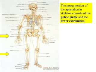
  • 1. The lower portion of the appendicular skeleton consists of the pelvic girdle and the lower extremities .
  • 2. According to your text , the term “pelvic girdle” refers only to the left and right ossa coxae (each of which is composed of the ilium, ischium, and pubis). However, some texts also include the sacrum. Ilium Iliac crest Acetabulum
  • 3. The pelvis consists of the two ossa coxae (singular = os coxae), the sacrum, and the coccyx.
  • 4. The pelvic girdle protects and supports several abdominal organs
  • 5. Each os coxae consist of three bones fused together Acetabulum
  • 6. Each os coxae (hip bone) consists of the ilium, ischium, and pubis. These three bones fuse between 13-15 years of age. Acetabulum
  • 7. The ilium is the largest and uppermost of the pelvic bones
  • 8. The auricular surface is the portion of the ilium that articulates with the sacrum
  • 9. The iliac crest terminates anteriorly at the anterior superior iliac spine .
  • 10.  
  • 11. This person is actually pressing on the anterior superior iliac spine .
  • 12. The iliac crest terminates posteriorly at the posterior superior iliac spine
  • 13.  
  • 14.  
  • 15. Bone marrow biopsy in posterior portion of the iliac crest
  • 16. The ischium is the posterior-inferior component of the os coxae. It is the portion of the pelvis we sit on.
  • 17. Note the ischial tuberosity of the ischium on which we sit.
  • 18.  
  • 19. The pubis is the anterior component of the os coxae.
  • 20.  
  • 21.  
  • 22. WHAT PORTION OF YOUR PELVIS IS MOST PROXIMAL TO YOUR BELT? A COCCYX B SACRUM C ILIUM D ISCHIUM E PUBIS
  • 23. The false pelvis (shown in green) is the upper portion of the pelvic basin and is enclosed by the wing-like sides of the flared iliac bones.
  • 24. The true pelvis (shown in pink) is the lower portion of the pelvic basin and it encloses the pelvic cavity and forms a deep narrower region that contains the pelvic organs.
  • 25. During birth (parturition) the fetus passes through the true pelvis during a normal vaginal delivery
  • 26. Pelvimetry is the measurement of the size/shape of the pelvic outlet Inferior view
  • 27. Fetus’ head ready to pass through the true pelvis and pelvic outlet during birth
  • 28.  
  • 29. Are you comfortable and relaxed?
  • 30. The old ways are sometimes the best
  • 31. The squatting position can increase the diameter of the true pelvis
  • 32.
  • 33. Birth (parturition) in the squatting position with support from partner.
  • 35. During pregnancy there is a special hormone called relaxin that is released by the corpus luteum of the ovary
  • 36. Relaxin loosens the symphysis pubis and the sacroiliac joint to increase the diameter of the true pelvis and pelvic outlet to facilitate parturition.
  • 37. Male Female Note the female pelvis has wider flair of iliac crests, has a more spherical true pelvis, and the pubic arch is wider than the male
  • 38. Articular surface of pubic bones at symphysis pubis
  • 39. Articular surface of pubic bones at symphysis pubis
  • 40. The femur is the only bone of the thigh. However, we will also discuss the patella. The femur is the strongest bone in the body. It is a useful study technique to compare and contrast the bones of the lower extremity with their counterparts in the upper extremity.
  • 41. The proximal rounded head of the femur articulates with the acetabulum of the os coxae. Below the head is a constricted neck that is a common site for fractures of the femur in elderly persons.
  • 42. A tiny ligament connects the acetabulum to the fovea in the proximal head of the femur
  • 43. shaft The constricted neck joins the shaft of the femur..
  • 44. The shaft of the femur has a slight medial bow to bring the knee joints more in line with the body’s plane of gravity. This degree of convergence is more pronounced in females.
  • 45. Because of a female’s wider hips the medial bow of the femur is more noticeable and the knees are in closer proximity to each other.
  • 46. It is technically incorrect to say that an elderly person, usually a female, has broken her “hip”. It is more accurate to state that they broke the neck of their femur .
  • 47. The greater trochanter , which develops because of pulling stress on the periosteum, is found on the proximal lateral portion of the femur.
  • 49. The patella is a sesamoid bone positioned on the anterior surface of the femur. It does NOT articulate with the tibia.
  • 50. Patella on femur
  • 51. Note largest facet is lateral when apex is inferior.
  • 52. WHICH OF THE FOLLOWING DOES NOT ARTICULATE WITH THE FEMUR? A TIBIAL TUBEROSITY B PUBIS C SESAMOID BONE D ILIUM E ISCHIUM
  • 53. The lower leg , or crural region, contains two bones: the tibia , which is a weight-bearing bone, and the fibula , which not a weight-bearing bone but is important for muscle attachment. posterior anterior
  • 55. The tibia is the larger and more medial of the two bones of the crural region. At its proximal end the tibia articulates with the femur. At its distal end the tibia articulates with the talus.
  • 56. The intercondylar eminence of the tibia is just a small stabilizing bump in the center of the tibial plateau. The medial and lateral condyles of the tibia articulate with the medial and lateral condyles of the distal femur. Anterior Posterior
  • 57. Anterior view The fibula and the tibia at their proximal ends form the superior tibiofibular joint
  • 58. The tibial tuberosity of the tibia is the distal attachment site of the patellar ligament.
  • 59. About one finger’s width below the tibial tuberosity, and just medial, is a flat surface on the anterior proximal portion of the tibia where interosseous infusions of life-saving fluids can be administered by punching a large needle through the bone into the medullary cavity. This is easiest in children under five years of age.
  • 60. An interosseous infusion needle used for punching through bone
  • 62. Interosseous infusion of life-saving fluid works best in children under five years of age . Unlike veins, which may collapse when blood pressure falls, the medullary cavity of the tibia ALWAYS stays open.
  • 63. The anterior crest of the tibia can be easily palpated. The medial malleolus is at the distal end of the tibia and forms a medial knob that is also easily palpated. It is erroneously referred to as an ankle bone. Anterior Posterior
  • 64. A child receiving life-saving fluid via interosseous infusion into the medial malleolus of the tibia. As was stated previously, such I.O. infusions work best in children under five years of age.
  • 65. for interosseous (I.O.) infusion of life-saving fluids So both the proximal AND distal ends of the tibia can be used for I.O. infusions.
  • 66. The inferior portions of the tibia and fibula articulate to form the inferior tibiofibular joint
  • 67. The fibula does not articulate with the femur but it does with the talus. The distal end of the fibula forms the lateral malleolus, which is easily palpated on the lateral side of the ankle. It is erroneously referred to as an ankle bone.
  • 68.  
  • 69. There are seven tarsal bones that contribute to the ankle. We will only discuss two of these. The talus is the most superior of the tarsal bones and it articulates with the malleoli of the tibia and fibula. The calcaneus forms the prominence of the heel.
  • 70. The calcaneus forms the prominence of the heel. It also serves as an attachment site for large calf muscles.
  • 71. The foot contains five metatarsal bones, with #1 leading to the great toe. All of the toes (digits) contain three bones , except for the great toe (digit #1), that only contains two. These bones in the toes are called phalanges .
  • 72. There are three arches that support the weight of the body and provide leverage when walking: transverse arch, medial longitudinal arch, and lateral longitudinal arch .
  • 73. The ligamentous medial and lateral longitudinal arches and the transverse arch of the foot are maintained primarily by the foot bones themselves.
  • 74. Talipes or clubfoot is a congenital malformation in which the sole of the foot is twisted medially. Read about other foot pathologies in the clinical view in the text.
  • 75. ACCORDING TO THE CLINICAL VIEW IN YOUR TEXT ON PATHOLOGIES OF THE FOOT, WHICH OF THE FOLLOWING CONDITIONS IS OFTEN SEEN IN PERSONS WITH POLIOMYELITIS ? A PES PLANUS B TALIPES EQUINOVARUS C PES CAVUS D METATARSAL STRESS FRACTURE E CLUBFOOT
  • 76. Fracture of the pelvis in a child caused by blunt trauma. Open or closed fractures of the pelvis are associated with the risk of life-threatening blood loss.
  • 77. Indirect forces can fracture the pelvis
  • 78. Closed fractures of the pelvis may occur in elderly persons who suffer from osteoporosis.
  • 79. Since it is a complete circle, stress to the pelvis usually results in fractures in two places when stress is applied.
  • 80. Fractures of the pelvis can be associated with life-threatening blood loss. Umbilicus Bleeding into tissues
  • 81. Rapidly restoration of normal anatomy to control pelvic fracture bleeding is essential.
  • 82. What many persons refer to as a “hip fracture” is actually a fracture of the neck of the femur .
  • 83. Fractures of the neck of the femur most commonly occur in elderly post-menopausal females with osteoporosis.
  • 84. Risk-taking young adults can also fracture the neck of the femur in traumatic accidents.
  • 85. Teenage male slipped on ice and suffered break in neck of femur
  • 86. Repair of the teenage male’s femoral neck fracture with stabilizing screws.
  • 87. Risk-taking male snowboarder’s chest X-ray after fracture of his femoral neck. Diaphragm Heart ?
  • 88. Fracture of the neck of the femur in risk-taking male snowboarder.
  • 89. This has to be painful!
  • 90. His repair with a screwed on plate to the femoral shaft with an attached screw into the femoral neck.
  • 91. Close-up of screw into femoral neck and head
  • 92. Severe trauma is needed to fracture the femoral shaft in young adults.
  • 93. Since severe trauma is needed to fracture the massive femur in young adults, such fractures are often open.
  • 94. Open or closed femoral fractures can be associated with life-threatening blood loss .
  • 95. Excessive internal bleeding in femoral shaft fracture knee Thigh
  • 96. Femoral shaft fractures are of special concern because the large elastic muscles which span the femur can pull jagged ends of the fractured bone through the muscles, blood vessels, and nerves causing on-going damage after the original fracture!
  • 97. A Hare Traction Splint can stop this on-going trauma by pulling on the leg.
  • 98. The Hare Traction Splint can be removed in the Operating Room after the femoral fracture is repaired with a metal plate and screws. These plates and screws can be left in place for the patient’s lifetime, or removed after about a year.
  • 99.  
  • 100. X-ray of comminuted patellar fracture
  • 101. Repair of comminuted patellar fracture with pins and wire.
  • 102.  
  • 103. Note runner (see his helmet) coming in leading with his knee The baseman is experiencing a tibia/fibula fracture and doesn’t know it yet. He is still trying to make the play.
  • 104. Close-up of collusion of knee with baseman’s tibia/fibula.
  • 105. A former student of mine, who was playing as a baseman in a coed softball game behind the HYPER building while wearing cleats, suffered a similar tibia/ fibula fracture.
  • 106. The tibial nail or rod was inserted into his medullary cavity just below his tibial tuberosity so it extended through the fracture site and was screwed in place.
  • 107. The nail or rod was inserted into the tibial medullary cavity through a hole drilled just below the tibial tuberosity.
  • 108. Tibial rod or nail in place, inserted past the site of fracture, and screwed in place. Note that the fibula is left as is as it is not a weight-bearing bone.
  • 109. Fixation of the tibial rod or nail in place with screws.
  • 110. View of the healed tibia and fibula of the male softball player mentioned earlier who sustained a tibia/fibula fracture behind the HYPER building. This is one year after the injury. He opted to have the rod (nail) removed the next week. The rod is removed by screwing a device into the proximal end of the rod and they using a slap hammer to pull it out. It is essential that all the screws are removed before the hammering begins!
  • 111. Long jumper experiencing right tibial fracture on takeoff.
  • 112. Close-up of right tibial fracture that occurred on takeoff
  • 113. Open fracture of tibia upon landing
  • 114. WHICH OF THE FOLLOWING IS TYPICALLY ASSOCIATED WITH POTENTIALLY FATAL HYPOVOLEMIC SHOCK ? A ILIAC FRACTURE B POTTS FRACTURE C CRURAL FRACTURE D COLLES FRACTURE E CRUCIFIXION
  • 115. Fractures in the ankle region were common before better boots were designed. Now skiers are more likely to suffer boot-top fractures of the tibia/fibula or dislocate the knee.
  • 116. A Pott’s fracture occurs when a side-ways force causes both malleoli to fracture.
  • 119. Repair of calcaneal fracture
  • 120. All gangrenous (dead) tissue must be surgically amputated. However, a surgeon can do it with style.
  • 121. Note that at the end of the surgery the foot has retained its symmetry and aesthetics.