Russian Call Girls in Goa Samaira 7001305949 Independent Escort Service Goa
GI bleeding.pdf
1. Sapir Goldshtein
GI bleeding
Preface
GIB presents as either overt or occult bleeding;
• Overt GIB: can manifested by –
✓ Hematemesis - vomitus of bright red blood. Suggests that upper GI bleeding which is more acute or more severe.
✓ “Coffee-grounds” – occurs from slow bleeds that are partially digested in stomach.
✓ Melena - black, tarry stool. Due to blood digestion within stomach and small bowel (present for ≥14 h, and as long as 3–5 days). Typically caused by bleeding proximal to ligament of
Treitz (in the duodenojejunal flexure). Iron supplements can cause dark stools that imitate GI bleeding.
✓ Hematochezia - passage of red, fresh blood from the rectum, usually due to lower GI bleeding, but 10% results from high volume brisk upper GI bleeding).
• Occult GIB: in the absence of overt bleeding. The source is usually small intestinal. Can manifested by –
✓ Present with symptoms of blood loss such as lightheadedness, syncope, angina, or dyspnea.
✓ Iron-deficiency anemia.
✓ Positive fecal occult blood test on routine testing. GIB is
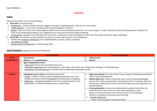
Upper GI bleeding (Esophagus, Stomach and Duodenum)
Variceal bleeding Non-variceal bleeding
Clinical
manifestations
• Hematemesis.
• Melena or even hematochezia.
• Coffee ground.
• Melena.
• Signs of hypovolemic shock:
✓ Tachycardia → more than 15% blood volume loss.
✓ Orthostatic hypotension (fall in blood pressure > 20mmHg or rise in heart rate > 20 bpm with standing) → 15-30% blood loss.
✓ Hypotension, cool and moist skin → 30-40% blood loss (potentially hypovolemic shock).
Etiology • Esophageal varices rupture (portocaval anastomosis):
✓ History - of Hep C, cirrhosis, ascites, engorged paraumbilical veins, may
already have history of EGD (Esophagogastroduodenoscopy ) and ligation.
✓ Mechanism - engorged esophageal varices from increased pressure in the
portal system → ulcerate overlying mucosa → hemorrhage.
• Peptic ulcer disease: the most common cause of upper GI bleeding (accounting for
~50% of UGIB hospitalizations).
• Mallory-Weiss tear: a linear mucosal tear near or across the gastroesophageal
junction that is often associated with retching (
להקי ניסיון
א
) or vomiting. When the
tear disrupts a submucosal arteriole, brisk hemorrhage may result (especially in an
alcoholic patient).
• Erosive gastropathy: erosions are endoscopically visualized breaks which are
confined to the mucosa and do not cause major bleeding due to the
absence of arteries and veins in the mucosa. Erosions in the esophagus,
stomach, or duodenum commonly cause mild UGIB.
✓ Possible mechanisms –
o Sepsis → decrease gastric mucosal perfusion.
2. Sapir Goldshtein
o NSAIDS (most important cause), chronic alcohol use, chemotherapeutic agents,
stress → direct toxic effect on gastric mucosa.
• Aorto-enteric fistula: less common. Usually a complication of previous aorta surgery.
Can cause massive hemorrhage!
• Dieulafoy’s lesion: less common. An aberrant vessel (tortuous arteriole) in the
mucosa (most commonly in the stomach wall) that erodes and bleeds from a
pinpoint mucosal defect.
Diagnosis Lab
• Hb: may be normal in early acute bleeding (does not fall immediately with acute GIB, due to proportionate reductions in plasma and red cell volumes - people bleed
whole blood). Patients with slow, chronic GIB may have very low Hb values despite normal BP and HR.
• Coagulation labs (PT/INR, PTT) and platelets: coagulopathy can be caused by warfarin, liver disease or high-volume blood transfusion (iatrogenic due to dilution of clotting
factors). fresh frozen plasma (FFP) is transfused for elevated INR.
• Increased BUN: blood undergo through the GI tract → blood’s proteins absorbed in the small intestine → digested into urea → reabsorb → build up → encephalopathy
(especially in patients with cirrhosis and already malfunctioning liver).
EGD
Performed after patient stabilization, preferably within 24 h of presentation. If EGD for suspected upper GI bleeding is negative, colonoscopy should be performed to assess for
lower GI bleeding (video capsule endoscopy - VCE is performed to assess small bowel for bleeding, if EGD and colonoscopy are both negative).
Angiography
Can diagnose and treat GI bleeding in unstable patients (or if EGD and colonoscopy are negative in setting of severe bleeding)
Complications • Rebleeding: define as bleeding occurs more than 5 days after admission
provided that the initial bleeding stops for 48 h (bleeding occurs especially in
the first 48h of therapy – meaning therapy failed).
✓ Risk factor for rebleeding- severe bleeding at admission, renal failure, large
varices or age > 60 years.
Management Esophageal varices bleeding
Have poorer outcomes than patients with other sources of UGIB. In contrast to
other causes of bleeding stop spontaneously only in 50% of cases, making
intervention necessary!
• Urgent endoscopy (within 12 h): endoscopic ligation is performed.
• IV vasoactive medication: e.g. octreotide (somatostatin analogue) or
somatostatin) is given for 2–5 days.
✓ Mechanism - splanchnic vasoconstriction → decreases splanchnic blood flow
→ reduces blood flow to varices → slows hemorrhage.
• Prophylactic abx: cirrhotic patients with variceal bleeding are at high risk of
infections (the most common infections are UTI and SBP).
• Intubation: airways protection in moderate to severe hematemesis which has
high risk for aspiration and pulmonary complications.
Peptic ulcers bleeding
• High dose IV PPI (proton pump inhibitors): stabilize clots and stop bleeding due to
reduced acidity. Decreases further bleeding and mortality in patients with high-risk
ulcers when given after endoscopic therapy.
• Endoscopic therapy: heater probe (direct thermal coagulation) or clips.
• Prevention of recurrent bleeding: focuses on the 3 main factors in ulcer
pathogenesis; Helicobacter pylori eradication, NSAIDs discontinuation and
counteract acid with PPI (approximately 10–50% of patients with bleeding ulcers will
rebleed within the next year if no preventive strategies are employed).
3. Sapir Goldshtein
• Blood product resuscitation: to maintain BP and correct Hb (caution to avoid
fluid overload which can cause rebleeding due to dilution of clotting factors).
Transfusion is recommended when the hemoglobin drops below 7 g/dL. Hb
should be maintained > 9 in esophageal variceal hemorrhage (vs general
hemorrhage > 7).
• Transfuse platelets: to keep platelets > 50K (acute variceal bleeding may
decrease platelets and cirrhotic may already have very low platelet count).
• Long term treatment: endoscopic ligation + nonselective beta blockers
(unopposed alpha activity → vasoconstriction of splanchnic vessels → reduced
portal blood flow → less variceal bleeding).
• TIPS (Transjugular intrahepatic portosystemic shunt): catheter is inserted
through the right jugular vein and creates a shunt btw the portal vein and the
hepatic vein → reduce pressure in portal system. Recommended in patients
who have persistent or recurrent bleeding despite endoscopic and medical
therapy. TIPS should also be considered in the first 1–2 days of hospitalization
for acute variceal bleeding in patients with advanced liver disease (e.g. Child-
Pugh class C with Child-Pugh score 10–13). It increases the amount of blood
that bypass the liver without detoxifying → can lead to hepatic
encephalopathy.
• Surgery: for portocaval shunt or esophageal transection with variceal removal.
If TIPS fail or is contraindicated.
• Discharge from the emergency room with outpatient management:
suggested for patients with a Glasgow-Blatchford score (possible range 0–23)
of 0–1 or 0–2 among patients <70 years.
Mallory-Weiss tear bleeding
• Endoscopic therapy: with epinephrine injection, band ligation, or hemoclips. Unlike
peptic ulcer, a Mallory-Weiss tear rarely rebleeds and thus does not necessitate
endoscopic therapy (usually stops spontaneously in 80–90% of patients and recurs in
only 0–10%).
5. Sapir Goldshtein
Lower GI bleeding (beyond the Duodenum)
Small intestinal source of bleeding Colonic source of bleeding
Clinical
manifestations
• Usually obscure bleeding: patients without a source of GIB identified on upper
endoscopy and colonoscopy were previously labeled as having obscure GIB.
With the advent of improved diagnostic modalities, ~75% of GIB previously
labeled obscure is now estimated to originate in the small intestine beyond the
extent of a standard upper endoscopic exam.
• Can present as melena or hematochezia.
• Can present as melena or hematochezia.
Etiology In adults > 40 years
• Vascular ectasias: an uncommon cause of chronic GIB or iron deficiency
anemia. The condition is associated with dilated small blood vessels.
• Neoplasm: e.g. GI stromal tumor, carcinoid, adenocarcinoma, lymphoma,
metastases.
• NSAID-induced erosions and ulcers.
In patients <40 years
• Meckel’s diverticulum: an outpouching in the lower part of the small intestine
(leftover of the umbilical cord. It’s the most common congenital defect of the
GI and the most common cause of significant small-intestinal GIB in children
(decreasing in frequency as a cause of bleeding with age).
• Crohn’s disease.
• Polyposis syndromes.
• Neoplasm.
• Local anal processes: hemorrhoids are probably the most common cause of LGIB,
while anal fissures also cause minor bleeding and pain.
• Diverticulosis: the most common cause of LGIB in adults (the condition of having
multiple pouches – “diverticula” in the colon that are not inflamed. When diverticula
become inflame it calls diverticulitis).
• Vascular ectasias: especially in the proximal colon of patients >70 years.
• Neoplasms (primarily adenocarcinoma).
• Colitis: ischemic, infectious, Crohn’s or ulcerative colitis, NSAID induced colitis or
ulcers.
• Postpolypectomy bleeding.
• Radiation proctopathy.
Management • In case of hematochezia and hemodynamic instability → upper endoscopy, to rule out an upper GI source before evaluation of the lower GI tract.
• In patients with massive bleeding suspected to be from the small intestine →
angiography is the initial test.
• If second-look procedures (repeat upper and lower endoscopy) are negative →
evaluation of the entire small intestine is performed, usually with video
capsule endoscopy.
• Colonoscopy: the procedure of choice in most patients admitted with LGIB. Unless
bleeding is too massive, in which case angiography is recommended.