SlideShare ist ein Scribd-Unternehmen logo
1 von 21
SHOULDER
DISLOCATION
CLINICAL
PRESENTATION
History
• Age
• 2nd & 3rd decades instability
• 4th & 5th decades impingement, frozen shoulder,
inflammatory joint disease
• 6th decade onwards rotator cuff tears , degenerative joint
disease
• Hand dominance
• Occupation
Pain
•ACJ pain usually well localised
•Neck pain, pain over trapezius or medial border of
scapula usually cervical in origin
•Associated pain in wrist or hand +/- parasthesiae
usually neurogenic
•Poorly localised pain from deltoid region usually
subacromial and rotator cuff pathology
Pain
•Night pain often rotator cuff disease,
glenohumeral arthritis and frozen shoulder
•Sudden onset excruciating pain is typical of
acute calcific tendonitis
•Pain occurring in part of the range of
shoulder abduction is termed painful arc
Instability
• Any history of trauma?
• Was shoulder dislocated?
• How many dislocations since then?
• Was dislocation spontaneous? If atraumatic
dislocation is there history of joint laxity?
• Painless clicks in the shoulder are common and
usually have no significance
Weakness
•Following traumatic event - important to
exclude brachial plexus injury
•May be due to pain (would examine
with local anaesthetic joint examination
in shoulder clinic)
Stiffness
•Restriction of both passive and active
movements
•Usually associated with frozen shoulder,
osteoarthritis, rheumatoid arthritis,
chronic dislocation and cuff tear
Inspection (Look)
•Undressed to the waist
•Observe for difficulty getting undressed
•Scars
•Asymmetry or deformity of sternoclavicular
joints
•Outline and contour of clavicles and ACJ
compared
Inspection (Look)
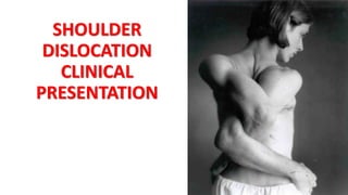
• Squaring off of the shoulder profile from anterior dislocation,
deltoid wasting or erosive arthritis with medialisation of
humeral head
• Bulk of pectoral and trapezius muscles should be compared
• “Pop-Eye” appearance of biceps might signify rupture of long
head of biceps
• Winging of scapula caused by injury to long thoracic nerve
Palpation (Feel)
•Start with sternoclavicular joint medially
•Move along clavicle to ACJ
•Tenderness over ACJ associated with
degenerative change (common and not nec.
abnormal) and traumatic subluxation
Palpation (Feel)
• Palpation lateral and inferior to coracoid assoc with
inflammatory arthropathy or primary frozen shoulder
• Palpation should continue anterolaterally to
intertubercular sulcus on humerus. Pain here may
suggest biciptal tendonitis
• Palpation of posterior joint line, where pain is more
typical of osteoarthritis
Movement
•Assess cervical spine to see if neck movements
recreate shoulder symptoms
•In full extension of C spine nose parallels the
floor and in full flexion chin should rest on chest
•Lateral rotation approx 80o
•Lateral flexion 40o
Movement
• Active and Passive range of movements of shoulder
• Forward elevation (0-170)
• Abduction (0-170)
• External rotation with abduction (0-90)
• Internal rotation (behind the back)
• Comparison with contralateral side
Neurovascular assessment
•Sensation dermatomes C4 toT2
•Power around elbow, wrist and hand
•Shoulder power tested separately
•Test peripheral nerves, esp. Axillary nerve
•Biceps and triceps reflexes
•Radial pulses
Special Tests
• Deltoid
• Arm in 90o abduction, neutral rotation
• With resistance deltoid can be felt to contract
• Supraspinatus
• “empty cans test”
• Subscapularis
• “Push-off test”
• Infraspinatus
• “Swinging doors test”
Empty Cans Test
Supraspinatus
• The purpose of this test is to assist the
integrity of the rotator cuff muscles.
• It specifically targets the supraspinatus
muscles.
• Aids in the diagnosis of rotator cuff tears
and shoulder impegment syndrome (SIS).
1. The patient is standing facing the
examiner
2. In this test, the examiner resists
abduction with the arm of the
patient eleveted to 90˚ combined
with internally rotation and 30˚ in
the forward plane.
3. If the patient gives away, the test is
considered positive.
Internal rotation lag
sign test
The purpose of this test is to assess tears of
the subscapularis muscle.
1. The patient is asked to bring the arm
behind the back with the palm facing
outwards.
2. The arm is held in near maximum
internal rotation and with the hand
away from the back by approximately
20˚ of extension.
3. The patient is asked to hold the position
while the examiner supports the elbow
but releases the wrist hold.
4. If the patient is unable to hold the
position, the lag sign is positive.
Swinging Doors Test
Infraspinatus
Other tests.
Painful arc/Impingement test
 Internally rotated arm passively abducted in scapula plane (20-30 deg
off coronal). Pain usually elicited in arc between 70-120o
Scarf test
 ACJ injury
Anterior apprehension test
 Arm externally rotated and shoulder abducted to 90 and elbow flexed.
With gentle external pressure on back of humeral head, arm is
externally rotated further
Scarf Test ACJ
1. The examiner forward flexes the involved
arm of the patient to 90˚ (with elbow to 90˚)
2. A flexed arm is then passively adducted
across the body putting an adduction stress
on the acromioclavicular joint.
3. This position results in compression of the
medial acromial facet against the distal
clavicle to provoke symptoms at the
acromioclavicular joint.
4. A positive test is indicated by reproduction
of the patients symptoms.
Anterior
Apprehension Test
This test assists with the diagnosis of
glenohumeral joint instability which can
be caused by pathology to the labrum,
rotator cuff muscles or joint capsule.
This test evaluates specifically anterior
joint instability.
• As the shoulder is moved passively
into maximum external rotation in
abduction and forward pressure is
applied to the posterior aspect of
the humeral head, the patient
complains of pain or instability.

Weitere ähnliche Inhalte

Was ist angesagt?

Tuberculosis of hip
Tuberculosis of hipTuberculosis of hip
Tuberculosis of hipHardik Pawar
 
Rotator cuff Tear and its management
Rotator cuff Tear and its managementRotator cuff Tear and its management
Rotator cuff Tear and its managementRohan Vakta
 
Legg calve perthes disease
Legg calve perthes disease Legg calve perthes disease
Legg calve perthes disease Ratan Khuman
 
4 knee assessment - History
4 knee assessment - History4 knee assessment - History
4 knee assessment - HistorySaurab Sharma
 
Shoulder examination ppt
Shoulder examination pptShoulder examination ppt
Shoulder examination pptAadarsh Bhadel
 
Shoulder dislocation: Types and Management Methods of Reduction
Shoulder dislocation: Types and Management Methods of ReductionShoulder dislocation: Types and Management Methods of Reduction
Shoulder dislocation: Types and Management Methods of ReductionUzair Siddiqui
 
Clinical Examination Of Shoulder
Clinical Examination Of Shoulder Clinical Examination Of Shoulder
Clinical Examination Of Shoulder Maley Deepak kumar
 
Clavicle fracture
Clavicle fractureClavicle fracture
Clavicle fractureToey Sutisa
 
Rotator cuff injuries
Rotator cuff injuriesRotator cuff injuries
Rotator cuff injuriesrajusvmc
 
examination of foot and ankle
examination of foot and ankleexamination of foot and ankle
examination of foot and anklemanoj das
 
Spinal orthosis dr.aliaa
Spinal orthosis  dr.aliaaSpinal orthosis  dr.aliaa
Spinal orthosis dr.aliaaAliaa El-hady
 
Spine Examination And Scoliosis
Spine Examination And ScoliosisSpine Examination And Scoliosis
Spine Examination And Scoliosisdrkmliau
 
A practical guide to Rotator Cuff Rehab
A practical guide to Rotator Cuff RehabA practical guide to Rotator Cuff Rehab
A practical guide to Rotator Cuff RehabThe Arm Clinic
 
Wrist and hand examination
Wrist and hand examinationWrist and hand examination
Wrist and hand examinationzahramp
 
Shoulder examination
Shoulder examinationShoulder examination
Shoulder examinationAhmad Sulong
 

Was ist angesagt? (20)

Tuberculosis of hip
Tuberculosis of hipTuberculosis of hip
Tuberculosis of hip
 
Rotator cuff Tear and its management
Rotator cuff Tear and its managementRotator cuff Tear and its management
Rotator cuff Tear and its management
 
Fracture of talus ppt
Fracture of talus pptFracture of talus ppt
Fracture of talus ppt
 
Humeral shaft fractures
Humeral shaft fracturesHumeral shaft fractures
Humeral shaft fractures
 
Legg calve perthes disease
Legg calve perthes disease Legg calve perthes disease
Legg calve perthes disease
 
4 knee assessment - History
4 knee assessment - History4 knee assessment - History
4 knee assessment - History
 
Shoulder examination ppt
Shoulder examination pptShoulder examination ppt
Shoulder examination ppt
 
Shoulder dislocation: Types and Management Methods of Reduction
Shoulder dislocation: Types and Management Methods of ReductionShoulder dislocation: Types and Management Methods of Reduction
Shoulder dislocation: Types and Management Methods of Reduction
 
Clinical Examination Of Shoulder
Clinical Examination Of Shoulder Clinical Examination Of Shoulder
Clinical Examination Of Shoulder
 
Clavicle fracture
Clavicle fractureClavicle fracture
Clavicle fracture
 
Wrist pain
Wrist painWrist pain
Wrist pain
 
Rotator cuff injuries
Rotator cuff injuriesRotator cuff injuries
Rotator cuff injuries
 
examination of foot and ankle
examination of foot and ankleexamination of foot and ankle
examination of foot and ankle
 
Wrist and hand examination
Wrist and hand examinationWrist and hand examination
Wrist and hand examination
 
Spinal orthosis dr.aliaa
Spinal orthosis  dr.aliaaSpinal orthosis  dr.aliaa
Spinal orthosis dr.aliaa
 
Spine Examination And Scoliosis
Spine Examination And ScoliosisSpine Examination And Scoliosis
Spine Examination And Scoliosis
 
A practical guide to Rotator Cuff Rehab
A practical guide to Rotator Cuff RehabA practical guide to Rotator Cuff Rehab
A practical guide to Rotator Cuff Rehab
 
Examination of hip joint
Examination of hip jointExamination of hip joint
Examination of hip joint
 
Wrist and hand examination
Wrist and hand examinationWrist and hand examination
Wrist and hand examination
 
Shoulder examination
Shoulder examinationShoulder examination
Shoulder examination
 

Ähnlich wie 02. shoulder examination

Seminar rotator cuff
Seminar  rotator cuffSeminar  rotator cuff
Seminar rotator cuffChapma9
 
Clinical Examination of shoulder joint
Clinical Examination of shoulder jointClinical Examination of shoulder joint
Clinical Examination of shoulder jointAbdullahIhsaas
 
Shoulder Joint Examinitaion
Shoulder Joint ExaminitaionShoulder Joint Examinitaion
Shoulder Joint ExaminitaionBharathk79
 
SHOULDER PAIN Diff. Diagnosis RB.pptx
SHOULDER PAIN Diff. Diagnosis RB.pptxSHOULDER PAIN Diff. Diagnosis RB.pptx
SHOULDER PAIN Diff. Diagnosis RB.pptxRohit Balyan
 
Shoulder dislocation
Shoulder dislocationShoulder dislocation
Shoulder dislocationvadcares
 
Rotator Cuff Injuries - Dr.CHINTAN N. PATEL
Rotator Cuff Injuries - Dr.CHINTAN N. PATELRotator Cuff Injuries - Dr.CHINTAN N. PATEL
Rotator Cuff Injuries - Dr.CHINTAN N. PATELDrChintan Patel
 
regonal pain (1) (1).pptx..............
regonal  pain (1) (1).pptx..............regonal  pain (1) (1).pptx..............
regonal pain (1) (1).pptx..............azzaelnenaey
 
Assessment of shoulder injuries in primary care
Assessment of shoulder injuries in primary care Assessment of shoulder injuries in primary care
Assessment of shoulder injuries in primary care Monis Khan
 
Assessment of shoulder joint
Assessment of shoulder jointAssessment of shoulder joint
Assessment of shoulder jointtahirakram13
 
Shoulder orthopedic disorders (dr.farouk)
Shoulder orthopedic disorders  (dr.farouk)Shoulder orthopedic disorders  (dr.farouk)
Shoulder orthopedic disorders (dr.farouk)FarouqAbdulkareem
 
Rheumatological examination
Rheumatological examinationRheumatological examination
Rheumatological examinationAshraf Okba
 
Injuries around the shoulder(maheswari)
Injuries around the shoulder(maheswari)Injuries around the shoulder(maheswari)
Injuries around the shoulder(maheswari)Yeswanth Mohan
 
SHOULDER 2222.pptx
SHOULDER 2222.pptxSHOULDER 2222.pptx
SHOULDER 2222.pptxAnujaJacob5
 
Motor System Examination
Motor System ExaminationMotor System Examination
Motor System ExaminationVib Kaul
 
Emergency management of common dislocations
Emergency management of common dislocationsEmergency management of common dislocations
Emergency management of common dislocationsMakafui Yigah
 
Clinical approch to rheumatological examination
Clinical approch to rheumatological examinationClinical approch to rheumatological examination
Clinical approch to rheumatological examinationAshraf Okba
 
Ankylosing spondylitis UG lecture
Ankylosing spondylitis UG lectureAnkylosing spondylitis UG lecture
Ankylosing spondylitis UG lectureDhananjaya Sabat
 

Ähnlich wie 02. shoulder examination (20)

Seminar rotator cuff
Seminar  rotator cuffSeminar  rotator cuff
Seminar rotator cuff
 
Clinical Examination of shoulder joint
Clinical Examination of shoulder jointClinical Examination of shoulder joint
Clinical Examination of shoulder joint
 
Shoulder Joint Examinitaion
Shoulder Joint ExaminitaionShoulder Joint Examinitaion
Shoulder Joint Examinitaion
 
SHOULDER PAIN Diff. Diagnosis RB.pptx
SHOULDER PAIN Diff. Diagnosis RB.pptxSHOULDER PAIN Diff. Diagnosis RB.pptx
SHOULDER PAIN Diff. Diagnosis RB.pptx
 
Shoulder dislocation
Shoulder dislocationShoulder dislocation
Shoulder dislocation
 
Rotator Cuff Injuries - Dr.CHINTAN N. PATEL
Rotator Cuff Injuries - Dr.CHINTAN N. PATELRotator Cuff Injuries - Dr.CHINTAN N. PATEL
Rotator Cuff Injuries - Dr.CHINTAN N. PATEL
 
regonal pain (1) (1).pptx..............
regonal  pain (1) (1).pptx..............regonal  pain (1) (1).pptx..............
regonal pain (1) (1).pptx..............
 
Shoulder
ShoulderShoulder
Shoulder
 
Shoulder examination
Shoulder examinationShoulder examination
Shoulder examination
 
Assessment of shoulder injuries in primary care
Assessment of shoulder injuries in primary care Assessment of shoulder injuries in primary care
Assessment of shoulder injuries in primary care
 
Assessment of shoulder joint
Assessment of shoulder jointAssessment of shoulder joint
Assessment of shoulder joint
 
Shoulder orthopedic disorders (dr.farouk)
Shoulder orthopedic disorders  (dr.farouk)Shoulder orthopedic disorders  (dr.farouk)
Shoulder orthopedic disorders (dr.farouk)
 
Rheumatological examination
Rheumatological examinationRheumatological examination
Rheumatological examination
 
Shoulder Disorders
Shoulder DisordersShoulder Disorders
Shoulder Disorders
 
Injuries around the shoulder(maheswari)
Injuries around the shoulder(maheswari)Injuries around the shoulder(maheswari)
Injuries around the shoulder(maheswari)
 
SHOULDER 2222.pptx
SHOULDER 2222.pptxSHOULDER 2222.pptx
SHOULDER 2222.pptx
 
Motor System Examination
Motor System ExaminationMotor System Examination
Motor System Examination
 
Emergency management of common dislocations
Emergency management of common dislocationsEmergency management of common dislocations
Emergency management of common dislocations
 
Clinical approch to rheumatological examination
Clinical approch to rheumatological examinationClinical approch to rheumatological examination
Clinical approch to rheumatological examination
 
Ankylosing spondylitis UG lecture
Ankylosing spondylitis UG lectureAnkylosing spondylitis UG lecture
Ankylosing spondylitis UG lecture
 

Mehr von Fahad Zakwan (20)

Version
VersionVersion
Version
 
Us in obstretics
Us in obstreticsUs in obstretics
Us in obstretics
 
Rupture of the uterus
Rupture of the uterusRupture of the uterus
Rupture of the uterus
 
Rpl
RplRpl
Rpl
 
Pueperium
PueperiumPueperium
Pueperium
 
Pueperal sepsis
Pueperal sepsisPueperal sepsis
Pueperal sepsis
 
Ptl
PtlPtl
Ptl
 
Prom
PromProm
Prom
 
Prolonged labour
Prolonged labourProlonged labour
Prolonged labour
 
Pph
PphPph
Pph
 
Placenta praevia
Placenta praeviaPlacenta praevia
Placenta praevia
 
Pih
PihPih
Pih
 
Pid
PidPid
Pid
 
Ovarian tumors
Ovarian tumorsOvarian tumors
Ovarian tumors
 
Ovarian cysts
Ovarian cystsOvarian cysts
Ovarian cysts
 
Obtetrics terms
Obtetrics termsObtetrics terms
Obtetrics terms
 
Multiple pregnancy
Multiple pregnancyMultiple pregnancy
Multiple pregnancy
 
Malaria in prgnancy
Malaria in prgnancyMalaria in prgnancy
Malaria in prgnancy
 
Labour and delivery
Labour and deliveryLabour and delivery
Labour and delivery
 
Iugr
IugrIugr
Iugr
 

02. shoulder examination

  • 2. History • Age • 2nd & 3rd decades instability • 4th & 5th decades impingement, frozen shoulder, inflammatory joint disease • 6th decade onwards rotator cuff tears , degenerative joint disease • Hand dominance • Occupation
  • 3. Pain •ACJ pain usually well localised •Neck pain, pain over trapezius or medial border of scapula usually cervical in origin •Associated pain in wrist or hand +/- parasthesiae usually neurogenic •Poorly localised pain from deltoid region usually subacromial and rotator cuff pathology
  • 4. Pain •Night pain often rotator cuff disease, glenohumeral arthritis and frozen shoulder •Sudden onset excruciating pain is typical of acute calcific tendonitis •Pain occurring in part of the range of shoulder abduction is termed painful arc
  • 5. Instability • Any history of trauma? • Was shoulder dislocated? • How many dislocations since then? • Was dislocation spontaneous? If atraumatic dislocation is there history of joint laxity? • Painless clicks in the shoulder are common and usually have no significance
  • 6. Weakness •Following traumatic event - important to exclude brachial plexus injury •May be due to pain (would examine with local anaesthetic joint examination in shoulder clinic)
  • 7. Stiffness •Restriction of both passive and active movements •Usually associated with frozen shoulder, osteoarthritis, rheumatoid arthritis, chronic dislocation and cuff tear
  • 8. Inspection (Look) •Undressed to the waist •Observe for difficulty getting undressed •Scars •Asymmetry or deformity of sternoclavicular joints •Outline and contour of clavicles and ACJ compared
  • 9. Inspection (Look) • Squaring off of the shoulder profile from anterior dislocation, deltoid wasting or erosive arthritis with medialisation of humeral head • Bulk of pectoral and trapezius muscles should be compared • “Pop-Eye” appearance of biceps might signify rupture of long head of biceps • Winging of scapula caused by injury to long thoracic nerve
  • 10. Palpation (Feel) •Start with sternoclavicular joint medially •Move along clavicle to ACJ •Tenderness over ACJ associated with degenerative change (common and not nec. abnormal) and traumatic subluxation
  • 11. Palpation (Feel) • Palpation lateral and inferior to coracoid assoc with inflammatory arthropathy or primary frozen shoulder • Palpation should continue anterolaterally to intertubercular sulcus on humerus. Pain here may suggest biciptal tendonitis • Palpation of posterior joint line, where pain is more typical of osteoarthritis
  • 12. Movement •Assess cervical spine to see if neck movements recreate shoulder symptoms •In full extension of C spine nose parallels the floor and in full flexion chin should rest on chest •Lateral rotation approx 80o •Lateral flexion 40o
  • 13. Movement • Active and Passive range of movements of shoulder • Forward elevation (0-170) • Abduction (0-170) • External rotation with abduction (0-90) • Internal rotation (behind the back) • Comparison with contralateral side
  • 14. Neurovascular assessment •Sensation dermatomes C4 toT2 •Power around elbow, wrist and hand •Shoulder power tested separately •Test peripheral nerves, esp. Axillary nerve •Biceps and triceps reflexes •Radial pulses
  • 15. Special Tests • Deltoid • Arm in 90o abduction, neutral rotation • With resistance deltoid can be felt to contract • Supraspinatus • “empty cans test” • Subscapularis • “Push-off test” • Infraspinatus • “Swinging doors test”
  • 16. Empty Cans Test Supraspinatus • The purpose of this test is to assist the integrity of the rotator cuff muscles. • It specifically targets the supraspinatus muscles. • Aids in the diagnosis of rotator cuff tears and shoulder impegment syndrome (SIS). 1. The patient is standing facing the examiner 2. In this test, the examiner resists abduction with the arm of the patient eleveted to 90˚ combined with internally rotation and 30˚ in the forward plane. 3. If the patient gives away, the test is considered positive.
  • 17. Internal rotation lag sign test The purpose of this test is to assess tears of the subscapularis muscle. 1. The patient is asked to bring the arm behind the back with the palm facing outwards. 2. The arm is held in near maximum internal rotation and with the hand away from the back by approximately 20˚ of extension. 3. The patient is asked to hold the position while the examiner supports the elbow but releases the wrist hold. 4. If the patient is unable to hold the position, the lag sign is positive.
  • 19. Other tests. Painful arc/Impingement test  Internally rotated arm passively abducted in scapula plane (20-30 deg off coronal). Pain usually elicited in arc between 70-120o Scarf test  ACJ injury Anterior apprehension test  Arm externally rotated and shoulder abducted to 90 and elbow flexed. With gentle external pressure on back of humeral head, arm is externally rotated further
  • 20. Scarf Test ACJ 1. The examiner forward flexes the involved arm of the patient to 90˚ (with elbow to 90˚) 2. A flexed arm is then passively adducted across the body putting an adduction stress on the acromioclavicular joint. 3. This position results in compression of the medial acromial facet against the distal clavicle to provoke symptoms at the acromioclavicular joint. 4. A positive test is indicated by reproduction of the patients symptoms.
  • 21. Anterior Apprehension Test This test assists with the diagnosis of glenohumeral joint instability which can be caused by pathology to the labrum, rotator cuff muscles or joint capsule. This test evaluates specifically anterior joint instability. • As the shoulder is moved passively into maximum external rotation in abduction and forward pressure is applied to the posterior aspect of the humeral head, the patient complains of pain or instability.