59y Female Case Review: Adult Idiopathic Scoliosis, Spondylolisthesis, Spinal Stenosis Corrected
•
2 gefällt mir•1,415 views

59 year old female presented with Progressive Adult Idiopathic Scoliosis, Spondylolisthesis, Flatback Deformity, and Stenosis. The patient was treated with a spinal fusion,

Empfohlen
Empfohlen
Weitere ähnliche Inhalte
Was ist angesagt?
Was ist angesagt? (20)
Andere mochten auch
Andere mochten auch (17)
Ähnlich wie 59y Female Case Review: Adult Idiopathic Scoliosis, Spondylolisthesis, Spinal Stenosis Corrected
Ähnlich wie 59y Female Case Review: Adult Idiopathic Scoliosis, Spondylolisthesis, Spinal Stenosis Corrected (19)
Mehr von Robert Pashman
Mehr von Robert Pashman (10)
Kürzlich hochgeladen
Kürzlich hochgeladen (20)
59y Female Case Review: Adult Idiopathic Scoliosis, Spondylolisthesis, Spinal Stenosis Corrected
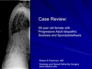
- 1. Case Review: 59 year old female with Progressive Adult Idiopathic Scoliosis and Spondylolisthesis Robert S Pashman, MD Scoliosis and Spinal Deformity Surgery www.eSpine.com
- 2. Patient History 59-year-old female Kim/SRP type II curve adult idiopathic progressive scoliosis Significant forward decompensation Spondylolisthesis Flat back Syndrome induced by critical spinal stenosis of the lumbar spine. The patient has failed conservative therapy.
- 3. Pre-op X-rays The patient has critical spinal stenosis at approximately L3-L4, L4- L5, maybe L5-S1; spondylolisthesis at L3-L4, possible spondylolisthesis at L5-S1 and what appears to be total lack of lumbar lordosis, resulting in a significant sagittal plane displacement of greater than 10 cm, C7 plumbline anterior to S1. Moreover, the patient has significant adult idiopathic scoliosis and therefore, the combination of loss of lumbar lordosis, spondylolisthesis at two levels, critical spinal stenosis and adult idiopathic scoliosis warrant major reconstruction of the lumbar spine, if any surgical solution is to be applied.
- 6. Indications for Surgery 1. KIM/SRP type 2 Adult Idiopathic Scoliosis. 2. Spondylolisthesis L3-4, L4-5. 3. Critical spinal stenosis L3-4, L4-5. 4. Significant lumbosacral obliquity. 5. Multiple co-morbidities 6. Failed conservative therapy. 7. Status post abdominal retroperitoneal approach in an anterior interbody fusion L4-5, L5-S1
- 7. Surgical Strategy – Stage 1 Abdominal retroperitoneal approach to the lumbosacral spine. Radical diskectomy at L4-5 and L5-S1. Interbody fusion at L4-5 and L5-S1 using Alphatec PEEK device with rh- BMP centrally. Spondylolisthesis reduction at L4-5. Anterior screw fixation of the lumbar spine with fully threaded screw over a washer. Intraoperative somatosensory evoked potentials. Intraoperative fluoroscopy management.
- 8. Surgical Strategy – Stage 2 T10 to sacropelvic segmental spinal instrumentation using pedicle screw/rod instrumentation. Posterior spinal fusion T10 to sacral pelvis using locally harvested autogenous bone and recombinant human bone morphogenetic protein. Radical laminectomy L2-L4 under loop magnification high intensity with bilateral interlaminar laminotomy and lateral recess decompression. Neural foraminotomy L2-3, L3-4, L4-5 for removal of severe spinal stenosis. Intraoperative spinal osteotomy L1-2, L2-3, L3-4, L4-5 for induction of sagittal plane correction from kyphosis to lordosis. These are Smith- Petersen osteotomies. Exposure iliac crest through separate incision for placement of pelvic instrumentation and local autogenous bone graft harvesting, left pelvis. Intraoperative O-Arm Stealth Navigation management with intraoperative C- arm and fluoro navigation. Intraoperative somatosensory and motor evoked potential management.
- 9. Post-Op Films The patient did well post-operatively, and is happy with her outcome.
- 11. Pre-Op/Post-op Comparison The patient is in saggittal balance, and her head is now balanced over her hips.