Case Review #11: Adult Idiopathic Scoliosis
•
1 gefällt mir•1,026 views

A 60 year old male presented with a 50+ degree curvature. He was status post lumbar fusion from L4-S1, and continued to have significant low back pain.

Empfohlen
Empfohlen
Weitere ähnliche Inhalte
Was ist angesagt?
Was ist angesagt? (20)
Andere mochten auch
Andere mochten auch (15)
Ähnlich wie Case Review #11: Adult Idiopathic Scoliosis
Ähnlich wie Case Review #11: Adult Idiopathic Scoliosis (18)
Mehr von Robert Pashman
Mehr von Robert Pashman (17)
Kürzlich hochgeladen
Kürzlich hochgeladen (20)
Case Review #11: Adult Idiopathic Scoliosis
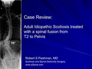
- 1. Case Review: Adult Idiopathic Scoliosis treated with a spinal fusion from T2 to Pelvis 70° Robert S Pashman, MD Scoliosis and Spinal Deformity Surgery www.eSpine.com
- 2. Patient History 60-year-old female. Adult Idiopathic Scoliosis, 50+ degree curvature. Status post anterior interbody fusion at L4-5 and L5-S1. The patient has significant thoracic kyphosis and thoracolumbar kyphosis. She reports increased pain over this area. The patient has significant left lumbar fullness. She is decompensated to the left. The patient is losing height. Intermittent radiculopathy
- 3. Pre-op X-rays She has a severe kyphoscoliosis with a 70° thoracolumbar curve. It is a fixed deformity including frontal and sagittal plane 70° decompensation.
- 4. Indications for Surgery 1. Adult idiopathic scoliosis, thoracolumbar curve greater than 70 degrees. 2. Fixed lumbosacral kyphosis. 3. Degeneration obliquity. 4. Thoracic kyphosis due to kyphoscoliosis. 5. Severe low back pain, leg pain due to the above diagnoses with lateral recess stenosis, L1-2 to L5-S1, due to curvature. 6. Failed conservative therapy. 7. Status post anterior abdominal retroperitoneal fusion, L4- 5, L5-S1
- 5. Surgical Strategy 1. Segmental spinal instrumentation, T2 to sacral pelvis. This is a 16- level fusion using Cotrel-Dubousset Legacy 5.5 stainless steel screw rod construct with pelvic fixation. 2. Sacropelvic fixation. 3. Posterior spinal fusion, T2 to the pelvis, using locally harvested autogenous bone as well as RHBMP. 4. Spinal osteotomy for mobilization of rigid kyphoscoliosis, including Smith-Peterson osteotomies, T4 to L3, with bilateral facet removal and interlaminar decompression. This is an 11- level osteotomy. 5. Interlaminar decompression, T1 to L5-S1 bilaterally. 6. Interlaminar decompression for stenosis using Loup magnification. 7. Intraoperative motor evoked potentials and somatosensory evoked potentials. 8. Intraoperative fluoroscopy.
- 6. Post-Op Films At 7 months post-op the patient is doing very well. No specific pain but she has some fatigue which is normal. 38°
- 7. Pre-Op/Post-op Comparison The patient’s curvature was reduced by 84% 70° 38°
- 8. Pre-Op and Post-op Comparison