Atelectasis
•Als PPTX, PDF herunterladen•
216 gefällt mir•86,493 views

short description on atelectasis

Empfohlen
Weitere ähnliche Inhalte
Was ist angesagt?
Was ist angesagt? (20)
Ähnlich wie Atelectasis
Ähnlich wie Atelectasis (20)
Mehr von education4227
Mehr von education4227 (20)
Kürzlich hochgeladen
Kürzlich hochgeladen (20)
Atelectasis
- 1. Atelectasis
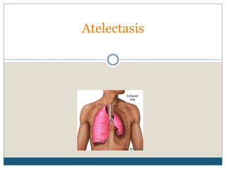
- 2. Introduction Atelectasis is defined as the collapse or closure of the lung resulting in reduced or absent gas exchange. It may affect part or all of one lung Atelectasis is the collapse of alveoli or lung tissue. It develops when the alveoli becomes airless from absorption of their air without replacement of the air with breathing.
- 4. Contd… Atelectasis may be acute or chronic The most commonly described atelectasis is acute atelectasis, which occurs frequently in the postoperative setting or in people who are immobilized and have a shallow, monotonous breathing pattern.
- 5. Etiology Obstruction of an airway Diminished distention of alveoli
- 6. Contd Airway foreign body Extrinsic compression on an airway (eg, compression due to an enlarged or aberrant vessel) Enlarged lymph nodes that compress the airway Masses in the chest that compress the airway or alveoli Cardiomegaly or enlarged pulmonary vessels that compress adjacent airways
- 7. Etiology of atelectasis Altered breathing patterns Retained secretions Pain, alterations in small airway function Anesthesia or sedation Increased abdominal pressure Reduced lung volumes due to musculoskeletal (Severe scoliosis) or neurologic disorders Pain from upper abdominal surgery
- 8. Contd… Restrictive defects, and specific surgical procedures (eg, upper abdominal, thoracic, or open heart surgery). Persistent low lung volumes Secretions or a mass obstructing or impeding airflow and compression of lung tissue Bronchospasm, airway secretions and airway inflammation in patients with asthma Abnormal airway secretions in cystic fibrosis
- 9. Contd…. Abnormal airway clearance, such as with ciliary dyskinesia syndrome Airway foreign body Excessive pressure on the lung tissue (pleural effusion, pneumothorax, hemothorax) Tumor growth within the thorax, or an elevated diaphragm
- 10. Pathophysiology Reduced alveolar ventilation or any type of blockage Impedes the passage of air The trapped alveolar air becomes absorbed into the bloodstream, but outside air cannot replace the absorbed air because of the blockage Isolated portion of the lung becomes airless and the alveoli collapse.
- 11. Excessive pressure on the lung tissue Restricts normal lung expansion on inspiration Becomes airless for prolong period Alveolar colapse
- 12. Clinical Manifestations Cough, sputum production, and low-grade fever. Marked respiratory distress Dyspnea, tachycardia, Tachypnea, pleural pain, and central cyanosis Difficulty breathing in the supine position Anxious
- 13. Assessment and Diagnostic Findings Chest x-ray : patchy infiltrates or consolidated areas. Pulse oximetry: (SpO2) (less than 90%) or a (PaO2). Physical examination: Decreased breath sounds and crackles are heard over the affected area.
- 15. Prevention Frequent turning, early mobilization, Strategies to expand the lungs and to manage secretions. Deep-breathing maneuvers (at least every 2 hours) The use of incentive spirometry or voluntary deep breathing Directed cough, suctioning, aerosol nebulizer treatments followed by chest physical therapy Postural Drainage and chest percussion, or bronchoscopy
- 16. Contd.. Change patient’s position frequently, especially from supine to upright position, to promote ventilation and prevent secretions from accumulating. Encourage early mobilization from bed to chair followed by early ambulation. Encourage appropriate deep breathing and coughing to mobilize secretions and prevent them from accumulating.
- 17. Contd… Administer prescribed Opioids and sedatives judiciously to prevent respiratory depression. Perform postural drainage and chest percussion, if indicated. Institute suctioning to remove tracheobronchial secretions, if indicated.
- 18. Management The goal in treating the patient with atelectasis is to improve ventilation and remove secretions In patients who do not respond to first-line measures or who cannot perform deep-breathing exercises, other treatments such as positive expiratory pressure (PEP therapy ) If the cause of atelectasis is bronchial obstruction from secretions, the secretions must be removed by coughing or suctioning to permit air to re-enter that portion of the lung
- 19. Chest physical therapy (chest percussion and postural drainage) Nebulizer treatments with a bronchodilator Medication or sodium bicarbonate may be used to assist the patient in the expectoration of secretions. If respiratory care measures fail to remove the obstruction, a bronchoscopy is performed. Endotracheal intubation and mechanical ventilation may be necessary for respiratory failure
- 20. Contd… Thoracentesis, removal of the fluid by needle aspiration, or insertion of a chest tube if cause is compression Bronchoscopy
- 21. Nursing diagnosis Ineffective breathing pattern related to collapse of lung tissue Activity intolerance
- 22. Thank You