![Joints (Articulations) ,[object Object],[object Object],[object Object],[object Object]](data:image/gif;base64,R0lGODlhAQABAIAAAAAAAP///yH5BAEAAAAALAAAAAABAAEAAAIBRAA7)
Empfohlen
Weitere ähnliche Inhalte
Was ist angesagt?
Was ist angesagt? (20)
Andere mochten auch
Andere mochten auch (20)
Ähnlich wie Joints and movements in the human body
Ähnlich wie Joints and movements in the human body (20)
Kürzlich hochgeladen
Kürzlich hochgeladen (20)
Joints and movements in the human body
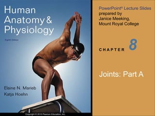
- 1. 8 Joints: Part A
- 7. Figure 8.1a Dense fibrous connective tissue Suture line (a) Suture Joint held together with very short, interconnecting fibers, and bone edges interlock. Found only in the skull.
- 9. Figure 8.1b Fibula Tibia Ligament (b) Syndesmosis Joint held together by a ligament. Fibrous tissue can vary in length, but is longer than in sutures.
- 11. Figure 8.1c Root of tooth Socket of alveolar process Periodontal ligament (c) Gomphosis “ Peg in socket” fibrous joint. Periodontal ligament holds tooth in socket.
- 14. Figure 8.2a Epiphyseal plate (temporary hyaline cartilage joint) Sternum (manubrium) Joint between first rib and sternum (immovable) (a) Synchondroses Bones united by hyaline cartilage
- 16. Figure 8.2b Fibrocartilaginous intervertebral disc Pubic symphysis Body of vertebra Hyaline cartilage (b) Symphyses Bones united by fibrocartilage
- 21. Figure 8.3 Periosteum Ligament Fibrous capsule Synovial membrane Joint cavity (contains synovial fluid) Articular (hyaline) cartilage Articular capsule
- 25. Figure 8.4b Coracoacromial ligament Subacromial bursa Cavity in bursa containing synovial fluid Bursa rolls and lessens friction. Humerus head rolls medially as arm abducts. (b) Enlargement of (a), showing how a bursa eliminates friction where a ligament (or other structure) would rub against a bone Humerus resting Humerus moving
- 27. Figure 8.4a Acromion of scapula Joint cavity containing synovial fluid Synovial membrane Fibrous capsule Humerus Hyaline cartilage Coracoacromial ligament Subacromial bursa Fibrous articular capsule Tendon sheath Tendon of long head of biceps brachii muscle (a) Frontal section through the right shoulder joint
- 32. Table 8.2 (1 of 4)
- 33. Table 8.2 (2 of 4)
- 34. Table 8.2 (3 of 4)
- 35. Table 8.2 (4 of 4)
- 39. Figure 8.5a Gliding (a) Gliding movements at the wrist
- 41. Figure 8.5b (b) Angular movements: flexion, extension, and hyperextension of the neck Hyperextension Extension Flexion
- 42. Figure 8.5c Hyperextension Flexion Extension (c) Angular movements: flexion, extension, and hyperextension of the vertebral column
- 43. Figure 8.5d Extension Extension Flexion Flexion (d) Angular movements: flexion and extension at the shoulder and knee
- 45. Figure 8.5e Abduction Adduction (e) Angular movements: abduction, adduction, and circumduction of the upper limb at the shoulder Circumduction
- 47. Figure 8.5f Lateral rotation Medial rotation Rotation (f) Rotation of the head, neck, and lower limb
- 49. Figure 8.6a Supination (radius and ulna are parallel) (a) Pronation (P) and supination (S) Pronation (radius rotates over ulna)
- 51. Figure 8.6b Dorsiflexion Plantar flexion Dorsiflexion Plantar flexion (b) Dorsiflexion and plantar flexion
- 53. Figure 8.6c Eversion Inversion (c) Inversion and eversion
- 55. Figure 8.6d Protraction of mandible Retraction of mandible (d) Protraction and retraction
- 57. Figure 8.6e Elevation of mandible Depression of mandible (e) Elevation and depression
- 59. Figure 8.6f (f) Opposition Opposition