Radiology 5th year, 8th & 9th lectures (Dr. Nasrin Alatrushi)
•Als PPTX, PDF herunterladen•
8 gefällt mir•2,149 views

The lecture has been given on Mar. 29th & Apr. 5th, 2011 by Dr. Nasrin Alatrushi.

Empfohlen
Weitere ähnliche Inhalte
Was ist angesagt?
Was ist angesagt? (20)
Andere mochten auch
Andere mochten auch (20)
Ähnlich wie Radiology 5th year, 8th & 9th lectures (Dr. Nasrin Alatrushi)
Ähnlich wie Radiology 5th year, 8th & 9th lectures (Dr. Nasrin Alatrushi) (20)
Mehr von College of Medicine, Sulaymaniyah
Mehr von College of Medicine, Sulaymaniyah (20)
Kürzlich hochgeladen
Kürzlich hochgeladen (20)
Radiology 5th year, 8th & 9th lectures (Dr. Nasrin Alatrushi)
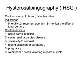
- 1. Hysterosalpingography ( HSG ) Contrast study of uterus , fallopian tubes . Indications 1- Infertility .2- recurrent abortion .3- monitor the effect of tubal surgery . Contraindication 1- acute pelvic infection . 2- sever renal or cardiac disease . 3- sensitivity to contrast . 4- recent dilatation or curettage . 5- pregnancy . 6- week prior & week following menstrual cycle .
- 2. HSG Complications : 1- pain . 2- Intravasation . 3- exacerbation of infection . Normal HSG . Congenital anomalies : 1- uterus didelphys . 2- uterus bicornis bicollis . 3- uterus bicornuate unicolies . 4- septate uterus ( arcuate uterus ) & complete septation .. 5- infantile uterus . 6- Unicornis unicollis uterus . Fibroid can be detected by HSG . Abnormalities in the fallopian tubes 1- hydrosalpinx 2- TB.
- 4. obstetrics' US in pregnant women T-abdominal. T-vaginal X-Ray ( Radiation Hazards ) . US in first trimester GS CRL is the longest fetal length within the GS .( this reliable method of dating from 7th Wks till 12th Wks. , Nuchal translucency US in the second & thited trimester Fetal maturity can be assessed by measuring the BPD & FL . Accurate dating based on BPD carried out before 24 – 26 Wks .After 24 -25 Ws the best neasurment of head size is head circomferance . Cerebral ventricles should be measured ,to exclude hydrocephalouas At 18 – 20 Wks of gestation the fetus is well formed and this is an approperiate time to obtain an accurate biparital diameter to estimate fetal maturation & also this is a sutable time to examin the fetus for fetal anomalies , so enabiling for medical abortion if fetal abnormlity is seen . Accurate dating is very important in late pregnancy for the obstatrician to know if induction of the labour or ceasarian section is necessory Obtaining sereal BPD & HC for fetal head growth , how ever the growth restriction may be manifestated first by lack of fetal body growth this estemated by maesuring the abd circomferanc . Sex of the fetus ( for those where there is risk of sex – linked inherited disease ) .
- 11. The Placenta Is usually evaluated at 9th Wks , as it mature it undergoes successive changes which is of no significance. Hemorrhage from placenta praevia ,the condition in which the placenta encroaches on the lower uterine segment,is a common cause of bleeding in the third trimester, occurring in about 0,5 %, sonography permits accurate delination of the placental position & its relation ship to the presenting part of the fetus . During the second trimester 1/3 – 1/2 of all placentas are low laying.thus raising the question of placenta praevia thus followed through the term the majority of these do not turn out to be true placenta praevia, because of diffirential growth rate of the uterus . The diagnosis of marginal placenta praevia befot 36th Ws should be confirmed with repeat the scan befor delivary . . Abruptio placenta ( accidental hemorrhage ) occur in 1 – 2 % of pregnancies .the majority of cases presented with vag bleeding but some are concealed . US show collection of blood separating the amniotic membrain from the uterine surface ,and also retromembranous hemorrahge can be diagnosed .
- 12. Large for date Causes 1- Mistake in calculating the date of conception . 2- Multiple pregnancy ( fetal abnormality is more frequent in in multiple pregnancy ) . 3- Trophoblastic disease : is spectrum of pathology ranging from benign hydatidiform mole to a malignant choriocarcinoma, Enlarged uterus filled with multiple vesicular ( cystic ) structure & in most cases no fetal parts . Rarely invasion of myometerium seen, but very rarely trophoblastic disease exist with a living pregnancy . . Benign and malignant form are indistinguishable by US In about 1/3 of cases multilocular cysts called theca lutein cysts may be identified, due to high out put of FSH.
- 13. Large for date 4 – uterine tumours in about 1% of cases The most common is fibroid, enlarged under the hormonal influnces, location of fibroid . Rarely undergoes cystic degeneration. Ovarian tumours usually corpus luteum cyst, thin wall . 5 - Polyhydramniosmay associated with : A -- Maternal abnormalities as in diabetes . B -- Fetal abnormality due to a- neural tube defect.due to production of CSF. b- obstruction of alimentary tract. Due to impaired circulation of swallowed amniotic fluid . C – poly occurs with normal multiple pregnancy .
- 14. Small for date Due to error in calculating the age from the menstrual history . Growth restriction : can be divided in to tow groups 1- Symmetrical in early pregnancy & it is associated with genetic factors & early IU infections . 2- Asymmetrical in late pregnancy affect fetal body before brain . Asymmetrical growth restriction occurs in third trimester & is associated with placental insufficiency either due to a – primary placental diseases . b - Maternal causes ( hypertension , diabetes ). Standarts are available for head circumference & body circumference in order that the fetal growth may be assessed. Fetal monitoringDoppler US of the unbilical artery enables the blood flow in the umbillical artery to be studied, in a normal preg placental resistance is low and a large portion of fetal cardiac out put flows through the placenta. There is therefor a high diastolic flow show by the flow velocity waveform in the umbilical artery .
- 16. Type III. Reverse Blood Flow During DiastoleWhen the resistance in the placenta increases further, absent diastolic flow becomes reverse diastolic flow in which the Doppler waveform is observed to be below the baseline. When the fetus develops this type of abnormality, intense surveillance is required if the fetus is less than 32 to 34 weeks and delivery if it is greater than 32 to 34 weeks. The surveillance that is currently recommended is evaluation of the ductus venosus and/or inferior vena cava, in addition to traditional antepartum testing. The following illustrates reverse diastolic flow during diastole (blue circle). When this occurs there is abnormal resistance in the placenta which results in a marked decrease in blood flow from the fetus to the placenta.
- 17. Fetal abnormality Early detection of fetal abnormality help us for appropriate management . The commonest abnormality is neural tube defects particularly spina bifida & anencephaly both identified by 18 ws, these may associated with elevated serum and amniotic alpha fetoprotien levels ,sever cases may associated with meningocele or meningomyelocele .these may associated with polyhydramnios & since polyhydramnios is due to fetal anomaly in 20%. Tumours associated with spine can be diagnosed the most common is teratoma. & may contain calcification. Most of fetal abnormality can identified in utero .
- 20. Fetal abnormality Head hydroceplalus frequently associated with spina bifida. The fetal ventricles . .Cyst of choroid plexus.Anencephaly may be diagnosed after 12 ws Chest : Pulmonary hypoplasia . Congenital diaphragmatic hernia . Heart the four-chamber view is the most rialable view. GIT : Duodenal atresia . Omphalocele & gastroschiasis . Urinary tract : the kidneys contribute significantly to the amniotic fluid volium . The presence of profound oligohydramnios although most frequently due to premature rupture of the membranes, should raise the possibility of the renal abnormality . Renal agenisis. ,the comonest are congenital hydronephrosis and dysplastic, multicystic kidney. Chronic bladder out let obstruction . Skeleton , dwarfism .the lethal form are usally associated with polyh
- 27. Us for karyotyping Three main techniques & all require US to guide the needle to required position . 1 – chorion villus sampling which carried out between 10th – 14th Wks sample from placenta . 2 – Amniocentesis carried out at 16th Wks in order to a – analysis for chromosomal abnormality & b – for alpha fetoprotein level . 3 – cordocentesis .puncturing the umbilical vein. Amniocentesis is the simplest method but it may take up two Wks for karyotyping . While villus sampling & cordocentesis the results may be available within two to three days .
- 28. Obstatrices Fetal death . A blighted ovum. Ectopic pregnancy : patient present with acute abdominal pain & pregnancy + Ve with absent IU GS. US appear as Complex adnexal mass No GS in utero. Free fluid in pouch of Douglas . DD Pelvic inflammatory diseases . Ruptured adnexal cyst . Various neoplasm . Perperium . RPOC Abdominal problem in pregnancy ( Doppler US for suspect venous thrombosis .). For detection of IUCD .