Action potentials animal systems
•Als PPTX, PDF herunterladen•
2 gefällt mir•5,215 views

Action Potential

Empfohlen
Weitere ähnliche Inhalte
Was ist angesagt?
Was ist angesagt? (20)
Ähnlich wie Action potentials animal systems
Ähnlich wie Action potentials animal systems (20)
Mehr von Yukti Sharma
Kürzlich hochgeladen
Kürzlich hochgeladen (20)
Action potentials animal systems
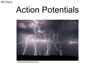
- 1. MO Figure Action Potentials Mike Hollingshead/Science Source
- 2. Figure 1 Membrane ion channels Include sodium (Na+), potassium (K+), and calcium (Ca2+) ion channels.
- 3. Figure 2 Membrane potential Electrical potential difference across the cell membrane caused by different concentrations of K+, Na+, and Cl- ions on each side of the membrane. Membrane potential of neurons is usually between -60 and -80 mV.
- 4. Figure 2a
- 5. Figure 2b
- 6. Figure 3 The action potential Small changes in membrane potential (graded potentials) can be depolarizing or hyperpolarizing. A depolarizing potential that exceeds a threshold becomes an action potential.
- 7. Figure 4 The action potential During an action potential, membrane potential changes as a result of ion flow through voltage-gated Na+ channels, voltage-gated K+ channels, and the Na+/K+ pump.
- 8. Figure 4
- 9. Figure 5 The action potential The membrane depolarization during an action potential triggers action potentials in adjacent regions of an axon. Depolarization spreads down the length of the axon as a result.
- 10. Figure 5a
- 11. Figure 5b
- 12. Figure 5c
- 13. Figure 6 Saltatory conduction Some axons are myelinated by insulating glial cells. Ion channels are only present at the nodes of Ranvier between glia. Electrical current jumps from node to node, increasing the speed of neural transmission.
- 14. Figure 6
- 15. Figure 6a
- 16. Figure 6b
- 17. MO Figure Neurons and Synaptic Communication Photo Researchers, Inc./Science Source
- 18. Figure 1
- 19. Figure 2 Postsynaptic potentials A signal from a presynaptic neuron may induce an inhibitory (IPSP) or excitatory (EPSP) potential in the postsynaptic neuron. An IPSP causes a hyperpolarization and makes a new action potential less likely to form. An EPSP causes a depolarization and increases the likelihood of a new action potential.
- 21. Table 2b
- 22. Table 2c
- 23. Figure 3 GABA g-aminobutyric acid (GABA) is an inhibitory neurotransmitter that triggers opening of Cl- channels in the postsynaptic neuron, which hyperpolarizes its membrane.
- 24. MO Figure Structure and Function of the Vertebrate Nervous System Bartolommeo Eustachi, Tabulae Anatomicae, 2nd edition. Amsterdam, 1722.
- 25. Figure 1 The nervous system Subdivided into the central nervous system (CNS) and peripheral nervous system (PNS).
- 26. Figure 2 White and gray matter CNS contains both white and gray matter. White matter consists of myelinated axons. Gray matter consists of unmyelinated axons, dendrites, and cell bodies.
- 27. Figure 2a
- 28. Figure 2b
- 29. Figure 3 The peripheral nervous system (PNS) Includes sensory and motor neurons and the autonomic nervous system (ANS).
- 30. Figure 4 The autonomic nervous system Contains antagonistic sympathetic and parasympathetic divisions.
- 31. Figure 5 Regions of the brain Subdivided into forebrain, midbrain, and hindbrain regions.
- 32. Figure 6 The cerebral cortex Includes the frontal, parietal, temporal, and occipital lobes.
- 33. Figure 7
- 34. MO Figure The Human Brain: Language, Memory, and fMRI Photo via Wikimedia Commons. Originally published by Fowlers & Wells.
- 35. Figure 1 Brain language centers Broca’s area is important for speech. Wernicke’s area is important for language comprehension.
- 36. Figure 2 Functional MRI (fMRI) © 2008 Nature Publishing Group deCharms, R. Applications of real-time fMRI. Nature Reviews Neuroscience 9, 720–729 (2008) doi:10.1038/nrn2414. Used with permission. Colors indicate sites of brain activity.
- 37. Figure 3 Functional MRI (fMRI) These images were collected during a visual memory test. Red/yellow areas indicate regions with increased activity, and blue indicates decreased activity. Yellow indicates moderate activity. © 2009 Nature Publishing Group Dickerson, B. and Eichenbaum, H. The Episodic Memory System: Neurocircuitry and Disorders. Neuropsychopharmacology 35, 86–104 (2009) doi:10.1038/npp.2009.126. Used with permission.
- 38. Figure 4 Protein and brain activity fMRI revealed that high-protein breakfasts (HP, dark blue) reduce brain activity in regions associated with food motivation and reward pathways compared to normal-protein breakfasts (NP, light blue).
- 39. Figure 5 Aplysia Martin Shields/Science Source. This sea slug with large, easily accessible neurons, is a good model organism in neurobiology to study the links between specific neurons and behavior.
Hinweis der Redaktion
- This interactive can be found as a separate file in your downloaded folder. This slide is a placeholder only. The image is a screen shot taken from the digital book.