

Empfohlen
Weitere ähnliche Inhalte
Was ist angesagt?
Was ist angesagt? (19)
Andere mochten auch
Andere mochten auch (16)
Ähnlich wie 11c emotion stress and relaxation
Ähnlich wie 11c emotion stress and relaxation (20)
Mehr von PS Deb
Mehr von PS Deb (20)
Kürzlich hochgeladen
Kürzlich hochgeladen (20)
11c emotion stress and relaxation
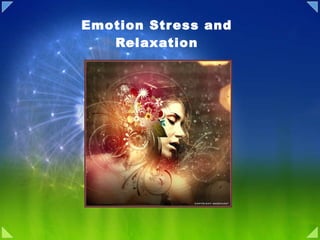
- 1. Emotion Stress and Relaxation
- 2. What is Emotion? What kind of an emotion of fear would be left if the feeling neither of quickened heart-beats nor of shallow breathing, neither of trembling lips nor of weakened limbs, neither of goose-flesh nor of visceral stirrings, were present, it is quite impossible for me to think … I say that for us emotion dissociated from all bodily feeling is inconceivable.” William James, 1893 ( Psychology : p. 379.) Stimulus Cognition Awareness Conation Urge to take action Affect Feeling Response
- 3. Emotional Arousal and Performance
- 4. Model of the basic neural systems control of emotions Neocortical processing Subcortical processing Skeletomotor and Autonomic control Periphery Stimulus Effectors Filtering and Evaluation
- 5. Cognitive Theory of Emotion <1880 Stimulus Cognition Conation/Affect Response 1 2 3 A conscious, emotional event initiates reflexive autonomic responses in the body
- 8. Cannon Bard: Sham Rage Animal
- 9. The Hypothalamus Coordinates the Peripheral Expression of Emotional States : Stephen Ranson 1932, Walter Hess 1940
- 10. Hypothalamic control of ANS
- 11. Schachter Singer Cognitive Theory of Emotion (1960) “ the variety of emotion, mood and feeling states are by no means matched by an equal variety of visceral patterns.” This “rather ambiguous situation” led the them to conclude “that cognitive factors may be major determinants of emotional states.”
- 12. Arnold Appraisal Theory of Emotion Stimulus Subconscious Appraisal Response Affect Conscious Appraisal
- 14. The Search for Cortical Representation of Feeling Has Led to the Limbic System
- 16. Papez Circuit
- 17. Papez Circuit of Emotional Response
- 20. Seat of Emotion: Amygdala
- 21. Learned Emotional Responses Are Processed in the Amygdala
- 22. Auditory emotional conditioning pathway
- 23. Model of associative learning in the amygdala
- 24. The Amygdala Mediates Both the Autonomic Expression and the Cognitive Experience of Emotion
- 25. The Amygdala Is the Part of the Limbic System Most Specifically Involved With Emotional Experience
- 26. The Amygdala May Be Involved in Both Pleasurable and Fearful Responses to Stimuli
- 27. Two Pathway of Emotion
- 30. The Frontal, Cingulate, and Parahippocampal Cortices Are Involved in Emotion
- 31. Emotional Expressions: Pyramidal and Extrapyramidal Contributions
- 32. Nervous system that organize emotional experience and expression
- 34. :Plutchik Stress and Relaxation
- 37. Physiology of Stress Stress
- 41. Immune response
- 42. Other responses
- 49. Smoking , Alcohol and Stress
- 54. Food and Mood
- 55. Exercise
- 56. Establish a Support Network
- 60. Meditation
- 61. Biofeedback
- 62. Thank you
Hinweis der Redaktion
- Emotional States and Feelings Susan Iversen Irving Kupfermann Eric R. Kandel PLEASURE, ELATION, EUPHORIA, ecstasy, sadness, despondency, depression, fear, anxiety, anger, hostility, and calm—these and other emotions color our lives. They contribute to the richness of our experiences and imbue our actions with passion and character. Moreover, as we shall learn in Chapter 61, disorders of emotion contribute importantly to several major psychiatric illnesses. An emotional state has two components, one evident in a characteristic physical sensation and the other as a conscious feeling—we sense our heart pounding and we consciously feel afraid. To maintain the distinction between these two components, the term emotion sometimes is used to refer only to the bodily state (ie, the emotional state) and the term feeling is used to refer to conscious sensation. Like perception and action, emotional states and feelings are mediated by distinct neuronal circuits within the brain. In fact, many drugs that affect the mind—ranging from addictive street drugs to therapeutic agents—do so by acting on specific neural circuits concerned with emotional states and feelings.
- Emotions Overview The subjective feelings and associated phsyiological states known as emotions are essential features of normal human experience. Moreover, some of the most devastating psychiatric problems involve emotional (affective) disorders. Although everyday emotions are as varied as happiness, surprise, anger, fear, and sadness, they share some common characteristics: All emotions are expressed through both visceral motor changes and stereotyped somatic motor responses, especially movements of the facial muscles. These responses accompany subjective experiences that are not easily described, but which are much the same in all human cultures. Emotional expression is closely tied to the visceral motor system, and therefore entails the activity of all of the central brain structures that govern the preganglionic neurons in the brainstem and spinal cord. Historically, the neural centers that coordinate emotional responses have been grouped under the rubric of the limbic system. More recently, however, several brain regions in addition to the classical limbic system have been shown to play a pivotal role in emotional processing, including the amygdala and several cortical areas in the orbital and medial aspects of the frontal lobe. This broader constellation of cortical and subcortical regions encompasses not only the central components of the visceral motor system but also regions in the forebrain and diencephalon that motivate lower motor neuronal pools concerned with the somatic expression of emotional behavior. Effectively, the concerted action of these diverse brain regions constitutes an emotional motor system. Physiological Changes Associated with Emotion The most obvious signs of emotional arousal involve changes in the activity of the visceral motor (autonomic) system (see Chapter 21). Thus, increases or decreases in heart rate, cutaneous blood flow (blushing or turning pale), piloerection, sweating, and gastrointestinal motility can all accompany various emotions. These responses are brought about by changes in activity in the sympathetic, parasympathetic, and enteric components of the visceral motor system, which govern smooth muscle, cardiac muscle, and glands throughout the body. As discussed in Chapter 21, Walter Cannon argued that intense activity of the sympathetic division of the visceral motor system prepares the animal to fully utilize metabolic and other resources in challenging or threatening situations. Conversely, activity of the parasympathetic division (and the enteric division) promotes a building up of metabolic reserves. Cannon further suggested that the natural opposition of the expenditure and storage of resources is reflected in a parallel opposition of the emotions associated with these different physiological states. As Cannon pointed out, “The desire for food and drink, the relish of taking them, all the pleasures of the table are naught in the presence of anger or great anxiety.” Activation of the visceral motor system, particularly the sympathetic division, was long considered an all-or-nothing process. Once effective stimuli engaged the system, it was argued, a widespread discharge of all of its components ensued. More recent studies have shown that the responses of the autonomic nervous system are actually quite specific, with different patterns of activation characterizing different situations and their associated emotional states. Indeed, emotion-specific expressions produced voluntarily can elicit distinct patterns of autonomic activity. For example, if subjects are given muscle-by-muscle instructions that result in facial expressions recognizable as anger, disgust, fear, happiness, sadness, or surprise without being told which emotion they are simulating, each pattern of facial muscle activity is accompanied by specific and reproducible differences in visceral motor activity (as measured by indices such as heart rate, skin conductance, and skin temperature). Moreover, autonomic responses are strongest when the facial expressions are judged to most closely resemble actual emotional expression and are often accompanied by the subjective experience of that emotion! One interpretation of these findings is that when voluntary facial expressions are produced, signals in the brain engage not only the motor cortex but also some of the circuits that produce emotional states. Perhaps this relationship helps explain how good actors can be so convincing. Nevertheless, we are quite adept at recognizing the difference between a contrived facial expression and the spontaneous smile that accompanies a pleasant emotional state (Box A). This evidence, along with many other observations, indicates that one source of emotion is sensory drive from muscles and internal organs. This input forms the sensory limb of reflex circuitry that allows rapid physiological changes in response to altered conditions. However, physiological responses can also be elicited by complex and idiosyncratic stimuli mediated by the forebrain. For example, an anticipated tryst with a lover, a suspenseful episode in a novel or film, stirring patriotic or religious music, or dishonest accusations can all lead to autonomic activation and strongly felt emotions. The neural activity evoked by such complex stimuli is relayed from the forebrain to autonomic and somatic motor nuclei via the hypothalamus and brainstem reticular formation, the major structures that coordinate the expression of emotional behavior (see next section). In summary, emotion and motor behavior are inextricably linked. As William James put it more than a century ago: What kind of an emotion of fear would be left if the feeling neither of quickened heart-beats nor of shallow breathing, neither of trembling lips nor of weakened limbs, neither of goose-flesh nor of visceral stirrings, were present, it is quite impossible for me to think … I say that for us emotion dissociated from all bodily feeling is inconceivable. William James, 1893 ( Psychology : p. 379.) Conation: Motivation, will, drive, desire, impulse, volition Cognition: Information Processing In this chapter we examine how emotion is represented in the brain. A neural analysis of emotion must take into account at least four issues. First, we must understand how stimuli acquire emotional significance and what roles conscious cognitive processes and automatic unconscious processes have in determining whether a particular stimulus at a particular moment will have emotional significance (Figure 50-1). Second, we must understand how certain autonomic and skeletomotor responses are triggered once a stimulus acquires emotional significance. Third, we must identify the circuits in the cerebral cortex responsible for feelings. Finally, we need to understand how somatic emotional states and conscious feeling states interact—how feedback from peripheral, autonomic, and skeletomotor systems to the cerebral cortex shapes emotional experience. As we will see, various theories of emotion largely differ in their emphasis on the importance of this feedback. They involve cognition, an awareness of the sensation and usually its cause; affect, the feeling itself; conation, the urge to take action; and physical changes such as hypertension, tachycardia, and sweating. The hypothalamus and limbic systems are intimately concerned with emotional expression and with the genesis of emotions.
- Figure 50-2 Performance is affected by arousal level. An intermediate level of arousal is optimal; performance is less adequate at both very high and very low levels of arousal. The Peripheral Components of Emotion Prepare the Body for Action and Communicate Our Emotional States to Other People The peripheral, skeletomotor, and autonomic aspects of emotion have preparatory and communicative functions. The preparatory function involves both general arousal , which prepares the organism as a whole for action, and specific arousal , which prepares the organism for a particular behavior. For example, sexual arousal involves an increase of heart rate, a change that prepares the organism generally for physical exertion. In addition, it involves more localized changes, such as tumescence, that are specific to sexual behavior. The mechanisms of generalized and specific arousal act synergistically to prepare the periphery (muscles, glands, blood vessels) and the cerebral cortex for ongoing or upcoming events. Unless it is extreme, arousal enhances intellectual and physical performance (Figure 50-2). The peripheral component of emotion also communicates emotion to others. In humans social communication of emotion is mediated primarily by the skeletomotor system, in particular by the muscles that control facial and postural expressions.
- Figure 50-1 Model of the basic neural systems that control emotions. Emotions are typically elicited by a specific stimulus. The stimulus affects both neocortical and subcortical structures, such the amygdala. In turn, cortical structures and the amygdala and other subcortical structures regulate the systems that mediate the peripheral manifestations of emotional behaviors. The particular emotion experienced is a function of cross-talk between neocortical and subcortical structures, as well as feedback from peripheral receptors. Conscious feeling is mediated by the cerebral cortex, in part by the cingulate cortex and by the frontal lobes. Emotional states are mediated by a family of peripheral, autonomic, endocrine, and skeletomotor responses. These responses involve subcortical structures: the amygdala, the hypothalamus, and the brain stem. When frightened we not only feel afraid but also experience increased heart rate and respiration, dryness of the mouth, tense muscles, and sweaty palms, all of which are regulated by subcortical structures. To understand an emotion such as fear we therefore need to understand the relationship between cognitive feeling represented in the cortex and the associated physiological signs orchestrated in subcortical structures.
- A Theory of Emotion Must Explain the Relationship of Cognitive and Physiological States Until the late nineteenth century the traditional view of the evocation and expression of emotion consisted of the following sequence. First, an important event is recognized—for example, you see your house on fire. This recognition in turn produces in the cerebral cortex a conscious emotional experience—fear—that triggers signals to peripheral structures including the heart, blood vessels, adrenal glands, and sweat glands. According to this traditional view, a conscious, emotional event initiates reflexive autonomic responses in the body.
- Support James Lange theory Specific pattern of autonomic response correlate with emotional type After spinal cord section emotional response reduced Against Emotional state outlast physiological changes In the James-Lange Theory Emotions Are Cognitive Responses to Information From the Periphery In 1884 the American psychologist William James rejected the traditional view that emotions are initiated by cognitive activity. In an article entitled What Is Emotion ? James proposed that the cognitive experience of emotion is secondary to the physiological expression of emotion. He suggested that when we encounter a potentially dangerous situation—for example, a bear sitting in the middle of our path—the evaluation of the bear's ferocity does not itself generate a consciously experienced emotional state. We do not experience fear until after we have run away from the bear. That is, we act instinctively by running away and then consciously explain our action and the changes in our body (the increase in heart rate and respiration) as “driven by fear.” Based on this idea, James and the Danish psychologist Carl Lange proposed an alternative hypothesis: The feeling state, the conscious experience of emotion, occurs after the cortex receives signals about changes in our physiological state. Feelings are preceded by certain physiological changes—an increase or decrease in blood pressure, heart rate, and muscular tension. Thus, when you see a fire you feel afraid because your cortex has received signals about your racing heart, knocking knees, and sweaty palms. James wrote: “We feel sorry because we cry, angry because we strike, afraid because we tremble and not that we cry, strike or tremble because we are sorry, angry or fearful as the case may be.” According to this view, emotions are cognitive responses to information from the periphery There is now experimental support for aspects of the James-Lange theory. For example, objectively distinguishable emotions can be correlated with specific patterns of autonomic, endocrine, and voluntary responses. Furthermore, patients in whom the spinal cord has been accidentally severed so that they lack feedback from the autonomic nervous system appear to experience a reduction in the intensity of their emotions. However, the James-Lange theory fails to explain certain aspects of emotional behavior. For example, one often continues to be emotionally aroused even after the physiological changes have subsided. Were physiological feedback the only controlling factor, the emotions should not outlast the physiological change. Yet a person can sustain a feeling of fear long after a threat has abated. Conversely, some feelings arise faster than the changes in bodily state normally associated with those feelings. Thus there may be more to emotions than just cortical interpretation of feedback information from the periphery.
- The James-Lange theory went largely unchallenged until the 1920’s when Walter Cannon, an experimental physiologist at Harvard, and psychologist Philip Bard presented a competing theory. Considering the James-Lange theory in a 1927 review Cannon compiled key inadequacies with the previous theory. Firstly, Cannon noted that deafferentation of the viscera (in canines) produced no alteration of emotional behavior. Secondly, similar visceral changes and autonomic activation seem to occur across a spectrum of both emotional and non-emotional states and are thus “too uniform to offer a satisfactory means of distinguishing emotions… very different in subjective quality.” Cannon also observed that “in the nerves distributed to the viscera the afferent (sensory) fibers may be only one-tenth as numerous as the efferent.” In addition, changes in visceral organs were noted to “respond with relative sluggishness” to potential changes in emotional state. [2] Taking into account the abovementioned critiques, Cannon and his associate Bard set out to provide an alternative theory in which “emotional expression results from action of the subcortical centers.” [2] Citing the nullifying effect of removing the thalamus in decorticated “sham rage” animal subjects and the proposed top-down mechanisms of alcohol and ether intoxication, Cannon presented “the optic thalamus as a region in which resides the neural organization for the different emotional expression.” [2] Thus in the Cannon-Bard theory, first an individual’s sensory organs take in the emotional stimulus. Next the stimulus is relayed to the cortex where “impulses… [are] associated with conditioned processes which determine the direction of the response… [which] therefore stimulate the thalamic processes”. [2] As the thalamic centers “discharge precipitately and intensely” they excite both viscera and “afferent paths to cortex” causing bodily fluctuation and emotional experience “almost simultaneously.”
- Figure 29.1. Midsagittal view of a cat's brain, illustrating the regions sufficient for the expression of emotional behavior. (A) Transection through the midbrain, disconnecting the hypothalamus and brainstem, abolishes “sham rage.” (B) The integrated emotional responses associated with “sham rage” survive removal of the cerebral hemispheres as long as the caudal hypothalamus remains intact. (After LeDoux, 1987.) The Integration of Emotional Behavior In 1928, Phillip Bard reported the results of a series of experiments that pointed to the hypothalamus as a critical center for coordination of both the autonomic and somatic components of emotional behavior (see Box A in Chapter 21). Bard removed both cerebral hemispheres (including the cortex, underlying white matter, and basal ganglia) in a series of cats. When the anesthesia had worn off, the animals behaved as if they were enraged. The angry behavior occurred spontaneously and included the usual autonomic correlates of this emotion: increased blood pressure and heart rate, retraction of the nictitating membranes (the thin connective tissue sheets associated with feline eyelids), dilation of the pupils, and erection of the hairs on the back and tail. The cats also exhibited somatic motor components of anger, such as arching the back, extending the claws, lashing the tail, and snarling. This behavior was called sham rage because it had no obvious target. Bard showed that a complete response occurred as long as the caudal hypothalamus was intact (Figure 29.1). Sham rage could not be elicited, however, when the brain was transected at the junction of the hypothalamus and midbrain (although some uncoordinated components of the response were still apparent). Bard suggested that whereas the subjective experience of emotion might depend on an intact cerebral cortex, the expression of coordinated emotional behaviors does not necessarily depend on cortical processes. He also emphasized that emotional behaviors are often directed toward self-preservation (a point made by Charles Darwin in his classic book on the evolution of emotion), and that the functional importance of emotions in all mammals is consistent with the involvement of phylogenetically older parts of the nervous system. Complementary results were reported by Walter Hess, who showed that electrical stimulation of discrete sites in the hypothalamus of awake, freely moving cats could also lead to a rage response, and even to subsequent attack behavior. Moreover, stimulation of other sites in the hypothalamus caused a defensive posture that resembled fear. In 1949, a share of the Nobel Prize in Physiology or Medicine was awarded to Hess “for his discovery of the functional organization of the interbrain [hypothalamus] as a coordinator of the activities of the internal organs.” Experiments like those of Bard and Hess led to the important conclusion that the basic circuits for organized behaviors accompanied by emotion are in the diencephalon and the brainstem structures connected to it. Furthermore, their work emphasized that the control of the involuntary motor system is not entirely separable from the control of the voluntary pathways. The routes by which the hypothalamus and other forebrain structures influence the visceral and somatic motor systems are complex. The major targets of the hypothalamus lie in the reticular formation, the tangled web of nerve cells and fibers in the core of the brainstem. This structure contains over 100 identifiable cell groups, including some of the nuclei that control the brain states associated with sleep and wakefulness described in the previous chapter. Other important circuits in the reticular formation control cardiovascular function, respiration, urination, vomiting, and swallowing, as described in Chapter 21. The reticular neurons receive hypothalamic input and feed into both somatic and autonomic effector systems in the brainstem and spinal cord. Their activity can therefore produce widespread visceral motor and somatic motor responses, often overriding reflex function and sometimes involving almost every organ in the body (as implied by Cannon's dictum about the sympathetic preparation of the animal for fight or flight). In addition to the hypothalamus, other sources of descending projections from the forebrain to the brainstem reticular formation contribute to the expression of emotional behavior. Collectively, these additional centers in the forebrain are considered part of the limbic system, which is described in the following section. These descending influences on the expression of somatic and visceral motor behavior arise outside of the classic motor cortical areas in the frontal lobe. Thus, the descending control of emotional expression entails two parallel systems that are anatomically and functionally distinct (Figure 29.2). The voluntary motor component described in detail in Chapters 17 through 19 comprises the classical motor areas of the frontal lobe and related circuitry in the basal ganglia and cerebellum. The descending pyramidal and extra-pyramidal projections from the motor cortex and brainstem ultimately convey the impulses responsible for voluntary somatic movements. In addition to the descending systems that govern volitional movements, several cortical and sub-cortical structures in the ventral forebrain, including related circuitry in the ventral part of the basal ganglia and hypothalamus, give rise to separate descending projections that run parallel to the pathways of the volitional motor system. These descending projections of the ventral forebrain terminate on visceral motor centers in the brainstem reticular formation, preganglionic autonomic neurons, and certain somatic premotor and motor neuronal pools that also receive projections from the volitional motor component. The two types of facial paresis illustrated in Box A underscore this dual nature of descending motor control. In short, the somatic and visceral activities associated with unified emotional behavior are mediated by the activity of both the somatic and visceral motor neurons, which integrate parallel, descending inputs from a constellation of forebrain sources. The remaining sections of the chapter are devoted to the organization and function of the forebrain centers that specifically govern the experience and expression of emotional behavior Perhaps the most serious challenge to the James-Lange theory came in the 1920s from Walter B. Cannon's study of peripheral responses to intense emotion. Cannon's work indicated that intense emotion triggered an emergency reaction—a fight-or-flight response —in anticipation of additional behavioral responses and expenditure of energy. Cannon suggested that this flight-or-fight response was mediated by the sympathetic component of the autonomic nervous system and that it acted as a whole, almost in an all-or-none way, independent of the specific emotionally significant stimuli that elicited it. He therefore proposed that the physiological responses to emotionally significant stimuli are too undifferentiated to convey to the cortex specific, detailed information about the nature of an emotional event. The Cnnon-Bard Theory Emphasizes the Role of the Hypothalamus and Other Subcortical Structures in Mediating Both the Cognitive and Peripheral Aspects of Emotion To deal with the shortcomings of the James-Lange theory, Cannon and Philip Bard suggested that two subcortical structures, the hypothalamus and the thalamus, have a key role in mediating emotions, including regulating the peripheral signs of emotion and providing the cortex with the information required for the cognitive processing of emotion. This idea was based on studies by Cannon and Bard using cats in which the whole cerebral cortex had been removed. Such animals retain fully integrated emotional responses, termed sham rage because the responses appear to lack elements of conscious experience that are characteristic of genuine, naturally occurring rage. Sham rage also differs from genuine rage because responses can be triggered by very mild stimuli, such as a weak touch, or can even occur spontaneously, without provocation. No matter how it is elicited, sham rage subsides very quickly once the stimulus is removed. In addition, sham rage is undirected, and the animals sometimes even bite themselves. When Bard analyzed sham rage by progressive transections he found that the coordinated response disappeared, leaving only isolated elements of the response, when the hypothalamus was included in the ablation (Figure 50-3). The question whether conscious feeling follows bodily changes (James-Lange) or bodily changes follow feeling continued to dominate modern discussions of emotional states for many years. Emotions are increasingly viewed as the outcome of a dynamic, ongoing interaction, perhaps at the level of the amygdala, of peripheral factors mediated by the hypothalamus and central factors mediated by the cerebral cortex. This synthesis of two theories, which now seems obvious, has emerged only slowly over the past three decades.
- Complementary results were reported by Walter Hess, who showed that electrical stimulation of discrete sites in the hypothalamus of awake, freely moving cats could also lead to a rage response, and even to subsequent attack behavior. Moreover, stimulation of other sites in the hypothalamus caused a defensive posture that resembled fear. In 1949, a share of the Nobel Prize in Physiology or Medicine was awarded to Hess “for his discovery of the functional organization of the interbrain [hypothalamus] as a coordinator of the activities of the internal organs.” Experiments like those of Bard and Hess led to the important conclusion that the basic circuits for organized behaviors accompanied by emotion are in the diencephalon and the brainstem structures connected to it. Furthermore, their work emphasized that the control of the involuntary motor system is not entirely separable from the control of the voluntary pathways. The Hypothalamus Coordinates the Peripheral Expression of Emotional States How does the hypothalamus regulate the physiologic expression of emotion? We now appreciate that the hypothalamus acts on the autonomic nervous system by modulating visceral reflex circuitry that is basically organized at the level of the brain stem. This was first shown in 1932 by Stephen Ranson in anesthetized animals, using stereotaxic methods that permit precise and reproducible placement of electrodes in the different regions of the hypothalamus. By stimulating these different hypothalamic regions Ranson evoked almost every conceivable autonomic reaction, including alterations in heart rate, blood pressure, and gastrointestinal motility, as well as erection of hairs and bladder contraction. In the 1940s Walter Hess extended Ranson's approach to awake, unanesthetized cats and found that different parts of the hypothalamus produce characteristic constellations of reactions that appear to be parts of organized responses characteristic of specific emotional states. For example, electrical stimulation in cats of the lateral hypothalamus and the fibers of passage in this area (see Chapter 51) elicits autonomic and somatic responses characteristic of anger: increased blood pressure, raising of the body hair, pupillary constriction, arching of the back, and raising of the tail. These observations provided the basis for the important conclusion that the hypothalamus is not only a motor nucleus for the autonomic nervous system. Rather, it is a coordinating center that integrates various inputs to ensure a well-organized, coherent, and appropriate set of autonomic and somatic responses. Since many of these responses resemble those seen during various types of emotional states, Hess suggested that the hypothalamus coordinates the peripheral expression of emotional states. This idea is supported by lesion studies that associate different hypothalamic structures with a wide range of emotional states. For example, animals with lesions in the lateral hypothalamus become placid, whereas animals with lesions of the medial hypothalamus are highly excitable and easily become aggressive.
- In 1962, following suite with the tide of the “cognitive revolution” in the field of psychology, researchers Stanley Schachter and Jerome E. Singer devised a new theory of emotion that took into account the influence of cognitive factors. In their analysis Schachter and Singer noted that, with reference to the pervious physiological based theories, there remained the question of accounting for the fact that “the variety of emotion, mood and feeling states are by no means matched by an equal variety of visceral patterns.” This “rather ambiguous situation” led the them to conclude “that cognitive factors may be major determinants of emotional states.” [3] Considering this implication the researchers formulated what is known as the Schachter-Singer or Two Factor of Emotion. The theory thus presents a model of emotional experience based on cognitive labels in response to physiological excitation. In this theory the individual senses the particular emotional object of situation through the sense organs. An induced form of autonomic arousal then follows this perception. Accompanying this “general pattern of sympathetic excitation” is a specific cognitive label, which allows one to interpret “this stirred-up state in terms of the characteristics of the precipitating situation and one’s apperceptive mass.” [3] The theory also addresses the salience of feedback mechanisms, as “past experience provide the framework within which one understands and labels his (sic) feelings.” [3] Following the implications of their theory Schachter and Singer also provided three important ancillary propositions: (1) In the event that an individual has no causal explanation for an arousal state he or she will label arousal in terms of available cognitions. (2) In the event that an individual has appropriate explanation for arousal alternative cognitive labeling will be unlikely. (3) Under identical “cognitive circumstances” an individual will only respond with emotional experience to the degree that he or she is physiologically excited. According to the Schachter Theory Feelings Are Cognitive Translations of Ambiguous Peripheral Signals The James-Lange view of emotion has been refined in important ways, first by Stanley Schachter and more recently by Antonio Damasio. In the 1960s Schachter began to emphasize that the cortex actually constructs emotion—much like it does vision—out of often ambiguous signals it receives from the periphery. According to the James-Lange theory emotional experience is the direct consequence of information arriving in the cerebral cortex from the periphery. Instead of this simple relation, Schachter proposed that the cortex actively translates peripheral signals, even nonspecific ones, into specific feelings. He suggested that the cortex creates a cognitive response to peripheral information consistent with the individual's expectations and social context. In one study Schachter injected volunteers with epinephrine; some subjects were informed of the side effects (eg, pounding heart), others were not. All of the subjects were then exposed either to annoying or amusing conditions. The subjects who had been warned about the side effects of epinephrine exhibited less anger or less pleasurable feelings. Schachter interpreted this finding as indicating that the informed subjects attributed their arousal to the drug, whereas the other group perceived their arousal as an emotional response—as strong anger or pleasant feelings depending on the conditions. More recent experiments have shown that the general arousal produced by exercise can lead to specific arousal, such as sexual arousal. Schachter's refinement of the James-Lange theory was elaborated even further by Damasio, who argues that the feeling state, responses are not as uniform and stereotyped as Cannon had originally believed. Different emotional states are typically accompanied by different patterns of autonomic responses, such as changes in blood flow or heart rate. the experience of emotion, is essentially a story that the brain constructs to explain bodily reactions. Indeed, recent studies indicate that autonomic
- Emotion is the product of unconscious evaluation of a situation as potentially harmful or beneficial, while feeling is the conscious reflection of the unconscious appraisal Feeling is therefore a tendency to respond in a particular way, not the response itself. Emotions differ from one another because they elicit different action tendencies In the Arnold Theory Autonomic Responses Are Not an Essential Component of Emotion Magda Arnold has advanced this line of thinking further. She argues that emotion is the product of unconscious evaluation of a situation as potentially harmful or beneficial, while feeling is the conscious reflection of the unconscious appraisal. Feeling is therefore a tendency to respond in a particular way, not the response itself. Emotions differ from one another because they elicit different action tendencies. Thus, unlike the James-Lange theory, Arnold's view does not require that we have an autonomic response to experience emotion. A consensus is emerging that Arnold's “appraisal” theory provides a good overall description of how emotions are generated: unconscious, implicit evaluation of a stimulus is followed by action tendencies, then peripheral responses, and finally conscious experience. A key finding supporting this idea is that we can have emotional reactions to subliminal stimuli. An important implication of Arnold's view is that emotions may have their own logic, one that is not derived from either conscious cognitive processes or somantic events associated with emotional states. To what degree do emotions require conscious and unconscious processes or feedback from peripheral organs? To answer these questions we must ground the study of emotion in neural science. During the past decade the neural pathways for the peripheral (autonomic) and central (evaluative components of emotion) have been identified with some precision. It is now clear from Cannon's work that the peripheral component involves the hypothalamus, while the central, evaluative component, both unconscious and conscious, involves the cerebral cortex, especially the cingulate and the prefrontal cortex. Central to both of these systems is the amygdala, a subcortical nuclear complex thought to coordinate the conscious experience of emotion and the peripheral expressions of emotions, in particular fear.
- Although emotional substrates cannot always be discerned in the behavior of nonhuman animals, many stimuli are experienced by people and animals alike and result in prototypical behavior followed by, generally, the reestablishment of an equilibruim state that might not have been achieved without the impulse precipitated by the inner state. In human experience it is common to use the term “emotion” to describe the feeling state, but in fact emotion is considerably more complex.
- Figure 50-4 The limbic system consists of the limbic lobe and deep-lying structures. (Adapted from Nieuwenhuys et al. 1988.) A. This medial view of the brain shows the prefrontal limbic cortex and the limbic lobe. The limbic lobe consists of primitive cortical tissue (blue) that encircles the upper brain stem as well as underlying cortical structures (hippocampus and amygdala). The Search for Cortical Representation of Feeling Has Led to the Limbic System Emotionally significant stimuli activate sensory pathways that trigger the hypothalamus to modulate heart rate, blood pressure, and respiration. (These observations are consistent with the James-Lange and Schachter-Damasio theories.) In turn, information about emotionally significant stimuli also is conveyed to the cerebral cortex both directly from the peripheral organs whose homeostatic state has been disturbed and indirectly from the hypothalamus, the amygdala, and related structures. How are feeling and emotion represented in the cortex? In 1937 James Papez proposed that the cortical machinery for feeling involves the limbic lobe, a region identified by Paul Broca. The limbic lobe comprises a ring of phylogenetically primitive cortex around the brain stem and includes the cingulate gyrus, the parahippocampal gyrus (which is the anterior and inferior continuation of the cingulate gyrus), and the hippocampal formation, which lies deep in the parahippocampal gyrus and is morphologically simpler than the overlying cortex (Figure 50-4). The hippocampal formation includes the hippocampus proper, the dentate gyrus, and the subiculum.
- For many years, these structures, along with the olfactory bulbs, were thought to be concerned primarily with the sense of smell. Papez, however, speculated that the function of the limbic lobe might be more related to the emotions. He knew from the work of Bard and Hess that the hypothalamus influences the expression of emotion; he also knew, as everyone does, that emotions reach consciousness, and that higher cognitive functions affect emotional behavior. Ultimately, Papez showed that the cingulate cortex and hypothalamus are interconnected via projections from the mammillary bodies (part of the posterior hypothalamus) to the anterior nucleus of the dorsal thalamus , which projects in turn to the cingulate gyrus. The cingulate gyrus (and a lot of other cortex as well) projects to the hippocampus. Finally, he showed that the hippocampus projects via the fornix (a large fiber bundle) back to the hypothalamus. Papez suggested that these pathways provided the connections necessary for cortical control of emotional expression, and they became known as the “Papez circuit
- Papez argued that sensory messages concerning emotional stimuli that arrive at the thalamus are then directed to both the cortex (stream of thinking) and the hypothalamus (stream of feeling). Papez proposed a series of connections from the hypothalamus to the anterior thalamus (1) and on to the cingulate cortex (2). Emotional experiences or feelings occur when the cingulate cortex integrates these signals from the hypothalamus with information from the sensory cortex. Output from the cingulate cortex to the hippocampus (3) and then to the hypothalamus (4) allows top–down cortical control of emotional responses. Modified, with permission, from Ref. 17 © (1996) Joseph Ledoux. Used by permission of Simon and Schuster. . Papez argued that, since the hypothalamus communicates reciprocally with areas of the cerebral cortex, information about the conscious and peripheral aspects of emotion affect each other. Papez proposed that the neocortex influences the hypothalamus by means of connections to the cingulate gyrus and from the cingulate gyrus to the hippocampal formation. According to this idea, the hippocampal formation processes information from the cingulate gyrus and conveys it to the mammillary bodies of the hypothalamus by way of the fornix (a fiber bundle that carries part of the outflow of the hippocampus; see Figure 50-4). In turn, the hypothalamus provides information to the cingulate gyrus by a pathway from the mammillary bodies to the anterior thalamic nuclei (the mammillothalamic tract) and from there to the cingulate gyrus (Figure 50-5). Consistent with this idea is the clinical observation that patients who have been infected with the rabies virus—which characteristically attacks the hippocampus—show profound changes in emotional state, including bouts of terror and rage.
- The Limbic System Attempts to understand the effector systems that control emotional behavior have a long history. In 1937, James Papez first proposed that specific brain circuits are devoted to emotional experience and expression (much as the occipital cortex is devoted to vision , for instance). In seeking to understand what parts of the brain serve this function, he began to explore the medial aspects of the cerebral hemisphere. In the 1850s, Paul Broca had used the term “limbic lobe” to refer to the part of the cerebral cortex that forms a rim ( limbus is Latin for rim) around the corpus callosum on the medial face of the hemispheres (Figure 29.3). Two prominent components of this region are the cingulate gyrus, which lies above the corpus callosum, and the hippocampus, which lies in the medial temporal lobe. The concept of the limbic system was later expanded by Paul MacLean to include parts of the hypothalamus, the septal area, the nucleus accumbens (a part of the striatum), neocortical areas such as the orbitofrontal cortex, and most important, the amygdala. Modern anatomical studies have also shown that there are extensive direct connections between neocortical areas, the hippocampal formation, and the amygdala (Figure 50-5). As we will see below, Papez was correct in attributing an important role to the cingulate cortex and the parahippocampal gyrus in the perception of feeling and emotion. He was incorrect, however, in thinking that the hippocampus coordinates the activity of the hypothalamus with these cortical areas: that coordinating role is carried out by the amygdala.
- The first clue to the representation of emotion in the limbic system was found in 1939, when Heinrich Klüver and Paul Bucy showed that bilateral removal of the temporal lobes in monkeys—including the amygdala and the hippocampal formation, as well as the nonlimbic temporal cortex—produced a dramatic behavioral syndrome that included a major change in emotional behavior. After the operation, the monkeys, which had been quite wild before the procedure, became tame and fearless and their emotions flattened. They also exhibited a variety of other behavioral changes not directly related to emotions. They put inedible objects into their mouths and exhibited an enormous increase in sexual behavior, including mounting inappropriate objects and species. Finally, the animals showed a compulsive tendency to observe and react to every visual stimulus but failed to recognize familiar objects. In 1937, Heinrich Klüver and Paul Bucy described a distinct, reproducible behavioral syndrome after bilateral temporal lobectomy in rhesus monkeys.[4] The animals developed a need to examine objects orally rather than with their hands, loss of normal anger and fear responses, and increased sexual activity, seen up to 4 weeks after temporal lobe resection. The animals were no longer able to recognize the objects of their surroundings. They developed an &quot;irresistible&quot; impulse to touch every object in sight and to examine all objects by mouth.[2] Klüver and Bucy believed this oral behavior was the result of a visual agnosia -- an inability to recognize objects by sight. Klüver-Bucy syndrome was first recognized in humans in 1955, when a patient with refractory seizures had bilateral temporal lobectomy.[5] He had symptoms similar to those seen in the animal model, without the need to examine objects by mouth. Since then, a number of features have been described as being part of this syndrome. Aphasia and amnesia have been described in nearly all patients, and most will exhibit one or more of the following: blunted affect, apathy, prosopagnosia or &quot;psychic blindness&quot; (the inability to distinguish among friends, relatives, and strangers), hypermetamorphosis manifested by consistent exploration of the environment and placement of objects into the mouth, bulimia, hyperactive oral behavior, and altered sexual behavior such as frequent sexual overtures and attempts at physical contact.[1] This cluster of symptoms was present in our patient who demonstrated both aphasia and amnesia, as well as blunted affect, increased oral activity, and a change in sexual behavior manifested by aggressive, flirtatious behavior toward male physicians in contact with her. Although the classic syndrome was described after bilateral temporal lobe resections, a number of nontraumatic and traumatic causes have been linked to the syndrome, including temporal lobe epilepsy,[5] herpes temporal encephalitis,[1,2] Alzheimer's and Pick's dementia,[1,6,7] cerebrovascular disease,[2] metabolic disturbances,[2,8] multicentric glioblastoma,[9] and traumatic brain injury.[1-3,10-13] Since certain aspects of human oral, affective, and sexual behavioral patterns have been localized to the temporal lobes, the etiology is thought to be significant damage to temporal lobes bilaterally.[9] More specifically, the sensory agnosia results from disruption of the temporal neocortex, while the oral behavior and the hypersexuality are caused by disturbances in the amygdala.[6] Classically, KBS has been associated with bitemporal lesions, though unilateral left temporal lobe resection has been noted to produce the syndrome as well.[9] Posttraumatic KBS occurs as a consequence of severe head trauma to bilateral temporal lobes. It typically occurs in patients with prolonged loss of consciousness and has been observed during the recovery or remission phase of traumatic brain injury.[2,3,10,13] There is even a suggestion that the occurrence of KBS is a positive prognostic factor, associated with a favorable outcome after severe head trauma.[13] The natural history of KBS is not known, but evidence suggests that in trauma, its course is temporary, ranging from 7 days to 1 year.[2,13] For this reason, therapy in traumatic cases may not be indicated. Saltuari and Gerstenbrand[13] noted that quick recovery from KBS correlated significantly with good prognosis. In nontraumatic cases, treatment with carbamazepine,[10,11] leuprolide,[7] and selective serotonin reuptake inhibitors[12] have all been used with various degrees of success. Perhaps the most striking aspect of our case is the mildness of the patient's presentation, as evidenced by her initial score of 14 on the Glasgow Coma Scale. Virtually all case reports have noted severe head injuries (GCS score of 3 to 7), with many patients requiring neurosurgical intervention.[3,10,12] We found no previous report of the combination of minor head trauma, unilateral temporal lobe involvement, and KBS in the literature. Furthermore, our patient's spontaneous recovery is consistent with the self-limiting nature of the syndrome. About the same time that Papez proposed that some of these structures were important for the integration of emotional behavior, Heinrich Klüver and Paul Bucy were carrying out a series of experiments on rhesus monkeys in which they removed a large part of both medial temporal lobes, thus destroying much of the limbic system. They reported a set of abnormal behaviors in these animals that is now known as the Klüver-Bucy syndrome (Box C). Among the most prominent changes was visual agnosia: The animals appeared to be unable to recognize objects, although they were not blind, a deficit similar to that seen in human patients following lesions of the temporal cortex (see Chapter 26). In addition, the monkeys displayed bizarre oral behaviors. For instance, these animals would put objects into their mouths that normal monkeys would not. They exhibited hyperactivity and hypersexuality, approaching and making physical contact with virtually anything in their environment; most importantly, they showed marked changes in emotional behavior. Because they had been caught in the wild, the monkeys had typically reacted with hostility and fear to humans before their surgery. Postoperatively, however, they were virtually tame. Motor and vocal reactions generally associated with anger or fear were no longer elicited by the approach of humans, and the animals showed little or no excitement when the experimenters handled them. Nor did they show fear when presented with a snake—a strongly aversive stimulus for a normal rhesus monkey. Klüver and Bucy concluded that this remarkable change in behavior was at least partly due to the interruption of the pathways described by Papez. Interestingly, a similar syndrome has been described in humans who have suffered bilateral damage of the temporal lobes. When it was later demonstrated that the emotional disturbances of the Klüver-Bucy syndrome could be elicited by removal of the amygdala alone, attention turned more specifically to the role of this structure in the control of emotional behavior.
- Sagittal view of the brain, illustrating the location of the amygdala in the temporal lobe. The line indicates the level of the section in (B). (B) Coronal section through the forebrain at the level of the amygdala. (C) Connections between the amygdala (specifically, the basolateral group of nuclei) and the orbital and medial prefrontal cortex. The amygdala participates in a “triangular” circuit linking the amygdala, the thalamic mediodorsal nucleus (directly and indirectly via the ventral parts of the basal ganglia), and the orbital and medial prefrontal cortex. These complex interconnections allow direct interactions between the amygdala and the prefrontal cortex, as well as indirect modulation via the circuitry of the ventral basal ganglia. The Amygdala Is the Part of the Limbic System Most Specifically Involved With Emotional Experience Because Papez's ideas were so influential, the whole Klüver-Bucy syndrome was for some years ascribed largely to the limbic system. It is now clear that the syndrome can be fractionated and that only some components involve the limbic system. For example, the visual deficits in Klüver-Bucy syndrome are mostly due to damage to the visual association areas of the inferior temporal cortex, the area concerned with, among other things, the recognition of faces and other complex visual forms (Chapter 28). Most important, the hippocampus, the mammillary bodies, and anterior thalamic nuclei, which were central to Papez's thinking about emotion, were found not to be involved in emotion but are critical for cognitive forms of memory storage (Chapter 62). Thus, with the exception of the cingulate and parahippocampal gyri, most parts of the limbic system as originally defined by Papez appear not to play a major role in the emotional components of the Klüver-Bucy syndrome or in emotion in general. Considerable evidence from both humans and experimental animals now indicates that the amygdala rather than the hippocampus intervenes between the regions concerned with the somatic expression of emotion (the hypothalamus and the brain stem nuclei) and the neocortical areas concerned with conscious feeling, especially fear (the cingulate, parahippocampal, and prefrontal cortices). For example, electrical stimulation of the amygdala in humans produces feelings of fear and apprehension. Conversely, damage to the amygdala in experimental animals produces tameness. Isolated lesions of the amygdala rarely occur in humans, but lesions of the amygdala occur as part of the widespread Urbach-Wiethe disease, a degenerative condition associated with calcium deposition in the amygdala. If the lesion occurs early in life, patients with bilateral amygdala damage fail to learn the cues that normal subjects use to discern fear in facial expressions and to discriminate fine differences in other facial expressions. Thus Urbach-Wiethe disease disrupts the unconscious processing of cues to fear in both real faces and imagined faces drawn from memory. The disease does not impair the conscious ability to discriminate complex visual stimuli such as faces. Indeed, patients can accurately identify familiar people from photographs. For example, one patient with degeneration of the amygdala was tested for her ability to rate the intensity of human facial expressions of happiness, surprise, fear, anger, disgust, and sadness. She rated fear, anger, and surprise as less intense than did any of the controls, although she was able to recognize the identity of familiar faces, some of which she had not seen for many years These results suggest that there are two anatomically separate neural systems. One, located in the inferotemporal cortex, is involved in the explicit memory of facial identity. The other, located in the amygdala, is concerned with the implicit memory of the appropriate cues that signal emotions expressed by faces. Consistent with this idea, studies using positron emission tomography (PET) and functional magnetic resonance imaging (fMRI) clearly show that recognition of emotional expression in faces involves the amygdala. When subjects were asked to view photographs of fearful or happy faces, the responses in the amygdala, especially in the amygdala of the left hemisphere, were significantly greater to fearful expressions than to happy expressions. Moreover, the response in the left amygdala increases with increasing fearfulness and decreases with increasing happiness (Figure 50-6). How does the amygdala participate in forming an emotional response to visual stimuli? Appropriate responses to the sight of emotionally charged signals are coded by the inferior temporal cortex. Neurons in the inferior temporal cortex respond to facial features, including the direction of gaze. Lesions in this area impair the ability to discriminate the direction of gaze in other faces. Since the amygdala receives input from the inferior temporal cortex and has strong connections to the autonomic nervous system, it can mediate emotional responses to complex visual stimuli. As Charles Darwin first pointed out in 1872, fearful, angry, and happy facial expressions are virtually universal and have not only personal but social significance. Indeed, the recognition of facial expressions is essential for successful social behavior in a complex social environment. Thus, the behavioral impairments resulting from damage to the amygdala suggest that the amygdala may be important for social cognition. The Anatomy of the Amygdala The amygdala is a complex mass of gray matter buried in the anterior-medial portion of the temporal lobe, just rostral to the hippocampus (figures A, B). It comprises multiple, distinct subnuclei and is richly connected to nearby cortical areas on the medial aspect of the hemispheric surface. The amygdala (or amygdaloid complex, as it is often called) contains three functional subdivisions, each of which has a unique set of connections with other parts of the brain (figure C). The medial group of subnuclei has extensive connections with the olfactory bulb and the olfactory cortex. The basolateral group, which is especially large in humans, has major connections with the cerebral cortex, especially the orbital and medial prefrontal cortex. The central and anterior group of nuclei is characterized by connections with the brainstem and hypothalamus and with visceral sensory structures, such as the nucleus of the solitary tract. The amygdala thus links cortical regions that process sensory information with hypothalamic and brainstem effector systems. Cortical inputs provide information about highly processed visual, somatic sensory, visceral sensory, and auditory stimuli. These pathways from sensory cortical areas distinguish the amygdala from the hypothalamus, which receives relatively unprocessed visceral sensory inputs. The amygdala also receives sensory input directly from some thalamic nuclei, the olfactory bulb, and the nucleus of the solitary tract in the brainstem. Physiological studies have confirmed this convergence of sensory information. Thus, many neurons in the amygdala respond to visual, auditory, somatic sensory, visceral sensory, gustatory, and olfactory stimuli. Moreover, highly complex stimuli (faces, for instance) are often required to evoke a response. In addition to sensory inputs, the prefrontal cortical connections of the amygdala give it access to more cognitive neocortical circuits, which integrate the emotional significance of sensory stimuli and guide complex behavior. Projections from the amygdala to the hypothalamus and brainstem (and possibly as far as the spinal cord) allow it to play an important role in the expression of emotional behavior by influencing activity in both the somatic and visceral motor efferent systems.
- Figure 50-7 Classical fear conditioning can be demonstrated by pairing a sound with a mild electric shock to the foot of a rat. In one set of trials the rat hears a sound (left panel), which has relatively little effect on the animal's blood pressure or patterns of movement. Next, the same sound is coupled with a foot shock (center). After several pairings the rat's blood pressure rises and the animal freezes; it does not move for an extended period when it hears the sound. The rat has been fear-conditioned. Now, when the sound alone is given, it evokes physiological changes in blood pressure and freezing similar to those evoked by the sound and shock together (right). (From LeDoux 1994.) Learned Emotional Responses Are Processed in the Amygdala The amygdala is a complex structure, consisting of about 10 distinct nuclei. The sensory inflow for various learned emotional states, particularly fear and anxiety, enters the amygdala by means of a particular set of nuclei: the basolateral complex. The amygdala mediates both inborn and acquired emotional responses. The best studied example of a learned emotional state is the classical conditioning of fear (Chapter 62). Bilateral lesions of the basolateral complex of the amygdala in experimental animals abolish this learned response to fear. In this form of learning an initially neutral stimulus, such as a sound that does not evoke autonomic responses, is paired with an electric shock to the feet, which produces pain, fear, and autonomic responses. After several pairings the sound itself elicits a fearful reaction, such as freezing in place or changes in heart rate or blood pressure (Figure 50-7).
- Figure 50-8 Some of the pathways involved in the processing of emotional information. Sensory information is transmitted to the thalamus via lemniscal pathways. The auditory input, for example, arrives in the ventral division of the medial geniculate nucleus. Other extralemniscal pathways deliver auditory information to other parts of the thalamus: the medial division of the medial geniculate nucleus and the posterior intralaminar nucleus. The lemniscal pathway of the ventral division of the medial geniculate nucleus projects only to the primary auditory cortex, but the extralemniscal auditory pathways of the medial geniculate nucleus and posterior intralaminar nucleus project to both the primary auditory cortex and auditory association cortex as well as to the basolateral nuclei of the amygdala. These pathways from the thalamus to the amygdala have been implicated in emotional learning. The anterior nucleus (not shown) projects widely to cortical areas and the central nucleus of the amygdala. The output nucleus of the amygdala, the central nucleus, makes extensive connections with brain stem areas involved in the control of emotional responses. It also projects to the nucleus basalis, which projects widely to cortical areas. The cholinergic projections from the nucleus basalis to the cortex have been implicated in cortical arousal. (Adapted from LeDoux 1992.) The sensory information about sound is conveyed to the basolateral complex from two sources: directly and rapidly from the auditory sensory nucleus in the thalamus, and indirectly and more slowly from the primary sensory areas of the cortex. For many types of emotions, particularly fear, information conveyed from the thalamus to the amygdala is especially important because it can initiate short-latency, primitive emotional responses that may be important in situations of immediate danger. This rapidly available information may also prepare the amygdala to receive more highly processed information from cortical centers, which project mainly into the basolateral nuclei but also to the accessory basomedial nuclei (Figure 50-8). Consistent with their role in memory storage, stimulation of either thalamic or cortical pathways produces long-lasting alterations of synaptic efficacy (long-term potentiation; see Chapter 63) in the basolateral complex. This pattern of responses to a once-neutral sound resembles human anxiety states, as we shall see in Chapter 61. For example, subjects presented repeatedly with a neutral sound together with an offensively loud noise soon show an emotional response—sweating hands, dry mouth, and facial perspiration—to the neutral sound alone. In contrast, patients with damage to the amygdala do not learn to fear the neutral sound even though most were consciously aware that the neutral sound and the offensive noise were paired together. In addition to simple conditioned fear, both experimental animals and people can also acquire fear-potentiated startle. People and experimental animals will startle more powerfully in response to a loud noise when they are frightened than if they are relaxed. For example, once a rat has learned fear by associating a neutral sound with a foot shock, it will startle much more forcefully to a loud noise heard with the conditioned sound (when the animal is fearful) than it would to the same noise in the absence of the conditioned sound (when the animal is relaxed). Bilateral destruction of the amygdala also eliminates this form of learned fear. Where is the memory for learned fear stored? One possibility is that emotional memories such as fear are directly stored in the amygdala itself since lesioning of the amygdala abolishes the emotional component of the learned response. Ablation of the amygdala, however, eliminates not only the learned response to fear, but also the innate (unconditioned) response to fear. It eliminates the very ability to express emotion. This leaves open the possibility that emotional memories are not stored in the amygdala directly but are stored in the cingulate and parahippocampal cortices, with which the amygdala is interconnected.
- Figure 29.6. Model of associative learning in the amygdala relevant to emotional function. Neutral sensory inputs are relayed to principal neurons in the amygdala by projections from “higher order” sensory processing areas that represent objects (e.g., faces), rather than elementary components of sensory stimuli. If these sensory inputs depolarize amygdalar neurons at the same time as inputs that represent other sensations with primary reinforcing value, then associative learning occurs by strengthening synaptic linkages between the previously neutral inputs and the neurons of the amygdala (see Chapter 25 for possible mechanisms). The output of the amygdala then informs a variety of integrative centers responsible for the somatic and visceral motor expression of emotion, and for modifying behavior relevant to seeking rewards and avoiding punishment. (After Rolls, 1999 .) The Relationship between Neocortex and Amygdala As these observations on the limbic system (and the amygdala in particular) make plain, understanding the neural basis of emotions requires understanding the role of the cerebral cortex. In animals like the rat, most behavioral responses are highly stereotyped. In more complex brains, however, individual experience is increasingly influential in determining responses to special and even idiosyncratic stimuli. Thus in humans, a stimulus that evokes fear or sadness in one person may have little or no effect on the emotions of another. Although the pathways underlying such responses are not well understood, the amygdala and its interconnections with an array of neocortical areas in the prefrontal cortex and several subcortical structures appear to be especially important in the higher order processing of emotion. In addition to its connections with the hypothalamus and brainstem centers that regulate autonomic function, the amygdala has significant connections with several cortical areas in the orbital and medial aspects of the frontal lobe (see Box B ). These cortical fields associate information from every sensory modality (including information about visceral activities) and can thus integrate a variety of inputs pertinent to moment-to-moment experience. In addition, the amygdala projects to the thalamus (specifically, the mediodorsal nucleus), which projects in turn to these same cortical areas. Moreover, the amygdala innervates neurons in the ventral portions of the basal ganglia that receive the major cortico-striatal projections from the regions of the prefrontal cortex thought to process emotions. Considering all these seemingly arcane anatomical connections, the amygdala emerges as a nodal point in a network that links together the cortical (and subcortical) brain regions involved in emotional processing. Clinical evidence concerning the significance of this circuitry linked through the amygdala has come from functional imaging studies of patients suffering from unipolar depression ( Box E ), in which this set of interrelated forebrain structures displayed abnormal patterns of cerebral blood flow, especially in the left hemisphere. More generally, the amygdala and its connections to the prefrontal cortex and basal ganglia are likely to influence the selection and initiation of behaviors aimed at obtaining rewards and avoiding punishments (recall that the process of motor program selection and initiation is an important function of basal ganglia circuitry; see Chapter 18 ). The parts of the prefrontal cortex interconnected with the amygdala are also involved in organizing and planning future behaviors; thus, the amygdala may provide emotional input to overt (and covert) deliberations of this sort (see “The Interplay of Emotion and Reason” below).
- Figure 50-9 The direct connections between the central nucleus of the amygdala and a variety of hypothalamic and brain stem areas that may be involved in different animal tests of fear and anxiety. ACTH = adrenocorticotropin; CER = conditioned emotional response; EEG = electroencephalographic; N = nucleus. (From Davis 1992.) The Amygdala Mediates Both the Autonomic Expression and the Cognitive Experience of Emotion The amygdala appears to be involved in mediating both the unconscious emotional state and conscious feeling. Consistent with this dual function of emotion, the amygdala has two projections. Many of the autonomic expressions of emotional states are mediated by the amygdala through its connections to the hypothalamus and the autonomic nervous system. The influence of the amygdala on conscious feeling is mediated by its projections to the cingulate gyrus and prefrontal cortex. The nuclei of the amygdala are reciprocally connected to the lateral hypothalamus, brain stem, hippocampus, thalamus, and neocortex. The basolateral nucleus of the amygdala receives important afferent information from all sensory modalities and relays this information to the major output region, the central nucleus, both directly and by way of the basolateral and accessory basal nuclei. The central nucleus is reciprocally connected to its target structure by means of two major efferent projections: the stria terminalis and the ventral amygdalofugal pathway (see Figure 50-4B). As one might expect from its dual role, the output of the amygdala influences both the autonomic and cognitive components of emotion. The stria terminalis projects to the hypothalamus as well as to the bed nucleus of the stria terminalis and the nucleus accumbens. The ventral amygdalofugal pathway projects to the brain stem, the dorsal medial nucleus of the thalamus, and the rostral cingulate gyrus of the cortex and the orbitofrontal cortex. Electrical stimulation of the central nucleus produces increases in heart rate, blood pressure, and respiration via its two output pathways to the lateral hypothalamic and brain stem regions (Figure 50-9). Conversely, lesions of this nucleus block these autonomic changes. The central nucleus also projects directly and indirectly (via the bed nucleus of the stria terminalis) to the paraventricular nucleus of the hypothalamus, which may be important in mediating neuroendocrine responses to fearful and stressful stimuli The central nucleus also plays an important role in arousal and the conscious perception of emotion. It does this by means of its projections to the association areas of the cortex, especially the rostral cingulate gyrus and the orbitofrontal cortex. Projections from the central nucleus are thought to mediate these aspects of arousal, not only by direct projections to various nuclei (Figure 50-9) but also through indirect projections to the nucleus basalis. In mice and other animals b-adrenergic mechanisms in the limbic system are known to be involved in the storage of emotional events. Lawrence Cahill, James McGaugh, and co-workers investigated the effect of propranolol, a b-adrenergic receptor blocker, on the long-term memory of an emotionally arousing short story or a closely matched, but more emotionally neutral, story. The b-adrenergic blocker selectively impaired the memory for the emotional story, suggesting that nonspecific effects of the drug on arousal or attention could not account for the result. Furthermore, the drug did not block the subjects' initial emotional reaction to the story when it was first presented, but selectively blocked the subjects' memory of it.
- Figure 50-6 Brain imaging studies demonstrate the role of the amygdala in emotional responses. (From Morris et al. 1996.) A. A series of faces shows a continuum of expression between happiness and fear. Activity in the brain of normal subjects was recorded as they viewed these faces. B. With the presentation of each of the faces only the left amygdala was found to vary in a systematic fashion. The region with activity that was correlated with the type of face that was shown is indicated in yellow and red. C. The mean regional cerebral blood flow (rCBF) for the predominantly happy and predominantly fearful expressions. These results are consistent with recording and ablation experiments on animals that suggest the amygdala has a critical role in emotions, particularly in fear. The Amygdala Is the Part of the Limbic System Most Specifically Involved With Emotional Experience Because Papez's ideas were so influential, the whole Klüver-Bucy syndrome was for some years ascribed largely to the limbic system. It is now clear that the syndrome can be fractionated and that only some components involve the limbic system. For example, the visual deficits in Klüver-Bucy syndrome are mostly due to damage to the visual association areas of the inferior temporal cortex, the area concerned with, among other things, the recognition of faces and other complex visual forms (Chapter 28). Most important, the hippocampus, the mammillary bodies, and anterior thalamic nuclei, which were central to Papez's thinking about emotion, were found not to be involved in emotion but are critical for cognitive forms of memory storage (Chapter 62). Thus, with the exception of the cingulate and parahippocampal gyri, most parts of the limbic system as originally defined by Papez appear not to play a major role in the emotional components of the Klüver-Bucy syndrome or in emotion in general. Considerable evidence from both humans and experimental animals now indicates that the amygdala rather than the hippocampus intervenes between the regions concerned with the somatic expression of emotion (the hypothalamus and the brain stem nuclei) and the neocortical areas concerned with conscious feeling, especially fear (the cingulate, parahippocampal, and prefrontal cortices). For example, electrical stimulation of the amygdala in humans produces feelings of fear and apprehension. Conversely, damage to the amygdala in experimental animals produces tameness. Isolated lesions of the amygdala rarely occur in humans, but lesions of the amygdala occur as part of the widespread Urbach-Wiethe disease, a degenerative condition associated with calcium deposition in the amygdala. If the lesion occurs early in life, patients with bilateral amygdala damage fail to learn the cues that normal subjects use to discern fear in facial expressions and to discriminate fine differences in other facial expressions. Thus Urbach-Wiethe disease disrupts the unconscious processing of cues to fear in both real faces and imagined faces drawn from memory. The disease does not impair the conscious ability to discriminate complex visual stimuli such as faces. Indeed, patients can accurately identify familiar people from photographs. For example, one patient with degeneration of the amygdala was tested for her ability to rate the intensity of human facial expressions of happiness, surprise, fear, anger, disgust, and sadness. She rated fear, anger, and surprise as less intense than did any of the controls, although she was able to recognize the identity of familiar faces, some of which she had not seen for many years These results suggest that there are two anatomically separate neural systems. One, located in the inferotemporal cortex, is involved in the explicit memory of facial identity. The other, located in the amygdala, is concerned with the implicit memory of the appropriate cues that signal emotions expressed by faces. Consistent with this idea, studies using positron emission tomography (PET) and functional magnetic resonance imaging (fMRI) clearly show that recognition of emotional expression in faces involves the amygdala. When subjects were asked to view photographs of fearful or happy faces, the responses in the amygdala, especially in the amygdala of the left hemisphere, were significantly greater to fearful expressions than to happy expressions. Moreover, the response in the left amygdala increases with increasing fearfulness and decreases with increasing happiness (Figure 50-6). How does the amygdala participate in forming an emotional response to visual stimuli? Appropriate responses to the sight of emotionally charged signals are coded by the inferior temporal cortex. Neurons in the inferior temporal cortex respond to facial features, including the direction of gaze. Lesions in this area impair the ability to discriminate the direction of gaze in other faces. Since the amygdala receives input from the inferior temporal cortex and has strong connections to the autonomic nervous system, it can mediate emotional responses to complex visual stimuli. As Charles Darwin first pointed out in 1872, fearful, angry, and happy facial expressions are virtually universal and have not only personal but social significance. Indeed, the recognition of facial expressions is essential for successful social behavior in a complex social environment. Thus, the behavioral impairments resulting from damage to the amygdala suggest that the amygdala may be important for social cognition.
- The Amygdala May Be Involved in Both Pleasurable and Fearful Responses to Stimuli In addition to its role in fear and other negative emotional reactions, the amygdala may also play a role in pleasure or other appetitive, emotional reactions. When a neutral discriminative stimulus such as a tone is paired with a positive reinforcing stimulus such as food, the tone can become associated either with rewarding attributes of the food (its taste) or with nonrewarding attributes (its visual appearance). Lesions of the basolateral nuclei leave intact the learned association between the tone and the nonrewarding aspects of the food, but they disrupt the association of the tone with rewarding attributes of the food. Recall that animals with Klüver-Bucy syndrome frequently take inedible (nonrewarding) objects into their mouths. Finally, the amygdala is required for a type of learning termed context conditioning , (or place-preference) by which an animal learns to increase its contact with environments in which it has previously encountered stimuli essential for survival and to minimize contact with environments that are aversive or dangerous. The positive preferences for place can be conditioned not only to food or sexual partners but also to drugs, such as stimulants. Place preference can be used to measure the rewarding properties of stimuli ranging from simple rewards (sweets) to complex ones (sexual partners). The constellation of stimuli that make up the distinctive environment in which a reward is obtained becomes associated with the reward. As a result, these place cues later take on positive values and increase the likelihood that the animal will again seek out this place and maintain contact with it, even in the absence of the primary reward. Presumably, place cues gain positive properties in part by means of classical conditioning (Chapter 62). There is considerable evidence that the amygdala, particularly the basolateral complex, which integrates incoming sensory input, is involved in associating place cues with reward value. Contextual conditioning also involves acquiring and binding together a variety of sensory information about place, a process that requires the hippocampus (Chapter 62).
- When the brain receives a sensory stimulus indicating a danger, it is routed first to the thalamus. From there, the information is sent out over two parallel pathways: the thalamo-amygdala pathway (the “short route”) and the thalamo-cortico-amygdala pathway (the “long route”). The short route conveys a fast, rough impression of the situation, because it is a sub-cortical pathway in which no cognition is involved. This pathway activates the amygdala which, through its central nucleus , generates emotional responses before any perceptual integration has even occurred and before the mind can form a complete representation of the stimulus. Subsequently, the information that has travelled via the long route and been processed in the cortex reaches the amygdala and tells it whether or not the stimulus represents a real threat . To provide this assessment, various levels of cortical processing are required. First, the various modalities of the perceived object are processed by the primary sensory cortex. Then the unimodal associative cortex provides the amygdala with a representation of the object. At an even higher level of analysis, the polymodal associative cortex conceptualizes the object and also informs the amygdala about it. This elaborate representation of the object is then compared with the contents of explicit memory by means of the hippocampus, which also communicates closely with the amygdala. The hippocampus is the structure that supports the explicit memory required to learn about the dangerousness of an object or situation in the first place. The hippocampus is also especially sensitive to the encoding of the context associated with an aversive experience. It is because of the hippocampus that not only can a stimulus become a source of conditioned fear, but so can all the objects surrounding it and the situation or location in which it occurs. The imminent presence of a danger then performs the task of activating the amygdala, whose discharge patterns in turn activate the efferent structures responsible for physical manifestations of fear, such as increased heart rate and blood pressure, sweaty hands, dry mouth, and tense muscles.
- There are two basic pathways. The fastest is designed to take immediate defensive action, focusing on bodily responses. This happens unconsciously. The other path is a slower but more thoughtful one through our consciousness that allows us to become aware, feel the emotion, and comprehend its meaning. Events begin with our senses: sight, hearing, smell, taste, and somatic feedback from throughout our body, including touch. The fast track passes any sense of fear directly to our amygdala for action. The amygdala is “emotions central” within our brain. It is connected through our hypothalamus , pituitary glands, and adrenal cortex directly to our bodies. This initiates immediate physiological actions including freezing; muscular preparation for fleeing, or fighting; the distinctive facial expression of fear; stress hormones are released throughout our body; and the Autonomic Nervous System is energized to activate increased blood pressure, increased heart rate, piloerection (goose bumps), and sweating. If we suddenly see a long thin cylindrical object, this path prepares our bodies to defend against a snake attack even before we become conscious it is a snake. In parallel, the sensory thalamus passes the sensory information to the sensory cortex for analysis that results in perception. Here the sensed cylindrical object gets perceived as either a harmful snake or a harmless stick and is then represented by the corresponding mental symbol . The transition cortex combines the sensory information with long term memories to form a still more detailed and precise appraisal of what has been encountered. Does this sound and smell more like a stick or a snake? Is a snake or a sick dangerous? Finally, detailed long-term declarative memories from the hippocampus are accessed to aid in the appraisal. What happened the last time I encountered one? If it is a dangerous snake, the body has already prepared for action and the defensive strategy can continue. If it is a stick, you can decide to relax and catch your breath. However, in any case your body has tensed up from the fast-track defensive actions that have already occurred and you also become conscious of feeling fear. Consciousness is our awareness of what is in working memory—the activated long-term memories, short-term memories, and the associated decision processes. The amygdala alerts the brain as well as the body. It arouses the cortex and focuses the attention of working memory, sharpens the senses, and hastens retrieval of long-term memories relevant to this emotion or context. Working memory assesses all of this and makes us conscious that we feel afraid. Feelings arise when the activity of specialized emotional systems get recognized by our consciousness. Various types of memories are stored using a variety of different systems within the brain. Long term declarative memory, including the memory of an emotion, is stored in the hippocampus. But emotional memory, the raw recollection that something significant happened before, is stored below the level of consciousness in the amygdala. These are retrieved independently and perceived differently by us.
- The emotional stimulus is represented in one or more of the brain's sensory processing systems. This information, which can be derived from the environment or recalled from memory, is made available to the amygdala and orbitofrontal cortex, which are trigger sites for emotion. The sites of emotion execution include the hypothalamus, the basal forebrain and nuclei in the brainstem tegmentum. Only the visceral response is represented, although emotion includes endocrine and somatomotor responses as well. Visceral sensations reach the anterior insular cortex by passing through the brainstem. Feelings result from the re-representation of changes in the viscera in relation to the object or event that initiated them. The anterior cingulate cortex is a site of this second-order mapping. The Interplay of Emotion and Reason The experience of emotion—even on a subconscious level—has a powerful influence on the neural faculties responsible for making rational decisions. Evidence for this statement has come principally from studies of patients with damage to parts of the orbital and medial prefrontal cortex, as well as patients with injury or disease involving the amygdala (see Box D). Such patients have in common an impairment in emotional processing, especially emotions engendered by complex personal and social situations, and an inability to make advantageous decisions (see also Chapter 26). Antonio Damasio and his colleagues at the University of Iowa have suggested that such decision making entails the rapid evaluation of a set of possible outcomes with respect to the future consequences associated with each course of action. It seems plausible that the generation of conscious or subconscious mental images that represent the consequences of each contingency triggers emotional states that involve either actual alterations of somatic and visceral motor function, or the activation of neural representations of such activity. Whereas William James proposed that we are “afraid because we tremble,” Damasio and his colleagues suppose a vicarious representation of motor action and sensory feedback in the neural circuits of the frontal and parietal lobes. It is these vicarious states, according to Damasio, that give mental representations of contingencies the emotional valence that helps an individual to identify favorable or unfavorable outcomes. Experimental studies of fear conditioning have suggested just such a linking role for the amygdala in associating sensory stimuli with aversive consequences. Indeed, the patient described in Box D showed an inability to recognize and experience fear, together with impairment in rational decision making. Similar evidence of the emotional influences on decision making have also come from studies of patients with lesions in the orbital and medial prefrontal cortex. These clinical observations suggest that the amygdala and prefrontal cortex, as well as their striatal and thalamic connections, are not only involved in processing emotions, but also participate in the complex neural processing responsible for what we consider rational thinking Summary The word “emotion” covers a wide range of states that have in common the association of visceral motor responses, somatic behavior (e.g., facial expressions), and powerful subjective feelings. The visceral motor responses are mediated by the visceral motor nervous system, which is itself regulated by inputs from many other parts of the brain. The organization of the somatic motor behavior associated with emotion is governed by circuits in the limbic system, which includes the hypothalamus, the amygdala, and several regions of the cerebral cortex. Although a good deal is known about the neuroanatomy and transmitter chemistry of the different parts of the limbic system, there is still a dearth of information about how this complex circuitry mediates specific emotional states. Similarly, neuropsychologists and neurologists are only now coming to appreciate the important role of emotional processing in other complex brain functions, such as decision making. A variety of other evidence indicates that the two hemispheres are differently specialized for the governance of emotion, the right hemisphere being the more important in this regard. The prevalence and social significance of human emotions and their disorders ensure that the neurobiology of emotion will be an increasingly important theme in modern neuroscience.
- The Frontal, Cingulate, and Parahippocampal Cortices Are Involved in Emotion Electrical stimulation of the orbitofrontal cortex produces many autonomic responses (increases in arterial blood pressure, dilation of the pupils, salivation, and inhibition of gastrointestinal contractions), suggesting that this area is involved in generalized arousal. Lesions of the orbitofrontal cortex reduce the normal aggressiveness and emotional responsiveness of primates, and lesioned animals sometimes fail to show anger when they do not receive expected rewards in a training task. Lesions that include the anterior cingulate cortex also reduce chronic intractable pain, suggesting still another effect of the limbic cortex on emotional behavior. In 1935 John Fulton and Carlyle Jacobsen first reported that removing the frontal cortex ( lobotomy ) had a calming effect in chimpanzees. Within a few months of Fulton and Jacobsen's report, Egas Moniz, a Portuguese neuropsychiatrist, performed the first prefrontal lobotomy in humans. In an attempt to treat the emotional impairment that often accompanies severe mental illness, Moniz cut the limbic association connections, thereby isolating the orbital frontal cortex. The early results of frontal lobotomy appeared favorable; many patients seemed less anxious. However, later, more controlled studies led to abandonment of this procedure, in part because lobotomy was associated with a high incidence of complications, including the development of epilepsy and abnormal personality changes, such as a lack of inhibition or a lack of initiative and drive. In addition, the advent of effective psychotherapeutic drugs made radical surgical intervention unnecessary. The reciprocal connections between the amygdala and the neocortex could permit learning and experience to be incorporated into the cognitive aspects of emotion. Cortical mechanisms provide a means by which memory and imagination, not just external stimuli, can evoke emotional feelings and they enable us to use emotional information generally in cognitive processing. Cortical structures also provide the means by which conscious thought can suppress reflex emotional responses. Once we know that a “bear” is only a shadow that looks like a bear, the fear subsides. The ventromedial frontal cortex is thought to provide one source of cognitive control of emotional responses, but we still understand relatively little of the role of the forebrain in complex feeling states. Lesions to the ventral sector of the frontal lobe result in disinhibition of inappropriate behavior in social situations. This lack of restraint has frequently been noted in patients after psychosurgery to the frontal lobes. It was a prominent behavioral feature in the historical case of Phineas Gage, who survived a traumatic lesion to the anterior part of his brain when a metal bolt was blown through his skull in a mining accident. Gage made a remarkable recovery from this horrendous accident, but he was a changed person. He could no longer plan for the future, conduct himself according to the social rules he had followed previously, or decide on a course of action that would be most advantageous to his survival. At his death more than a decade later no autopsy was performed, but fortunately his skull, with the bolt hole, was kept in a museum. Medical detective work by Hannah Damasio using modern skull measurements led to the conclusion that the bolt almost certainly destroyed the ventromedial aspect of his frontal lobe (See Figure 19-2C). Rigorous neuropsychological tests have been used on patients with ventromedial frontal lobe damage to evaluate the influence of affective information on behavior. One such test is a “gambling experiment” in which a player sits in front of four decks of cards, labeled A, B, C, and D. The player is given a loan of $2000 (play money looking like the real thing) and is told that the game is about losing as little as possible and trying to make more money. Play consists of turning one card at a time from any of the four decks until the experimenter says “stop.” The player is told that turning every card results in earning more money, but occasionally a card will be turned that results in paying back money to the experimenter. No information is given about the size of the gains or losses or about the cards to be found in the different decks. Only when a card is turned does the player learn its value. No tally of gains and losses is available except in the subject's mind. The undisclosed rules are that A and B cards yield $100 but occasionally require the subject to repay $1250. Cards C and D yield $50 but only require repayment of small sums (less than $100). Normal people, lured by high rewards, initially play decks A and B, but gradually, usually within 30 of the designated 100 trials of the game, they switch to a preference for decks C and D. Thus normal subjects appear to develop a hunch that decks A and B are more “dangerous” than the others. Patients with frontal lesions behave in quite a different way. After an early general sampling of the card decks they prefer cards from decks A and B and, despite the high penalties and the need to borrow from the bank, they hold to this preference throughout the test. They clearly know which decks are riskier but they continue to behave in this inappropriate way even when retested at a later time. In patients with either amygdala or frontal lobe damage there is a clear dissociation between autonomic responses to emotive stimuli and cognitive evaluation of those stimuli. Lesions of the amygdala do not impair autonomic responses to aversive stimuli, but they do prevent the subject from learning to associate a particular stimulus with a negative consequence. Patients with frontal lesions have normal galvanomic skin responses (sweating measured electrically) to “startle” stimuli, such as unexpected loud noises or bright lights, indicating a normal autonomic response mechanism. However, when patients with frontal lobe lesions were presented with disturbing images interspersed among a series of slides showing bland scenes or abstract patterns, they failed to show the expected autonomic responses to these emotionally charged stimuli. These patients sometimes commented that they knew they should have been disturbed by certain pictures but found themselves unmoved Two clinical syndromes dramatically illustrate the dissociation between conscious processing of visual information and unconscious processing of emotional information associated with an image. Patients with prosopagnosia (Chapter 25) cannot consciously identify faces, even those of familiar associates and relatives. Yet they exhibit autonomic responses (eg, skin conductance change) to familiar faces but not to unfamiliar faces. Conversely, patients who suffer from the rare Capgras syndrome can readily recognize familiar faces but apparently do not have emotional responses to them. Remarkably, these patients report that the face shown to them is that of an imposter who looks identical to the person they know. The Hippocampus Has Only an Indirect Role in Emotion Early theories of the neural control of emotional states accorded the hippocampus a major role in coordinating the activity of the hypothalamus and cerebral cortex (see Figure 50-5). Subsequent experimental studies on both monkeys and humans showed that the coordinating role is carried out by the amygdala rather than the hippocampus. The hippocampal system is involved in explicit (declarative) memory (Chapter 62). The distinctive roles of the amygdala and the hippocampus were clearly demonstrated in a study of three patients with selective damage to the amygdala, the hippocampus, or both. These patients were shown monochromatic slides (green, blue, yellow, or red) and their autonomic responses were measured. After some of the colored slides a frightening loud horn was sounded. Patients with the amygdala lesion did not become conditioned to the associated color. Yet when asked how many different colors they observed and how many were followed by the loud horn, the patients responded correctly and had clearly acquired explicit knowledge about the testing situation. Patients with hippocampal damage, on the other hand, became conditioned to colors associated with the loud horn but did not learn how many colors were associated with the sound of the loud horn. Patients with lesions in both the amygdala and hippocampus showed neither autonomic conditioning nor knowledge of the testing situation. An Overall View The emotional experiences that we perceive as fear, anger, pleasure, and contentment reflect an interplay between higher brain centers and subcortical regions such as the hypothalamus and amygdala. This is illustrated dramatically in patients in whom the prefrontal cortex or the cingulate gyrus has been removed. These patients are no longer bothered by pain. They experience pain as a sensation and exhibit appropriate autonomic reactions, but the sensation is not felt as a powerful unpleasant experience. Thus, noxious and pleasurable stimuli have dual effects. First, they trigger autonomic and endocrine responses, integrated by subcortical structures, that immediately alter internal states, thereby preparing the organism for attack, flight, sex, or other adaptive behaviors. These behaviors are relatively simple to execute and require no conscious control. Thereafter a second set of mechanisms come into play, involving the cerebral cortex. Cortical processing of emotionally significant stimuli results in a conscious experience of emotion (feeling) as well as in signals to lower centers that can suppress or enhance the somatic manifestations of emotions. Many aspects of our primary emotional responses are learned, and during this learning visceral feedback probably has an important role. But with experience we depend increasingly on cognition to evaluate the significance of our environment, and visceral sensations probably play a less important role. The anatomical connections of the amygdala with the temporal (cingulate gyrus) and frontal (prefrontal) association cortices provide the means by which visceral sensations trigger a rich assortment of associations and narratives, the cognitive interpretation of emotional states. Nevertheless, emotional states may contribute to conscious feeling in a less direct way than originally proposed by William James. Antonio Damasio has suggested that when we think about the potential consequences of a behavior, the memory of our emotional state (visceral experiences) in similar circumstances may provide useful information for evaluating the behavior. The memory may activate ascending noradrenergic and cholinergic projections of the brain stem and basal forebrain, thereby activating the cortex and replicating the conscious sensations of the remembered emotional state, bypassing the feedback of the autonomic nervous system. This may be the basis of what we refer to as “gut feelings.” As discussed in the next chapter, emotions and feelings are closely linked to motivated behaviors such as feeding, drinking, and sexual behaviors. Appropriately motivated animals seek particular stimuli in the environment: food, water, warmth, and novelty. These stimuli are related to survival and consequently are particularly meaningful. Almost by definition they evoke pleasure and pain and generate emotional responses.
- (B) Left panels: Mouth of a patient with a lesion that destroyed descending fibers from the right motor cortex displaying voluntary facial paresis. When asked to show her teeth, the patient was unable to contract the muscles on the left side of her mouth (upper left), yet her spontaneous smile in response to a humorous remark is nearly symmetrical (lower left). Right panels: Face of a child with a lesion in the left forebrain that interrupted descending pathways from nonclassical motor cortical areas, producing emotional facial paresis. When asked to smile volitionally, the contractions of the facial muscles are nearly symmetrical (upper right). In spontaneous response to a humorous comment, however, the right side of the patient's face fails to express emotion (lower right). (C) The complementary deficits demonstrated in figure B are explained by selective lesions of one of two anatomically and functionally distinct sets of descending projections that motivate the muscles of facial expression. Facial Expressions: Pyramidal and Extrapyramidal Contributions In 1862, the French neurologist and physiologist G.-B. Duchenne de Boulogne published a remarkable treatise on facial expressions. His work was the first to systematically examine the contributions of small groups of cranial muscles to the expressions that communicate the rich experience of human emotion. Duchenne reasoned that “one would be able, like nature herself, to paint the expressive lines of the emotions of the soul on the face of man.” In so doing, he sought to understand how the coordinated contractions of groups of muscles express distinct, pan-cultural emotional states. To achieve this goal, he pioneered the use of transcutaneous electrical stimulation (called “faradization” after the British chemist and physicist Michael Faraday) to activate single muscles and small groups of muscles in the face, dorsal surface of the head, and neck. Duchenne also documented the faces of his subjects with another technological innovation: photography (see figure A). His seminal contribution was the identification of muscles and muscle groups, such as the obicularis oculi, that cannot be activated by force of the will, but only “put into play by the sweet emotions of the soul.” Duchenne concluded that the emotion-driven contraction of these muscle groups surrounding the eyes, together with the zygomaticus major, communicates the genuine experience of happiness, joy and laughter. The smile characteristic of these emotional states has therefore been termed the “Duchenne smile” by subsequent investigators. In normal individuals, such as the Parisian shoemaker illustrated here (figure A), the difference between a forced smile (produced by voluntary contraction or electrical stimulation of facial muscles) and a spontaneous, “emotional” smile testifies to the convergence of descending motor signals from different forebrain centers onto premotor and motor neurons in the brainstem that control the facial musculature. In contrast to the Duchenne smile, the contrived smile of volition (sometimes called a “pyramidal smile”) is driven by the motor cortex, which communicates with the brainstem and spinal cord via the pyramidal tracts. The Duchenne smile is motivated by accessory motor areas in the prefrontal cortex and ventral parts of the basal ganglia that access brainstem nuclei via multisynaptic, “extrapyramidal” pathways through the brainstem reticular formation. Studies of patients with specific neurological injury to these separate descending systems of control have further differentiated the forebrain centers responsible for control of the muscles of facial expression (figure B). Patients with unilateral facial paralysis due to damage of descending pathways from the motor cortex (upper motor neuron syndrome; see Chapter 17 ) are unable to move their lower facial muscles on one side, either voluntarily or in response to commands, a condition called voluntary facial paresis (figure B, left panels). Nonetheless, many such individuals produce symmetrical involuntary facial movements when they laugh, frown, or cry in response to amusing or distressing stimuli. In such patients, pathways from regions of the forebrain other than the classical motor cortex in the frontal lobe remain available to activate facial movements in response to stimuli with emotional significance. A much less common form of neurological injury, called emotional facial paresis, demonstrates the opposite set of impairments, i.e., loss of the ability to express emotions by using the muscles of the face without loss of volitional control (figure B, right panels). Such individuals are able to produce symmetrical pyramidal smiles, but fail to display spontaneous emotional expressions involving the facial musculature contralateral to the lesion. These two systems are diagrammed in figure C. (A) Duchenne and one of his subjects undergoing “faradization” of the muscles of facial expression (1). Bilateral electrical stimulation of the zygomaticus major mimicked a genuine expression of happiness (2), although closer examination shows insufficient contraction of the obicularis oculi (surrounding the eyes) compared to spontaneous laughter (3). Stimulation of the brow and neck produced an expression of “terror mixed with pain, torture … that of the damned” (4); however, the subject reported no discomfort or emotional experience consistent with the evoked contractions.
- Figure 29.2. Components of the nervous system that organize emotional experience and expression. (A) The neural systems that process emotion include the visceral motor system and forebrain centers that govern the nonvolitional expression of somatic motor behavior. (B) Diagram of the descending systems that control the relevant visceral and somatic motor effectors. Functionally and anatomically distinct centers in the forebrain govern the expression of emotional behavior. Motor cortical areas in the frontal lobe give rise to descending projections that, together with secondary projections arising in the brainstem, are organized into medial and lateral components, as described in Chapter 17 . “Limbic” centers in the ventral forebrain and hypothalamus also give rise to medial and lateral descending projections. For both systems of descending projections, the lateral components elicit specific behaviors (e.g., volitional digit movements and involuntary facial expressions), while the medial components provide support for the display of such behaviors. The descending projections of both systems terminate in several integrative centers in the brainstem reticular formation, as well as the motor neuronal pools of the brainstem and spinal cord. In addition, the limbic forebrain centers innervate components of the visceral motor system that govern preganglionic autonomic neurons in the brainstem and spinal cord.
- Cortical Lateralization of Emotional Functions Since functional asymmetries of complex cortical processes are commonplace (see Chapters 26 and 27), it should come as no surprise that the two hemispheres make different contributions to the governance of emotion. Emotionality is lateralized in the cerebral hemispheres in at least two ways. First, as discussed in Chapter 27, the right hemisphere is especially important for the expression and comprehension of the affective aspects of speech. Thus, patients with damage to the supra-Sylvian portions of the posterior frontal and anterior parietal lobes on the right side may lose the ability to express emotion by modulation of their speech patterns (recall that this loss of emotionalexpression is referred to as aprosody or aprosodia, and that similar lesions in the left hemisphere give rise to Broca's aphasia). Patients with aprosodia tend to speak in a monotone, no matter what the circumstances or meaning of what is said. For example, one such patient, a teacher, had trouble maintaining discipline in the classroom. Because her pupils (and even her own children) couldn't tell when she was angry or upset, she had to resort to adding phrases such as “I am angry and I mean it” to indicate the emotional significance of her remarks. The wife of another patient felt her husband no longer loved her because he could not imbue his speech with cheerfulness or affection. Although such patients cannot express emotion in speech, they nonetheless experience normal emotional feelings. A second way in which the hemispheric processing of emotionality is asymmetrical concerns mood. Both clinical and experimental studies indicate that the left hemisphere is more importantly involved with what can be thought of as positive emotions, whereas the right hemisphere is more involved with negative ones. For example, the incidence and severity of depression (see Box E) is significantly higher in patients with lesions of the left anterior hemisphere compared to any other location. In contrast, patients with lesions of the right anterior hemisphere are often described as unduly cheerful. These observations suggest that lesions in the left hemisphere result in the loss of positive feelings, facilitating depression, whereas lesions of the right hemisphere result in the loss of negative feelings, leading to inappropriate optimism. Hemispheric asymmetry related to emotion is also apparent in normal individuals. For instance, auditory experiments that introduce sound into one ear or the other indicate a right-hemisphere superiority in detecting the emotional nuances of speech. Moreover, when facial expressions are specifically presented to either the right or the left visual hemifield, the depicted emotions are more readily and accurately identified from the information in the left hemifield (that is, the hemifield perceived by the right hemisphere; see Chapters 12 and 27). Finally, kinematic studies of facial expressions show that most individuals more quickly and fully express emotions with the left facial musculature than with the right (recall that the left lower face is controlled by the right hemisphere, and vice versa) (Figure 29.7). Taken together, this evidence is consistent with the idea that the right hemisphere is more intimately concerned with both the perception and expression of emotions than is the left hemisphere. However, it is important to remember that, as in the case of other lateralized behaviors (language, for instance), both hemispheres participate in processing emotion
- Author’s three-dimensional circumplex model describes the relations among emotion concepts, which are analogous to the colors on a color wheel. The cone’s vertical dimension represents intensity , and the circle represents degrees of similarity among the emotions. The eight sectors are designed to indicate that there are eight primary emotion dimensions defined by the theory arranged as four pairs of opposites. In the exploded model the emotions in the blank spaces are the primary dyads—emotions that are mixtures of two of the primary emotions.
- What is Stress? Stress is a reaction to some change that upsets our balance. Stress is a reaction to physical or mental changes in our life. Some physical things that cause a stress reaction are: a cut, scrape or burn on your finger any illness or disease Things that upset our mental balance and cause the stress reaction are: driving in traffic when you are in a hurry to get to work having to finish patient care in time to attend an inservice class a conflict with a family member, your boss or your co-workers Some stress is good for us and some stress is harmful to us. Good stress helps us adjust to changes within and outside of our body. Human beings would not breathe if they were not stressed. The stress of rising carbon dioxide in our body makes us breathe. Breathing is automatic because of stress. Without good stress, human beings would not be able to breathe, learn or go to work. Stress is a natural way for us to adjust to changes so we can keep in balance. It also helps us to avoid danger. When the human race was living in caves, we had to escape dangers like wild lions and tigers. Stress helped us to escape when we were faced with these dangers. It made our: eyes more able to see the lions and tigers muscles tense and strong so we could run from the lions and tigers heart pump more oxygen so we could be stronger and able to run mind much more alert so we could plan a way to get back into our cave and not be killed by the lion or tiger Our body does the same thing today when it is stressed. There are no longer lions and tigers running in our streets, but when we get stressed while we are stuck in traffic we react the same way. We react as if tigers and lions were running after us, even when they are not. This reaction is not good. Our minds and bodies will suffer if we are under a lot of stress for a long period of time. We must manage stress. We have to learn how to change our lion and tiger reaction to one that is more healthy. Stress will never go away but we can change how we RESPOND to it. We must learn how to manage it before it manages us and makes us sick and unhappy. The key to coping with stress is to identify the causes of stress in your life and then learn healthy ways to deal with them. It's important to remember that stress comes from OUR responses to stressful events. Therefore, you have some control over stress and how it affects you. Understanding stress Stress can be short-term (acute) or long-term (chronic). Acute stress is a reaction to an immediate threat — either real or perceived. Chronic stress involves situations that aren't short-lived, such as relationship problems, workplace pressures, and financial or health worries. When you're unable to cope with the circumstances, a physical stress response occurs to meet the energy demands of the situation. First, the stress hormone adrenaline is released. Then your heart beats faster, your breathing quickens and your blood pressure rises. Your liver increases its output of blood sugar, and blood flow is diverted to your brain and large muscles. After the threat or anger passes, your body relaxes again. You may be able to handle an occasional stressful event, but when it happens repeatedly, as with chronic stress, the effects multiply and compound over time. &quot;The response to stress is highly individual,&quot; says Edward Creagan, M.D., an oncologist at Mayo Clinic, Rochester, Minn. &quot;It's like a football player who has repetitive trauma in the game. One hit and he'll survive. But add up week after week of hits in a season and he'll be hurting. He may not be able to handle it anymore.&quot; We are all familiar with the word &quot;stress&quot;. Stress is when you are worried about getting laid off your job, or worried about having enough money to pay your bills, or worried about your mother when the doctor says she may need an operation. In fact, to most of us, stress is synonymous with worry. If it is something that makes you worry, then it is stress. Your body, however, has a much broader definition of stress. TO YOUR BODY, STRESS IS SYNONYMOUS WITH CHANGE. Anything that causes a change in your life causes stress. It doesn't matter if it is a &quot;good&quot; change, or a &quot;bad&quot; change, they are both stress. When you find your dream apartment and get ready to move, that is stress. If you break your leg, that is stress. Good or bad, if it is a CHANGE in your life, it is stress as far as your body is concerned. Even IMAGINED CHANGE is stress. (Imagining changes is what we call &quot;worrying&quot;.) If you fear that you will not have enough money to pay your rent, that is stress. If you worry that you may get fired, that is stress. If you think that you may receive a promotion at work, that is also stress (even though this would be a good change). Whether the event is good or bad, imagining changes in your life is stressful. Anything that causes CHANGE IN YOUR DAILY ROUTINE is stressful. Anything that causes CHANGE IN YOUR BODY HEALTH is stressful. IMAGINED CHANGES are just as stressful as real changes. Let us look at several types of stress -- ones that are so commonplace that you might not even realize that they are stressful....... Stress is your body’s physical and psychological response to anything you perceive as overwhelming. This may be viewed as a result of life’s demands, pleasant or unpleasant, and your lack of resources to meet them. When stressed, your body creates extra energy to protect itself. This additional energy cannot be destroyed. If not used, it creates an imbalance within your system. Somehow the energy must be channeled into responses to regain a balance. Stress is a natural part of your life. Without some stress you would lose your energy for living. You will thrive on certain amounts; but too much or too little stress will limit your effectiveness. Ideally, you find your optimal level of stress—the balance at which you are most motivated. This home study program is designed to help you do that. What is stress? Stress is your reaction to something you consider a challenge or a threat. Some situations -- a car accident, for example -- are stressful for anyone. Others affect different people in different ways. For instance, haggling over a price is very stressproducing for some people, but enjoyable for others. When you are under stress, your body begins to 'gear up' for action. This makes you stronger and more alert, at least in the short term. In cases of extreme danger, this extra strength can save your life. Other times, it can help you get through a job or help you adjust to a major change, like the arrival of a new child.
- The foundation of all Neuro Group activities, including continued research and development and the commercialization of existing products and services, is based on a unique communication pathway, called the HPAS Axis , for H ypothalamus, P ituitary, A drenal and S ympathetic Nervous System. The HPAS Axis is a communication link between the brain, the endocrine system and the immune system. The Breakthrough Our researchers discovered how these systems work in synergy to protect the body following a stressful event, minimizing vulnerability to infection or inflammation, as well as to auto-immune diseases and psycho-affective disorders. Most importantly, they identified the chemical compounds, called neurotransmitters, that are released into the blood, via the HPAS Axis , during the protective process. A simple blood test can now detect neuroscience-related disorders at an early stage, helping physicians find conclusive solutions for the first time ever. Glucocorticoid-mediated cell death in neuronal cell lines: an alternative model system to study neurodegenerative processes. Stress is a physical or psychosocial challenge to homeostasis and leads to the activation of a hormonal cascade, which is under the control of the hypothalamus-pituitary-adrenal (HPA) axis (see figure 1). A stressful event - which can range broadly from a viral infection to the decease of a relative or friend - will lead to the release of several neuropeptides in the hypothalamus. This subsequently stimulates the release of the adenotropic hormone (ACTH) from the pituitary, which in turn will activate the cortex of the adrenals to produce glucocorticoid hormone, also called the stress hormone. In man, the main glucocorticoid is cortisol and in rodents it is corticosterone. Figure 1. The hypothalamus-pituitary-adrenal axis (HPA-axis). Stress activates the release of neuropeptides from the hypothalamus. These induce the release of ACTH in the blood circulation that in its turn induces the release of glucocorticoids from the adrenals. Glucocorticoids influence a broad range of tissues including the brain. In the brain, they affect cognition by binding to glucocorticoid receptors in the hippocampus. In addition, glucocorticoids have a negative feedback on their own release by inhibiting the release of ACTH. This feedback is hampered in depressed patients, which is known as negative feedback resistance. The release of adrenal glucocorticoids aims to restore homeostasis. For example, elevated glucocorticoids suppress insulin secretion and stimulate gluconeogenesis, thereby supplying energy, i.e. glucose, for an organism to cope with a stressor. They act on the immune system where the synthesis and release of inflammatory compounds is inhibited. Also, glucocorticoids have a negative feedback on the pituitary by inhibiting the synthesis and release of ACTH. In central brain areas, glucocorticoids have a profound effect on the hippocampus, a brain structure involved in learning and memory formation. All these effects are thought to maximize the ability to deal with a stressor, to improve an organism to adapt to its changing environment and thus can be qualified as beneficial for the organism. However, these 'good' effects of glucocorticoids can be transformed into 'bad' effects, i.e. disease, if acute stress changes to chronic stress, as is the case during chronic psychosocial stress. For example, 50% of patients with depression have chronically elevated levels of circulating glucocorticoids, which have been described as 'negative feedback resistance', i.e. glucocorticoids are no longer capable of inhibiting the release of ACTH. In addition, aging is also related with increasing amounts of glucocorticoids. Chronically elevated glucocorticoid levels have in particular deleterious, neurodegenerative effects on the hippocampus. Neurons of the CA3, a subfield of the hippocampus, exhibit features of atrophy and in other animal model systems high levels of the synthetic glucocorticoid, dexamethasone, induce apoptosis of neurons in the dentate gyrus, another sub-field of the hippocampus. On the other hand, glucocorticoids are also crucial for a healthy neuronal circuitry. Blocking the glucocorticoid input to the hippocampus by removal of the adrenals, also results in apoptosis in the dentate gyrus; a process that can be prevented by administration of low amounts of glucocorticoids. These glucocorticoid-induced neurodegenerative processes have been shown to induce aberrant functioning of the hippocampus i.e. learning and memory formation which is associated with certain diseases such as depression. Most of our present knowledge on the stress effects of glucocorticoids are based on animal models, but is mostly confirmed by the limited amount of human studies. Figure 2. Molecular action of glucocorticoids . Glucocorticoids bind to two types of intracellular receptors, the glucocorticoid receptor (GR) and the mineralocorticoid receptor (MR). Ligand binding leads to translocation to the nucleus were they affect the expression of specific target genes both positively and negatively. The action of these genes is thought to underlie the pleiotrophic glucocorticoid effects. Glucocorticoids bind to two types of intracellular receptors: the glucocorticoid receptor (GR) and the mineralocorticoid receptor (MR). The MR binds glucocorticoids with high affinity and as a consequence is permanently occupied, while the GR binds glucocorticoids with a lower affinity and thus is partially occupied at basal glucocorticoid levels and completely at stress levels. After binding glucocorticoids, both MR and GR are translocated to the nucleus where they affect the expression of specific target genes. Thus, the biological effects of glucocorticoids are mediated by the action of GR and MR responsive genes (see figure 2). Aberrant expression of these genes, due to malfunctioning of GR and/or MR may underlie diseases such as depression. Knowledge on their structure may reveal novel molecular targets and lead to novel drugs to treat diseases as depression. Most of our present knowledge on the stress effects of glucocorticoids is based on animal models. Given the many glucocorticoid targets and their pleiotropic effects, how can alternative approaches for animal models be designed to study glucocorticoid effects on brain function? The answer lies in their mode of action. An alternative approach is the use of neuronal cell lines expressing GR and MR, which are subsequently exposed to different concentrations of glucocorticoids. In 1995, a program was approved by the 'Dutch Platform Alternatives to Animal Experiments' to address this issue. The aim of the program was the identification of neuronal cell lines expressing GR and MR. Such a cell-line model has a number of advantages above an in vivo model. Firstly, cell lines are in general homogeneous, expressing a limited number of different RNA molecules. In contrast, brain tissue like the hippocampus consists of many different cell types resulting in a very complex RNA population. Secondly, in contrast to in vivo models, cell lines can be easily manipulated. All kinds of DNA constructs can be routinely expressed leading to insights of the biological role of unknown proteins. In addition, the effect of pharmacological compounds or antisense DNA molecules on the expression of putative MR and/or GR responsive genes can easily be monitored in culture. In contrast, application of these compounds in the brain is often severely hampered by the blood brain barrier. A number of cells ranging from mouse neuroblastoma cells, embryonic cells to human carcinoma cell lines have been screened for the presence of GR and MR. This was achieved at both the RNA level by application of the polymerase chain reaction (PCR) technique and at the protein level using MR and GR specific antibodies. In addition, all these cell lines were tested for their sensitivity for apoptosis-inducing compounds like staurosporin or serum-deprivation, which is a model to study the effect of growth factors on survival. In addition, all cell lines were tested for their potential to develop a neuronal phenotype. Figure 3. Differentiation of NS20Y cells into a neuronal phenotype by the dopamine agonist SKF38393 . A: NS20Y cells at beginning of exposure to 10-7 SKF38393 and, B: 24 hrs later. Arrows indicate dendritic and axon-like structure. All investigated cell lines express GR at both the RNA and protein level. Suprisingly, none of the investigated neuronal cell lines express MR at a detectable level. This finding was crucial for our aim to identify a cell line mimicking hippocampal neurons at the molecular level, i.e. expression both MR and GR. Also, critical examination of the literature confirmed our findings. Thus, unfortunately we have not been able to identify a neuronal (or non-neuronal) cell line model, which could replace hippocampal neurons, thereby making animal models redundant. However, the program has led to the characterization of a number of cell lines, such as the neuroblastoma cell lines NG108 and NS20Y, which develop several neuronal characteristics like dendrites and axon-like structures and express neuronal markers (see figure 3). Presently, these cells are routinely cultured in our laboratory to study the biological role of MR and/or GR responsive genes, in particular in relation to neurodegenerative processes. This research has been instrumental in the development of an experimental tool to understand at the molecular level how stress affects brain function and diseases like depression.
- Acute Stress. Acute stress is the reaction to an immediate threat, commonly known as the fight or flight response. The threat can be any situation that is experienced, even subconsciously or falsely, as a danger. Common acute stressors include: noise, crowding, isolation, hunger, danger, infection, and imagining a threat or remembering a dangerous event. Under most circumstances, once the acute threat has passed, the response becomes inactivated and levels of stress hormones return to normal, a condition called the relaxation response.
- Chronic Stress. Frequently, however, modern life poses on-going stressful situations that are not short-lived and the urge to act (to fight or to flee) must be suppressed. Stress, then, becomes chronic. Common chronic stressors include: on-going highly pressured work, long-term relationship problems, loneliness, and persistent financial worries.
- The Psychological Signs of Stress Ask yourself the following questions: You feel irritable. You have trouble sleeping — you're either sleepy all of the time or you can't sleep at all. You don't get any joy out of life. You lose your appetite or can't stop eating. You have relationship problems and no longer get along with friends and family members Brain OVERSTRESS Fatigue, aches and pains, crying spells, depression, anxiety attacks, sleep disturbance. Gastrointestinal Tract Ulcer, cramps and diarrhea, colitis, irritable bowel. Glandular System Thyroid gland malfunction. Cardiovascular High blood pressure, heart attack, abnormal heart beat, stroke. Skin Itchy skin rashes. Immune System Decreased resistance to infections and neoplasm. We have known for a long time that OVERSTRESS could cause physical damage to the gastrointestinal tract, glandular system, skin or cardiovascular system. But only recently have we learned that OVERSTRESS actually causes physical changes in the brain. One of the most exciting medical advances of our decade has been an understanding of how OVERSTRESS physically affects your brain. We now know that the fatigue, aches and pains, crying spells, depression, anxiety attacks and sleep disturbances of OVERSTRESS are caused by brain CHEMICAL MALFUNCTION. Here is how it works... How much stress should you be able to handle? &quot;You are too stressed when the five telltale signs appear,&quot; Dr. Creagan says. You feel irritable. You have trouble sleeping — you're either sleepy all of the time or you can't sleep at all. You don't get any joy out of life. You lose your appetite or can't stop eating. You have relationship problems and no longer get along with friends and family members. Too much stress may appear in the form of illness, infertility or fatigue. Chronic stress can damage your overall health, including: Your immune system. Stress can suppress your immune system, making you more susceptible to viral infections, such as influenza, and bacterial infections, such as tuberculosis. Your cardiovascular health. Stress causes a more rapid heartbeat and may bring on chest pain (angina) and irregular heart rhythms (arrhythmia). Stress may even lead to a heart attack or stroke. If you already have some existing health concerns, such as asthma or gastrointestinal problems, stress can make your symptoms worse. Acute stress can cause:Chronic stress can cause: Uneasiness and concern Anxiety and panic attacks Sadness Depression or melancholia Loss of appetite Anorexia or overeating Alertness and a heightened sense of energy Irritability Suppression of the immune system Lowered resistance to infections Increased metabolism and use of body fats Diabetes or hypertension Infertility Absence of menstruation (amenorrhea) or loss of sex drive Response by the Heart, Lungs, and Circulation to Acute Stress As the bear comes closer, the heart rate and blood pressure increase instantaneously. Breathing becomes rapid and the lungs take in more oxygen. Blood flow may actually increase 300% to 400%, priming the muscles, lungs, and brain for added demands. The spleen discharges red and white blood cells, allowing the blood to transport more oxygen.
- The Immune System's Response to Acute Stress The effect on the immune system from confrontation with the bear is similar to marshaling a defensive line of soldiers to potentially critical areas. The steroid hormones dampen parts of the immune system, so that infection fighters (including important white blood cells) or other immune molecules can be redistributed. These immune-boosting troops are sent to the body's front lines where injury or infection is most likely, such as the skin, the bone marrow, and the lymph nodes
- The Acute Response in the Mouth and Throat As the bear gets closer, fluids are diverted from nonessential locations, including the mouth. This causes dryness and difficulty in talking. In addition, stress can cause spasms of the throat muscles, making it difficult to swallow. The Skin's Response to Acute Stress The stress effect diverts blood flow away from the skin to support the heart and muscle tissues. (This also reduces blood loss in the event that the bear catches up.) The physical effect is a cool, clammy, sweaty skin. The scalp also tightens so that the hair seems to stand up. Metabolic Response to Acute Stress Stress shuts down digestive activity, a nonessential body function during short-term periods of physical exertion or crisis. The Relaxation Response: the Resolution of Acute Stress Once the threat has passed and the effect has not been harmful (ie, the bear has not eaten or seriously wounded the human), the stress hormones return to normal. This is known as the relaxation response. In turn, the body's systems also normalize
- Heart Disease Mental stress is as major a trigger for angina as physical stress. Incidents of acute stress have been associated with a higher risk for serious cardiac events, such as heart rhythm abnormalities and heart attacks, and even death from such events in people with heart disease. Stress activates the sympathetic nervous system (the automatic part of the nervous system that affects many organs, including the heart). Such actions and others may negatively affect the heart in several ways: Sudden stress increases the pumping action and rate of the heart and causes the arteries to constrict, thereby posing a risk for blocking blood flow to the heart. Emotional effects of stress alter the heart rhythms and pose a risk for serious arrythmias in people with existing heart rhythm disturbances. Stress causes blood to become stickier (possibly in preparation of potential injury), increasing the likelihood of an artery-clogging blood clot. Stress may signal the body to release fat into the bloodstream, raising blood-cholesterol levels, at least temporarily. In women, chronic stress may reduce estrogen levels, which are important for cardiac health. Stressful events may cause men and women who have relatively low levels of the neurotransmitter serotonin (and therefore a higher risk for depression or anger) to produce more of certain immune system proteins (called cytokines ), which in high amounts cause inflammation and damage to cells, including possibly heart cells. Recent evidence confirms the association between stress and hypertension (high blood pressure). People who regularly experience sudden increases in blood pressure caused by mental stress may, over time, develop injuries in the inner lining of their blood vessels. In one 20-year study, for example, men who periodically measured highest on the stress scale were twice as likely to have high blood pressure as those with normal stress. The effects of stress on blood pressure in women were less clear. More research is needed to confirm the actual harm of stress on the heart. For example, one study of people who work under demanding conditions suggested that heart disease, including high blood pressure, attributed to work stress may simply be due to the way people cope with the stress. People who are trying to deal with stress often resort to unhealthy habits including high-fat and high-salt diets, tobacco use, alcohol abuse, and a sedentary lifestyle. In one study, men were more apt to use alcohol or eat less healthily in response to stress, while women tended to have healthier ways of coping.
- Psychologic Effects of Stress Studies suggest that the inability to adapt to stress is associated with the onset of depression or anxiety. In one study, two-thirds of subjects who experienced a stressful situation had nearly six times the risk of developing depression within that month. Some evidence suggests that repeated release of stress hormone produces hyperactivity in the hypothalamus-pituitary-adrenal axis and disrupts normal levels of serotonin, the nerve chemical that is critical for feelings of well-being. Certainly, on a more obvious level, stress diminishes the quality of life by reducing feelings of pleasure and accomplishment, and relationships are often threatened
- Stroke One survey revealed that men who had a more intense response to stressful situations, such as waiting in line or problems at work, were more likely to have strokes than those who did not report such distress. In some people prolonged or frequent mental stress causes an exaggerated increase in blood pressure. In fact, a 2001 study has linked for the first time a higher risk for stroke in adult Caucasian men and elevated blood pressure during times of stress. Susceptibility to Infections Chronic stress appears to blunt the immune response and increase the risk for infections and may even impair a person's response to immunizations. A number of studies have shown that subjects under chronic stress have low white blood cell counts and are vulnerable to colds. And once any person catches a cold or flu, stress can exacerbate symptoms. People who harbor herpes or HIV viruses may be more susceptible to viral activation following exposure to stress. Even more serious, some research has found that HIV-infected men with high stress levels progress more rapidly to AIDS when compared to those with lower stress levels. (In some studies, stressful events most linked with a higher incidence of infections were interpersonal conflicts, such as those at work or in a marriage.) Immune Disorders The contradictory effects of stress on the immune system can have mixed effects on autoimmune diseases (which are those that are caused by inflammation and damage from immune attacks on the body). For example, eczema, lupus, and rheumatoid arthritis may demonstrate changes ranging from improvement to deterioration in response to stress. A 2001 study reported that short-term stress appears to have no negative effect on multiple sclerosis, but chronic stress is a major risk factor for flare-ups. Cancer Current evidence does not support the idea that stress causes cancer. Nevertheless, some animal studies suggest that lack of control over stress (not simply stress itself) had negative effects on immune function and contributed to tumor growth. And, two small studies on melanoma and breast cancer patients reported improved survival with therapies that offered emotional support. Other research has not detected similar survival benefits, but support groups still have great value in reducing stress in patients with terminal cancer. Gastrointestinal Problems The brain and the intestine are strongly related and mediated by many of the same hormones and nervous system. (Indeed, some research suggests that the gut itself has features of a primitive brain.) It is not surprising then that prolonged stress can disrupt the digestive system, irritating the large intestine and causing diarrhea, constipation, cramping, and bloating. Excessive production of digestive acids in the stomach may cause a painful burning. Irritable Bowel Syndrome. Irritable bowel syndrome (or spastic colon) is strongly related to stress. With this condition, the large intestine becomes irritated, and its muscular contractions are spastic rather than smooth and wave like. The abdomen is bloated and the patient experiences cramping and alternating periods of constipation and diarrhea. Sleep disturbances due to stress can further exacerbate irritable bowel syndrome. Peptic Ulcers. It is now well established that most peptic ulcers are either caused by the H. pylori bacteria or by the use of nonsteroidal anti-inflammatory (NSAID) medications (such as aspirin and ibuprofen). Nevertheless, studies still suggest that stress may predispose someone to ulcers or sustain existing ulcers. Some experts, in fact, estimate that social and psychologic factors play some contributing role in 30% to 60% of peptic ulcer cases, whether they are caused by H. pylori or NSAIDs. In any case, some experts believe that the anecdotal relationship between stress and ulcers is so strong that attention to psychological factors is still warranted. Inflammatory Bowel Disease. Although stress is not a cause of inflammatory bowel disease (Crohn's disease or ulcerative colitis), there are reports of an association between stress and symptom flare-ups. One study, for example, found that while short term (past month) stress did not significantly exacerbate ulcerative colitis symptoms, long term perceived stress tripled the rate of flare-ups compared to patients who did not report feelings of stress. Eating Problems Stress can have varying effects on eating problems and weight. Weight Gain. Often stress is related to weight gain and obesity. Many people develop cravings for salt, fat, and sugar to counteract tension and, thus, gain weight. Weight gain can occur even with a healthy diet, however, in some people exposed to stress. And the weight gained is often abdominal fat, a predictor of diabetes and heart problems. In a 2000 study, lean women who gained weight in response to stress tended to be less able to adapt to and manage stressful conditions. The release of cortisol, a major stress hormone, appears to promote abdominal fat and may be the primary connection between stress and weight gain in such people. Weight Loss. Some people suffer a loss of appetite and lose weight. In rare cases, stress may trigger hyperactivity of the thyroid gland, stimulating appetite but causing the body to burn up calories at a faster than normal rate. Eating Disorders . Anorexia nervosa and bulimia nervosa are eating disorders that are highly associated with adjustment problems in response to stress and emotional issues. Diabetes Chronic stress has been associated with the development of insulin-resistance, a condition in which the body is unable to use insulin effectively to regulate glucose (blood sugar). Insulin-resistance is a primary factor in diabetes. Stress can also exacerbate existing diabetes by impairing the patient's ability to manage the disease effectively. Pain Researchers are attempting to find the relationship between pain and emotion, but the area is complicated by many factors, including effects of personality types, fear of pain, and stress itself. Muscular and Joint Pain. Chronic pain caused by arthritis and other conditions may be intensified by stress. (According to a study on patients with rheumatoid arthritis, however, stress management techniques do not appear to have much effect on arthritic pain.) Psychologic distress also plays a significant role in the severity of back pain. Some studies have clearly associated job dissatisfaction and depression to back problems, although it is still unclear if stress is a direct cause of the back pain. Headaches. Tension-type headache episodes are highly associated with stress and stressful events. (Sometimes the headache doesn't even start until long after a stressful event is over.) Some research suggests that tension-type headache sufferers may actually have some biological predisposition for translating stress into muscle contraction. Among the wide range of possible migraine triggers is emotional stress (although the headaches often erupt after the stress has eased). One study suggested that women with migraines tend to have personalities that over-respond to stressful situations. Sleep Disturbances The tensions of unresolved stress frequently cause insomnia, generally keeping the stressed person awake or causing awakening in the middle of the night or early morning. Sexual and Reproductive Dysfunction Sexual Function. Stress can lead to diminished sexual desire and an inability to achieve orgasm in women. Stress response can also cause temporary impotence in men. Part of the stress response involves the release of brain chemicals that constrict the smooth muscles of the penis and its arteries. This constriction reduces the blood flow into and increases the blood flow out of the penis, which can prevent erection. Premenstrual Syndrome. Some studies indicate that the stress response in women with premenstrual syndrome may be more intense than in those without the syndrome. Fertility. Stress may even affect fertility. Stress hormones have an impact on the hypothalamus gland, which produces reproductive hormones. Severely elevated cortisol levels can even shut down menstruation. One interesting small study reported a significantly higher incidence of pregnancy loss in women who experienced both high stress and prolonged menstrual cycles. Another reported that women with stressful jobs had shorter periods than women with low-stress jobs. Effects on Pregnancy. Old wives' tales about a pregnant woman's emotions affecting her baby may have some credence. Maternal stress during pregnancy has been linked to a 50% higher risk for miscarriage. It is also associated with lower birth weights and increased incidence of premature births, both of which are risk factors for infant mortality. One study suggested that stress experienced by expectant mothers can even influence the way in which the baby's brain and nervous system will react to stressful events. Stress may cause physiologic alterations, such as increased adrenal hormone levels or resistance in the arteries, that may interfere with normal blood flow to the placenta. Memory, Concentration, and Learning Stress has significant effects on the brain, particularly on memory. The typical victim of severe stress suffers loss of concentration at work and at home and may become inefficient and accident-prone. In children, the physiologic responses to stress can clearly inhibit learning. Although some memory loss occurs with age, stress may play an even more important role than simple aging in this process. In one study older people with low stress hormone levels tested as well as younger people in cognitive tests: those with higher stress levels tested between 20% and 50% lower. Effect of Acute Stress on Memory. Studies indicate that the immediate effect of acute stress impairs short-term memory, particularly verbal memory. In one interesting 2000 study, subjects took pills containing either cortisone (a stress hormone) or a placebo (a dummy pill). Those taking the cortisone performed significantly worse on memorization tests than those taking the placebo pill did. In an earlier study, when individuals were subjected to four days of stress, verbal memory was also impaired. Fortunately, in such cases, memory is restored after a period of relaxation. Effect of Chronic Stress on Memory. Studies have strongly associated prolonged exposure to cortisol (the major stress hormone) to shrinkage in the hippocampus, the center of memory. For example, two studies reported that groups who suffered from post-traumatic stress disorder (Vietnam veterans and women who suffered from sexual abuse) displayed up to 8% shrinkage in the hippocampus. It is not yet known if this shrinkage is reversible. Other Disorders Allergies. Research suggests that stress, not indoor pollutants, may actually be a cause of the so-called sick-building syndrome, which produces allergy-like symptoms, such as eczema, headaches, asthma, and sinus problems, in office workers. Skin Disorders. Stress plays a role in exacerbating a number of skin conditions, including hives, psoriasis, acne, rosacea, and eczema. Unexplained itching may also be caused by stress. Unexplained Hair Loss (Alopecia Areata). Alopecia areata is hair loss that occurs in localized (or discrete) patches. The cause is unknown but stress is suspected as a player in this condition. For example, hair loss often occurs during periods of intense stress, such as mourning. Teeth and Gums. Stress has now been implicated in increasing the risk for periodontal disease, which is disease in the gums that can cause tooth loss. Self-Medication with Unhealthy Lifestyles People under chronic stress frequently seek relief through drug or alcohol abuse, tobacco use, abnormal eating patterns, or passive activities, such as watching television. The damage these self-destructive habits cause under ordinary circumstances is compounded by the physiologic effects of stress itself. And the cycle is self-perpetuating; a sedentary routine, an unhealthy diet, alcohol abuse, and smoking promote heart disease, interfere with sleep patterns, and lead to increased rather than reduced tension levels. Drinking four or five cups of coffee, for example, can cause changes in blood pressure and stress hormone levels similar to those produced by chronic stress. Animal fats, simple sugars, and salt are known contributors to health problems.
- WHO IS AT RISK FOR CHRONIC STRESS OR STRESS-RELATED DISEASES? General Factors that Increase Susceptibility At some point in their lives virtually everyone will experience stressful events or situations that overwhelm their natural coping mechanisms. In one poll, 89% of respondents indicated that they had experienced serious stress in their lives. Many factors influence susceptibility to stress. Conditions that Influence the Effects of Stress. People respond to stress differently depending on different factors: Early nurturing. (Abusive behavior towards children may cause long-term abnormalities in the hypothalamus-pituitary system, which regulates stress.) Personality traits. Certain people have personality traits that cause them to over-respond to stressful events. Genetic factors. Some people have genetic factors that affect stress, such as having more or less efficient relaxation response. One 2001 study found a genetic abnormality in serotonin regulation that was associated with a heightened reactivity of the heart rates and blood pressure in response to stress. (Serotonin is a brain chemical involved with feelings of well being.) Immune Regulated Diseases. Certain diseases that are associated with immune abnormalities (such as rheumatoid arthritis or eczema) may actual impair a response to stress. The Length and Quality of Stressors. Naturally the longer the duration and more intense the stressors, the more harmful the effects. Individuals at Higher Risk. Studies indicate that the following people are more vulnerable to the effects of stress than others: Younger adults. No one is immune to stress, however, and it may simply go unnoticed in the very young and old. Women in general. (Women, in fact, may be at higher risk than men are from stress-related chest pain, although men's hearts may be more vulnerable to adverse effects from long-term stress, such as from their jobs.) Working mothers. (Working mothers, regardless of whether they are married or single, face higher stress levels and possibly adverse health effects, most likely because they bear a greater and more diffuse work load than men or other women. This has been observed in women in the US and in Europe. Such stress may also have a domino and harmful effect on their children.) Less educated individuals. Divorced or widowed individuals. (A number of studies indicate that unmarried people generally do not live as long as their married contemporaries.) The unemployed. Isolated individuals. People who are targets of racial or sexual discrimination. Those without health insurance. People who live in cities. Effects in Childhood Animal studies report that rats that have been exposed to maternal grooming (ie, positive physical affection by the mother) have lower stress hormone levels in adulthood. Depressed or aggressive mothers are particularly powerful sources of stress in children, even more important than poverty or overcrowding. Children are frequent victims of stress because they are often unable to communicate their feelings accurately or their responses to events over which they have no control. Adolescent boys and girls experience equal amounts of stress, but the source and effects may differ. Girls tend to become stressed from interpersonal situations, and stress is more likely to lead to depression in girls than in boys. For boys, one study suggested events such as changing schools or poor grades are the most important sources of stress. Another indicated, however, that the probability of childhood behavioral difficulties in a boy is increased with the number and type of stressors encountered in the home. Stress in the Elderly As people age, the ability to achieve a relaxation response after a stressful event becomes more difficult. Aging may simply wear out the systems in the brain that respond to stress, so that they become inefficient. The elderly, too, are very often exposed to major stressors such as medical problems, the loss of a spouse and friends, a change in a living situation, and financial worries. Caregivers Caregivers of Family Members. Studies show that caregivers of physically or mentally disabled family members are at risk for chronic stress. Spouses caring for a disabled partner are particularly vulnerable to a range of stress-related health threats including influenza, depression, heart disease, and even poorer survival rates. Caring for a spouse with even minor disabilities can induce severe stress. (Intervention programs that are aimed at helping the caregiver approach the situation positively can be very helpful at reducing stress and helping the caregiver maintain a positive attitude.) Wives experience significantly greater stress from caregiving than husbands, and, according to a 2000 study, tend to feel more negative about their husbands than caregiving husbands feel about their wives. Specific risk factors that put caregivers at higher risk for severe stress or stress-related illnesses include the following: Having a low income. Being African American. African Americans tend to be in poorer physical health than Caucasians and so face greater stress as caregivers to their spouses than their Caucasian counterparts.) Living alone with the patient. Helping a highly dependent patient. Having a difficult relationship with the patient. Health Professional Caregivers. Caregiving among the health professionals is also a high risk factor for stress. One 2000 study, for example, found that registered nurses with low job control, high job demands, and low work-related social support experienced very dramatic health declines, both physically and emotionally.
- Work Risk Factors According to one survey, 40% of American workers describe their jobs as very stressful. Job-related stress is particularly likely to become chronic because it is such a large part of daily life. And, stress in turn reduces a worker's effectiveness by impairing concentration, causing sleeplessness, and increasing the risk for illness, back problems, accidents, and lost time. Work stress can lead to harassment or even violence while on the job. At its most extreme, stress that places such a burden on the heart and circulation may be fatal. The Japanese even have a word for sudden death due to overwork, karoushi . In fact, a number of studies are now suggesting that job-related stress is as great a threat to health as smoking or not exercising. Among the intense stressors at work are the following: Having no participation in decisions that affect one's responsibilities. Unrelenting and unreasonable demands for performance. Lack of effective communication and conflict-resolution methods among workers and employers. Lack of job security. Long hours. Excessive time spent away from home and family. Office politics and conflicts between workers. Wages not commensurate with levels of responsibility.
- An Absent or Inadequate Relaxation Response In some people, stress hormones remain elevated instead of returning to normal levels. This may occur in highly competitive athletes or people with a history of depression. Biologic Factors In a 1999 study scientists reported the discovery of a small protein in the brain (orphanin FQ/ nociceptin) that plays an important role in the stress response. Animals with a genetic deficiency in this protein are unable to manage stress response and exhibit over-anxious behaviour in response to new situations. Future research may reveal similar findings in humans.
- WHAT OTHER CONDITIONS HAVE THE SAME SYMPTOMS AS STRESS? Anxiety Disorders The physical symptoms of anxiety disorders mirror many of those of stress, including a fast heart rate; rapid, shallow breathing; and increased muscle tension. Anxiety is an emotional disorder, however, and is characterized by feelings of apprehension, uncertainty, fear, or panic. Unlike stress, the triggers for anxiety are not necessarily or even usually associated with specific stressful or threatening conditions. Some individuals with anxiety disorders have numerous physical complaints, such as headaches, gastrointestinal disturbances, dizziness, and chest pain. Severe cases of anxiety disorders are debilitating, and interfere with career, family, and social spheres. Depression Depression can be a disabling condition, and, like anxiety disorders, may result from untreated chronic stress. Depression also mimics some of the symptoms of stress, including changes in appetite, sleep patterns, and concentration. Serious depression, however, is distinguished from stress by feelings of sadness, hopelessness, loss of interest in life, and, sometimes, thoughts of suicide. Acute depression is also accompanied by significant changes in the patient's functioning. Professional therapy may be needed in order to determine if depression is caused by stress or if it is the primary problem. Post-Traumatic Stress Disorder Symptoms Post-traumatic stress disorder (PTSD) is a reaction to a very traumatic event: it is actually classified as an anxiety disorder. The event that precipitates PTSD is usually outside the norm of human experience, such as intense combat or sexual assault. The patient struggles to forget the traumatic event and frequently develops emotional numbness and event-related amnesia. Often, however, there is a mental flashback, and the patient re-experiences the painful circumstance in the form of intrusive dreams and disturbing thoughts and memories, which resemble or recall the trauma. Other symptoms may include lack of pleasure in formerly enjoyed activities, hopelessness, irritability, mood swings, sleep problems, inability to concentrate, and an excessive startle-response to noise.
- Cognitive-Behavioral Techniques Cognitive-behavioral methods are the most effective ways to reduce stress. They include identifying sources of stress, restructuring priorities, changing one's response to stress, and finding methods for managing and reducing stress. This approach my be particularly helpful when the source of stress is chronic pain or other chronic diseases. Identifying Sources of Stress. It is useful to start the process of stress reduction with a diary that keeps an informal inventory of daily events and activities. While this exercise might itself seem stress producing (and yet one more chore), it need not be done in painstaking detail. A few words accompanying a time and date will usually be enough to serve as reminders of significant events or activities. The first step is to note activities that put a strain on energy and time, trigger anger or anxiety, or precipitate a negative physical response (eg, a sour stomach or headache). Also note positive experiences, such as those that are mentally or physically refreshing or produce a sense of accomplishment. After a week or two, try to identify two or three events or activities that have been significantly upsetting or overwhelming. Questioning the Sources of Stress. Individuals should then ask themselves the following questions: Do these stressful activities meet their own goals or someone else's? Have they taken on tasks that they can reasonably accomplish? Which tasks are in their control and which ones aren't? Restructuring Priorities: Adding Stress Reducing Activities. The next step is to attempt to shift the balance from stress-producing to stress-reducing activities. Eliminating stress is rarely practical or feasible, but there are many ways to reduce its impact. One study indicated, in fact, that adding daily pleasant events has more positive effects on the immune system than reducing stressful or negative ones. In most cases, small daily decisions for improvement accumulate and reconstruct a stressed existence into a pleasant and productive one. Consider as many relief options as possible. Examples include the following: Take long weekends or, ideally, vacations. If the source of stress is in the home, plan times away, even if it is only an hour or two a week. Replace unnecessary time-consuming chores with pleasurable or interesting activities. Make time for recreation. (This is as essential as paying bills or shopping for groceries.) Discuss Feelings. The concept of communication and &quot;letting your feelings out&quot; has been so excessively promoted and parodied that it has nearly lost its value as good psychologic advice. Nevertheless, feelings of anger or frustration that are not expressed in an acceptable way may lead to hostility, a sense of helplessness, and depression. Expressing feelings does not mean venting frustration on waiters and subordinates, boring friends with emotional minutia, or wallowing in self-pity. In fact, because blood pressure may spike when certain chronically hostile individuals become angry, some therapists strongly advise that just talking, not simply venting anger, is the best approach, especially for these people. The primary goal is to explain and assert one's needs to a trusted individual in as positive a way as possible. Direct communication may not even be necessary. Writing in a journal, writing a poem, or composing a letter that is never mailed may be sufficient. Expressing one's feelings solves only half of the communication puzzle. Learning to listen, empathize, and respond to others with understanding is just as important for maintaining the strong relationships necessary for emotional fulfillment and reduced stress. Keep Perspective and Look for the Positive. Reversing negative ideas and learning to focus on positive outcomes helps reduce tension and achieve goals. The following steps using an example of a person who is alarmed at the prospect of giving a speech may be useful: First, identify the worst possible outcomes (forgetting the speech, stumbling over words, humiliation, audience contempt). Rate the likelihood of these bad outcomes happening (probably very low or that speaker wouldn't have been selected in the first place). Envision a favorable result (a well-rounded, articulate presentation with rewarding applause). Develop a specific plan to achieve the positive outcome (preparing in front of a mirror, using a video camera or tape recorder, relaxation exercises). Try to recall previous situations that initially seemed negative but ended well. Use Humor. Research has shown that humor is a very effective mechanism for coping with acute stress. Keeping a sense of humor during difficult situations is a common recommendation from stress management experts. Laughter not only releases the tension of pent-up feelings and helps keep perspective, but it appears to have actual physical effects that reduce stress hormone levels. It is not uncommon for people to recall laughing intensely even during tragic events, such as the death of a loved one, and to remember this laughter as helping them to endure the emotional pain 1First, make a list of the things that cause you stress. Try to include any 'problem behind the problems'. For example, if you feel life is passing you by, that will colour the way you see everything else. Also, make sure to include all of the little things, like doing business over the phone or hunting for the right size of bolt to fix an implement. After making the list, expect things you missed to crop up from time to time. As they do, simply add them to the end of your list. 2 Next, think about how serious a problem stress is for you. Do you feel under constant stress, or is it 'on and off'? If it is an occasional problem, is it something that hits several times a day, or just now and then? Also, think about how stress has hurt you. Has it affected your health or work? Has it changed the way you treat other people? 3 Finally, try to decide if you are under more stress now than you were a year or two ago. If you are, have the pressures changed, or just your attitude toward them?
- Restructuring Priorities: Adding Stress Reducing Activities Discuss Feelings Keep Perspective and Look for the Positive Use Humor Learn to handle stress Once you understand how stress is affecting you, you can begin to bring it under control. This will be a gradual process because, for the most part, it involves learning good habits and forgetting bad ones. 1 Talking about your problems is one good way of relieving stress. Choose someone you feel you can be open and honest with, and tell him or her about your problem(s). If there is no one close you feel you can talk to, consider someone like a clergyman or family doctor. 2 Learn to recognize when you are coming under stress. Everyone has a definite physical response, but it varies from person to person. In one, it might be tightening of the neck or shoulder muscles; in another, queasiness; in yet another, frowning. When you learn what your own stress signals are, try to respond to them by telling yourself to relax. Concentrating on something other than the problem -- for example, taking a deep breath or deliberately relaxing your muscles -- will often help. 3 Look at the list of things that cause you stress and think about how serious each of them really is. Also, pick out things that are basically beyond your control, such as prices and the weather. Then, when you feel under stress, evaluate the cause. Is it something minor, or something you have no ability to control? If so, is the stress actually causing you more harm than the problem itself? 4 When dealing with a major problem, try to break it down into smaller parts. For example, if you have a barn that needs a lot of repairs, pick out one job and concentrate on getting it done. Once that task is completed, pick out another, and so on. Gradually, the problem as a whole will begin to seem more manageable. 5 Schedule your time realistically. Don't try to squeeze more work into a day than you can actually complete. Also, leave room for the unexpected. Usually, there will be something (for example, an unexpected visitor) that will hold up your work. 6 Take occasional short breaks from your work. A person who works without breaks becomes steadily less effective during the course of the day. By contrast, a few minutes off will refresh you and give you a new start at the job. 7 Learn how to relax. One way is to practice doing certain things slowly (eating or walking, for example). Another is to just sit back in a chair and concentrate on relaxing your muscles. If you find this difficult, try alternately tensing and relaxing, until you become familiar with the difference. 8 Develop other interests that will help you forget about your problems for a while. Sports work for some people, reading, exercising or socializing for others. 9 Consider outside help, such as counselling or group 'clinics'. While this is a more public approach to your problems, it has the advantage of input from other people. Often, they can point out things you might never see for yourself. The focus is control Whatever you do, there is no way to completely eliminate stress. Instead, your goal should be to limit the amount of stress and to keep it under control. This requires a definite personal commitment, but the rewards should prove well worthwhile! Reducing Stress on the Job Many institutions within the current culture, while paying lip service to stress reduction, put intense pressure on individuals to behave in ways that promote tension. Some experts argue that employers should be held responsible for taking measures to prevent stress from work overload and should provide help to deal with work-related stress. Treating stress has a number of benefits, not only for the individual but also for the employer. In one study, for example, in which a company set up a two-year stress management educational program, the savings to the company in workmen's compensations costs were nearly $150,000, compared to the cost of the program which was only $150 per participant for a total of $6,000. A study in Japan indicated that the most popular approaches for reducing stress in the work place were educational and consultation programs for each individual worker. Stress prevention methods that only involved management were inadequate. In general, however, few workplaces offer stress management programs, and it is usually up to the employee to find their own ways to reduce stress. Here are some suggestions: Seek out someone in the Human Resources department or a sympathetic manager and communicate concerns about job stress. Work with them in a non-confrontational way to improve working conditions, letting them know that productivity can be improved if some of the pressure is off. Establish or reinforce a network of friends at work and at home. Restructure priorities and eliminate unnecessary tasks. Learn to focus on positive outcomes. If the job is unendurable, plan and execute a career change. Send out resumes or work on transfers within the company. If this isn't possible, be sure to schedule daily pleasant activities and physical exercise during free time. It may be helpful to keep in mind that the bosses are also victimized by the same stressful conditions they are imposing.
- Healthy Diet. A healthy lifestyle is an essential companion to any stress-reduction program. General health and stress resistance can be enhanced by a regular exercise, a diet rich in a variety of whole grains, vegetables, and fruits, and by avoiding excessive alcohol, caffeine, and tobacco. Diet and Rest A balanced diet that contains a variety of nutritious foods can help you to think clearly and as a result, cope with stress. It is important to eat essential nutrients as proteins, carbohydrates, vitamins, minerals and fibre, in order to maintain a well- balanced diet. Although some fat is required to keep your body healthy, a diet with too much fat leads to fatigue and lethargy. Here are some important food tips to help you fight stress: Begin the day with a glass of orange juice and drink plenty of water throughout the day. Your body requires extra vitamin C when under stress and plenty of water intake will counteract dehydration. Eat a good breakfast to get your day off to a good, energetic start. Don't consume too much food or beverages containing caffeine. Too much caffeine is stress-inducing and increases anxiety levels. Avoid sugary snacks. When we are under stress the body will crave sugar. Eating a bagel, whole grain bread or pasta will reduce your sugar cravings. Get some exercise. Exercise in any form is a significant stress reducer. Avoid drinking alcohol. Alcohol, like caffeine, is a stress inducer. Mood and food: Understand the relationship By Mayo Clinic staff Unexpected changes at work, going out for dinner, dining at a buffet — all can trigger urges to overeat. Mood, however, also can trigger overeating. For some people, eating may be a way of suppressing or soothing negative emotions, such as stress, anger, anxiety, boredom, sadness and loneliness. These negative states can be caused by everything from major life events to simple day-to-day hassles. Though the &quot;comfort foods&quot; turned to in times of trouble might provide short-term fixes, they can lead to an unhealthy long-term habit of eating in response to negative feelings, not hunger. Emotional eaters don't necessarily eat more foods, they eat more unhealthy foods, such as starchy, sweet, salty and fatty foods. Consequently, if stress or negative emotions are chronic, emotional eating can cause health problems such as weight gain and increased cardiovascular risk. The good news is that if you're prone to emotional eating, you can regain control of your eating habits. By understanding the reasons why stress and negative emotions may cause you to crave those unhealthy snacks, and how you can avoid indulging your cravings, you're well on your way to avoiding a dietary disaster. The connection between mood and food Major life events — such as unemployment, health problems, divorce and a shortage of emotional support — and daily-life hassles — such as a difficult commute to work, bad weather, and changes in your normal routine — are both thought to trigger emotional eating. But why do negative emotions lead to overeating? A physiologic connection How your body reacts to mood and food may play a role. Research indicates that some foods might have seemingly addictive qualities for many people. When you eat palatable foods, such as chocolate, your body releases trace amounts of mood- and satisfaction-elevating opiates. That &quot;reward&quot; may reinforce a preference for foods that are most closely associated with specific feelings. Scientists are also studying the possibility that sweet and fatty foods might actually relieve your anxiety. Preliminary research in animals indicates that during a stressful event, the adrenal gland increases production of stress hormones, including those known as glucocorticoids. When they're present at high-enough concentrations, glucocorticoids help restore calm by shutting down the stress-response system. But when stress is chronic, the system keeps moving. The stress hormones maintain the stress response, which encourages the formation of fat cells, and steers you in the direction of the unhealthy favorites you think you need to restore your emotional state. A psychologic connection From a mental standpoint, food also can be a distraction. If you're worried about an upcoming event, or rethinking a conflict from earlier in the day, eating comfort foods may distract you. But the distraction is only temporary. While you're eating, your thoughts may be focused on the pleasant taste of your comfort food. Unfortunately, when you're done overeating, your attention returns to your worries, and you may now bear the additional burden of feeling guilt about overeating. Managing mood and food: How to cope In the long run, stress-related eating is an unhealthy coping strategy. If you think you have a clinical disorder, such as depression, see your doctor. If you think you're experiencing stress, follow these tips to help you avoid the unhealthy consequences of emotional eating: Learn to recognize true hunger. Is your hunger physical or mental? If you ate just a few hours ago and don't have a rumbling stomach, you're probably not really hungry. Give the craving a few minutes to pass. Know your triggers. For the next several days, write down what you eat, how much you eat, when you eat, how you're feeling and how hungry you are. Over time, you may see patterns emerge that reveal negative eating patterns and triggers to avoid. Look elsewhere for comfort. Instead of unwrapping a candy bar, take a walk, treat yourself to a movie or call a friend. If you think that stress relating to a particular event is nudging you toward the refrigerator, try talking to someone about it to distract yourself. Plan enjoyable events for yourself. Don't keep unhealthy foods around. Avoid having an abundance of starchy, high-fat, high-calorie comfort foods in the house. If you feel hungry or blue, postpone the shopping trip for a few hours so that these don't influence your decisions at the store. Snack healthy. If you feel the urge to eat between meals, choose a low-fat, low-calorie food, such as fresh fruit, pretzels or unbuttered popcorn. Or test low-fat, lower-calorie versions of your favorite foods to see if they satisfy your craving. Eat a balanced diet. If you're not getting enough calories to meet your energy needs, you may be more likely to give in to emotional eating. Try to eat at fairly regular times. Include foods from the basic groups in your meals. Emphasize whole grains, vegetables and fruits, as well as low-fat dairy products and lean protein sources. When you fill up on the basics, you're more likely to feel fuller, longer.
- WHAT ARE SOME SPECIFIC STRESS REDUCTION METHODS? Healthy Lifestyle Exercise. Exercise in combination with stress management techniques is extremely important for many reasons: Exercise is an effective distraction from stressful events. Employees who follow an active lifestyle need fewer sick and disability days than sedentary workers. And most importantly, stress itself poses significantly less danger to overall health in the physically active individual. The heart and circulation are able to work harder for longer stretches of time, and the muscles, ligaments, bones, and joints become stronger and more flexible. Usually, a varied exercise regime is more interesting, and thus easier to stick to. Start slowly. Strenuous exercise in people who are not used to it can be very dangerous and any exercise program should be discussed with a physician. In addition, half of all people who begin a vigorous training regime drop out within a year. The key is to find activities that are exciting, challenging, and satisfying. The following are some suggestions: Sign up for aerobics classes at a gym. Brisk walking is an excellent aerobic exercise that is free and available to nearly anyone. Even short brisk walks can relieve bouts of stress. Swimming is an ideal exercise for many people including pregnant women, individuals with musculoskeletal problems, and those who suffer exercise-induced asthma. Yoga or Tai Chi can be very effective, combining many of the benefits of breathing, muscle relaxation, and meditation while toning and stretching the muscles. The benefits of yoga may be considerable. Numerous studies have found it beneficial for many conditions in which stress is an important factor, such as anxiety, headaches, high blood pressure, and asthma. It also elevates mood and improves concentration and ability to focus. As in other areas of stress management, making a plan and executing it successfully develops feelings of mastery and control, which are very beneficial in and of themselves. Start small. Just 10 minutes of exercise three times a week can build a good base for novices. Gradually build up the length of these every-other-day sessions to 30 minutes or more. [ See also Well-Connected Report #29, Exercise .] Exercise regularly. Your mood is more manageable and your body can more effectively fight stress when it's fit and well rested. Prevent relapse. If you give in to emotional eating, forgive yourself and try to learn from it. Make a plan for how you can prevent it in the future.
- Strengthen or Establish a Support Network Studies of people who remain happy and healthy despite many life stresses conclude that most have very good networks of social support. One study indicated that support even from strangers reduced blood pressure surges in people undergoing a stressful event. Many studies suggest that having a pet helps reduce medical problems aggravated by stress, including heart disease and high blood pressure. Make the most of friends and family Hug your family and friends. Call a friend and strengthen or establish a support network. Consider the value of pets. Their love is unconditional.
- Relaxation Techniques Since stress is here to stay, everyone needs to develop methods for invoking the relaxation response, the natural unwinding of the stress response. Relaxation lowers blood pressure, respiration, and pulse rates, releases muscle tension, and eases emotional strains. This response is highly individualized, but there are certain approaches that seem to work. Combinations are probably best. For example, in a study of children and adolescents with adjustment disorder and depression, a combination of yoga, a brief massage, and progressive muscle relaxation effectively reduced both feelings of anxiety and stress hormone levels. No one should expect a total resolution of stress from these approaches, but if done regularly, these programs can be very effective. [See Table.]
- Relaxation Methods Specific Procedure Deep reathing Exercises. During stress, breathing becomes shallow and rapid. Taking a deep breath is an automatic and effective technique for winding down. Deep breathing exercises consciously intensify this natural physiologic reaction and can be very useful during a stressful situation, or for maintaining a relaxed state during the day. Inhale through the nose slowly and deeply to the count of ten. Make sure that the stomach and abdomen expand but the chest does not raise up. Exhale through the nose, slowly and completely, also to the count of ten. To help quiet the mind, concentrate fully on breathing and counting through each cycle. Repeat five to ten times and make a habit of doing the exercise several times each day, even when not feeling stressed. Deep Breathing Exercise Deep breathing can be done anytime, anywhere. Deep breathing provides extra oxygen to the blood and causes the body to release endorphins, which are naturally occurring hormones that re-energize and promote relaxation. Slowly inhale through your nose, expanding your abdomen before allowing air to fill your lungs. Reverse the process as you exhale. Do this exercise for three to five minutes whenever you feel tense.
- Progressive Muscle Relaxation Stretching can promote relaxation and reduce stress. Stretch only until you feel a gentle stretch - then hold for eight to ten seconds. Don't bounce. Try to relax the muscles and in doing so, you will be able to (gently) stretch it a little bit further. Start by scrunching up your toes. Tighten them up and hold for five to ten seconds then let them relax and stretch them out. Move on to the next set of muscles; scrunch tightly then release and stretch. Go on to each set of muscles in turn till you reach the muscles you use to furrow your brows. By this time, your whole body will feel relaxed. Muscle Relaxation. Muscle relaxation techniques, often combined with deep breathing, are simple to learn and very useful for getting to sleep. In the beginning it is useful to have a friend or partner check for tension by lifting an arm and dropping it; the arm should fall freely. Practice makes the exercise much more effective and produces relaxation much more rapidly. • After lying down in a comfortable position without crossing the limbs, concentrate on each part of the body. Maintain a slow, deep breathing pattern throughout this exercise. Tense each muscle as tightly as possible for a count of five to ten and then release it completely. Experience the muscle as totally relaxed and lead-heavy. Begin with the top of the head and progress downward to focus on all the muscles in the body. Be sure to include the forehead, ears, eyes, mouth, neck, shoulders, arms and hands, fingers, chest, belly, thighs, calves, and feet. Once the external review is complete, imagine tensing and releasing internal muscles. .
- The Relaxation Response Sit in a comfortable position (keeping a straight spine) Close your eyes Progressively relax all the muscles in your body Begin to breath slowly, inhaling through your nose and exhaling through your mouth. As you exhale, repeat your chosen word or phrase. If thoughts keep intruding don't dwell on them, simply note them and continue to repeat your chosen word(s). Continue doing this for 10 to 20 minutes. Practice this technique early or late in the day for optimum results but wait at least two hours after eating a meal. In the 1970's when many people were taking up the practice of meditation, a group of doctors at Harvard's Thorndike Memorial Hospital and Beth Israel Hospital in Boston conducted studies on the affects of meditation on people with high blood pressure brought on by the everyday stress of living. As a result of these studies, Dr. Herbert Benson wrote his popular book, The Relaxation Response. The Relaxation Response is, in effect, the opposite of the &quot;fight or flight&quot; response to stressful or threatening situations which over time may produce hypertension, cardiac and other problems which may seriously affect our health. It was found that relaxing just 20 minutes each day can be beneficial to both your physical and mental health. The Relaxation Response can be practised by anyone, at any time. Here is what you need: A quiet environment This can be a quiet room at home or at the office, a place of worship, or a place outdoors where you can be completely alone with no distractions. A comfortable position Assume a comfortable position. Sitting with a straight spine is preferable, although you can also sit cross-legged or in the lotus position. Do not lie down as this may result in falling asleep. A point of focus This can be a special word or phrase which you repeat throughout the session. You can practise with your eyes closed or focus them on an object. A passive attitude Do not worry about your thought processes during a relaxation session. Distracting thoughts are difficult to eliminate. Just let them happen but continue to concentrate on your chosen point of focus. There are a variety of methods to relieve your stress and you may need to explore different techniques to discover which one best suits you. Once you have found a technique that works for you, it is important to take the time and effort to make such practice a regular routine, as benefits compound over time. Here are a few of the relaxation techniques you may wish to try, all of which are a variation on the relaxation response: Meditation. Meditation, used for many years in Eastern cultures, is now widely accepted in this country as a relaxation technique. The goal of all meditative procedures, both religious and therapeutic, is to quiet the mind (essentially, to relax thought). With practice, meditation reduces stress hormone levels and elevates mood. The practiced meditator can achieve a reduction in heart rate, blood pressure, adrenaline levels, and skin temperature while meditating. Some recommend meditating for no longer than 20 minutes in the morning after awakening and then again in early evening before dinner. Even once a day is helpful. (One should probably not meditate before going to bed: some people who meditate before sleep wake up in the middle of the night alert and unable to return to sleep.) New practitioners should understand that it can be difficult to quiet the mind, and should not be discouraged by lack of immediate results. A number of techniques are available. A few are discussed here. Mindfulness Meditation. Mindfulness is a common practice that focuses on breathing. It employs the basic technique used in other forms of meditation. Sit upright with the spine straight, either cross-legged or sitting on a firm chair with both feet on the floor, uncrossed. With the eyes closed or gently looking a few feet ahead, observe the exhalation of the breath. As the mind wanders, one simply notes it as a fact and returns to the &quot;out&quot; breath. It may be helpful to imagine one's thoughts as clouds dissipating away. Transcendental Meditation (TM). TM uses a mantra (a word that has a specific chanting sound but no meaning). The meditator repeats the word silently letting thoughts come and go. In one study, TM was as effective as exercise in elevating mood. Mini-Meditation. The method involves heightening awareness of the immediate surrounding environment. Choose a routine activity when alone. For example: While washing dishes concentrate on the feel of the water and dishes. Allow the mind to wander to any immediate sensory experience (sounds outside the window, smells from the stove, colors in the room). If the mind begins to think about the past or future, abstractions or worries, redirect it gently back. This redirection of brain activity from your thoughts and worries to your senses disrupts the stress response and prompts relaxation. It also helps promote an emotional and sensual appreciation of simple pleasures already present in a person's life.
- Biofeedback • During biofeedback, electric leads are taped to a subject's head. The person is encouraged to relax using methods such as those described above. Brain waves are measured and an audible signal is emitted when alpha waves are detected, a frequency which coincides with a state of deep relaxation. By repeating the process, subjects associate the sound with the relaxed state and learn to achieve relaxation by themselves