SlideShare ist ein Scribd-Unternehmen logo
1 von 30
Giant Cell Tumor of Bone 
Dr Milind Merchant
Overview 
– Definition 
– Epidemiology 
– location 
– Presentation 
– Imaging 
– DDx 
– Tx
Definition 
• 10 bone neoplasm 
• Generally benign 
• First described Cooper 1818 
• Nelton microscopic description 1923 
• Potential for : 
– Recurrence 
– Pulmonary metastasis 
– Frank malignancy
Epidemiology 
• 5% of all bone tumors 
• 30 % of all bone tumours in 
India 
• 21% benign bone tumors 
• F : M 1.5 : 1 
• 70-80% age 20-40 
• Epiphyseometaphyseal 
• Rare skeletally immature 
• Monostotic
Incidence 
• Ends of long bones 
• >50% about knee 
• Distal femur most common 
• 2nd MC is proximal tibia 
• 3rd MC is distal end radius 
• High local recurrence rate 
• Malignant GCT <1% 
• Rare polyostotic form <1%
Gross 
• Involves epiphysiometaphyseal region 
of long bones 
• Tumour extends upto adjuvant articular 
cartilage which remains intact and 
rarely when neglected may involve 
diaphysis. 
• Usually eccentric to long axis of bone 
• The overlying cortex undergoes 
resorption and the contour of bone is 
expanded by the tumour which is 
covered by thin shell of subperiosteal 
new bone. 
• Tumour is grey to reddish brown in 
colour with soft vascular friable tissue. 
• Firm grey yellow areas of fibrosis and 
collagenisation and osteoid formation.
Histology 
• Fibrohistiocytic origin 
• Osteoclast like multinucleated giant cells (40 to 60 nuclei) 
• Mononuclear stroma with single large nucleus, Round / ovoid 
/ spindle shape with Indistinct cell membrane 
• Mitoses and intravascular extension of tumour may not 
indicate malignancy.
Presentation 
• 3rd to 4th decade with 1.5 
times female 
preponderance 
• Pain x wks. – mths 
• Mass x wks. – mths 
• Pathologic # at presentation 
in 5 to 10% 
• Neuro deficit (spine / 
sacrum) 
• Less common sites are 
sacrum,distal tibia,prox 
humerus,prox femur,prox 
fibula 
• Small bones of hands 
feet,ribs and spine are rare
• Lytic lesion Radiology 
• Epiphysiometaphyseal location 
• Eccentric or central 
• Bulges beyond confines of cortex which 
has undergone varying degrees of 
resorption. 
• Occ. Cortical breakthrough 
• Thin shell of subperiosteal new bone 
• No periosteal rxns unless pathological # 
• Margins generally well defined or 
sometimes ill defined. 
• No sclerotic margin 
• Peripheral bony ridges give appearance of 
trabaculations 
• Soap bubble appearance on x ray
Other modalities 
CT 
– Integrity cortical rim 
– Interosseous ext 
– Subchondral relation 
MRI 
• Low intensity on T1 and high on T2 
• Intramedulary tumour best seen on T1W 
and extraosseous portion best seen on 
T2W 
• Best to see extraosseous extent and joint 
involvment 
Bone Scan 
– Suspect multicentric loci
DDx 
– Chondroblastoma 
– Brown tumor 
– ABC 
– Chondromyxoid fibroma (rare) 
– Fibrogenic/Telangiectatic Osteosarcoma 
– Multiple fibrous histiocytoma
Enneking Staging 
Stage 1 
(Latent) 
Stage 2 
(Active) 
Stage 3(local 
aggeressive) 
Pt % 10-15% ~70% 10-15% 
Symptoms asymp pain pain 
Radiograp 
h 
sclerotic 
rim 
expanded 
cortex 
cortical 
perforation 
Histology benign benign benign
Campanacci grading 
• Based on radiological appearance. 
• Grade I: well marginated border of thin rim of 
mature bone, cortex intact thinned but not 
deformed. 
• Grade II: weel defined margins,no radioopaque 
rim,combined cortex and rim of reactive bone is thin 
and moderately expanded but still present. 
• Grade III: Fuzzy borders sugg/o rapid growth, bulges 
into soft tissues
Biopsy 
• Necessary for Dx 
• Histologic grade not 
helpful 
• R/O 10 malignant GCT 
• Occ assoc. 
– ABC 
– Pagets
Tx 
• Local control w/o sacrificing joint function 
• Traditionally: 
– Intralesional curettage / resection & bone graft 
– Recurrence 60% 
• En Bloc resection 
– Recurrence ~10% 
– Multiple complications 
• Adjuvant
Curretage 
Adequate curretage? 
• Adequate exposure of lesion 
• Wide cortical window to access 
tumour to avoid having to 
curette under overhanging 
shelves or ridges of bones 
(windowing) 
• Head lamp or dental mirror use 
to see small pockets 
• high speed burr to break bony 
ridges 
• Pulatile jet lavage to expose raw 
cancellous bone and wash out 
tumour cells. 
Phenol in 5 to 80 pc concentration 
to decrease recurrence rates 
H2O2 may also be used as an 
adjuvant
Curettings
Adjuvant Tx 
• PMMA (polymethylmethacrylate), Liquid N2, 
Phenol, CO2 laser, Electrocautery 
– Local extension of margin 
– Kill residual foci 
• Radiation – for in-operable lesions of spine or 
pelvis 
~10% risk of sarcomatous degeneration
Cementation using 
methylmethacrylate 
• Fill tumor cavity 
• Exothermic reaction or 
chemical cytotoxic effect 
associated with 
polymerization 
• Heat kill the tumor cells 
• Cytotoxic agents like 
adriamycin and 
methotrexate added in 
cement 
• 8-26% recurrence 
• Easy recurrence detection
Advantages of bone graft 
• Undergoes remodelling along stress lines 
• Once incorpoated,reconstruction is 
permanent 
Drawbacks of bone graft 
• Autograft qty is limited 
• Donor site morbidity 
• Allograft is expensive, requires bone bank 
• Recurrance relatively difficult to spot
Advantages of cementing 
• PMMA monomer is cytotoxic 
• Thermal effect- hyperthermia extend the boundary 
of tumour kill 
• Xray detection of recurrance is easier 
• Immediate structural support and rapid weight 
bearing ambulation 
Drawbacks of cementing 
• Relatively weak in shear forces so when used in head 
and neck of femur,high chances of # thro cement 
• Chances of long term degen of articular cartilage in 
subchondral lesions an wt bearing areas.
To decrease the chances of late articular degeneration in 
subarticular lesions where residual suchondral bone after 
extended curretage is <1cm 
• Multilayer reconstruction technique used 
• Mixture of morsellised autograft packed adjacent to 
subarticular surface. 
• Layer of gelfoam layered over this. 
• Remaining cavity is packed with cement. 
• If recurrance occurs danger of damage to articular cartilage 
during cement removal is reduced.
Recurrence
Cryotherapy 
• 3 freeze thaw cycles 
• Irrigate cavity with liq 
nitrogen 
• Circumferential necrosis 
• Complications 
– Soft tissue injury 
– Late fractures 
– Delayed wound healing
Excision 
• For expendable bones like lower end ulna and 
upper and fibula
Marginal / wide local excision 
Reconstruction done using 
• Megaprosthetic joint replacement-prone to 
loosening,wear or breakage and revisions 
• Biologic reconstructions: 
1. Autograft arthrodesis with internal fixation 
(wrist,knee, shoulder) 
2. Live microvascular fibula reconstructions 
3. Illizarov method 
4. Osteo articular allografts
Reconstruction
Local recurrence in GCT 
• Characterised clinically by pain and radiologically by lysis of 
bone graft or adjacent cancellous bone. 
• Following curretage and cementation an osteolytic zone 
caused by thermal injury of 2mm surrounds the cement. This 
is bordered by thin outer sclerotic rim for 6 mths.lysis or failed 
development of this sclerotic rim may suggest recurrence. 
• Soft itssue recurrence seen on xray coz of peripheral 
calcification. 
• Total serum acid phosphatase used as a serum marker for 
monitoring response to t/t 
• Majority of recurrences occur in 1st 2 yrs. 
• High recurrence in grade 3 lesions 
• t/t like primary lesions
Phenol 
• Wash cavity 
• Alcohol rinse 
• 10-20% recurrence 
Embolization 
Biphosphonates act by targeting osteoclast like 
giant cells inducing apoptosis and limiting 
tumour progression
Thank you 
Have a nice day

Weitere ähnliche Inhalte

Was ist angesagt?

Giant cell tumor of bone
Giant cell tumor of boneGiant cell tumor of bone
Giant cell tumor of boneorthoprince
 
Bone tumors introduction and general principles
Bone  tumors introduction and general principlesBone  tumors introduction and general principles
Bone tumors introduction and general principlesBarun Patel
 
Bone tumours and principles of limb salvage surgery
Bone tumours and principles of limb salvage surgeryBone tumours and principles of limb salvage surgery
Bone tumours and principles of limb salvage surgeryPaudel Sushil
 
Haemophilia & orthopaedics
Haemophilia  & orthopaedicsHaemophilia  & orthopaedics
Haemophilia & orthopaedicsUmesh Yadav
 
Fracture healing,stages& Factors affecting fracture healing
Fracture healing,stages& Factors affecting fracture healingFracture healing,stages& Factors affecting fracture healing
Fracture healing,stages& Factors affecting fracture healingVenkat Vinay
 
Nonunion definition, causes, classification and management
Nonunion definition, causes, classification and managementNonunion definition, causes, classification and management
Nonunion definition, causes, classification and managementBipulBorthakur
 
Cartilage forming tumors
Cartilage forming tumorsCartilage forming tumors
Cartilage forming tumorsKemUnited
 
Genetics in orthopaedics
Genetics in orthopaedicsGenetics in orthopaedics
Genetics in orthopaedicsBipulBorthakur
 
Tumor mega prosthesis
Tumor mega prosthesisTumor mega prosthesis
Tumor mega prosthesisSrinath Gupta
 

Was ist angesagt? (20)

Giant cell tumor of bone
Giant cell tumor of boneGiant cell tumor of bone
Giant cell tumor of bone
 
Bone tumours
Bone tumoursBone tumours
Bone tumours
 
Multiple myeloma
Multiple myelomaMultiple myeloma
Multiple myeloma
 
OSTEOSARCOMA
OSTEOSARCOMAOSTEOSARCOMA
OSTEOSARCOMA
 
Bone tumours
Bone tumoursBone tumours
Bone tumours
 
Bone tumors introduction and general principles
Bone  tumors introduction and general principlesBone  tumors introduction and general principles
Bone tumors introduction and general principles
 
Hemophilic arthropathy
Hemophilic arthropathyHemophilic arthropathy
Hemophilic arthropathy
 
Osteosarcoma ppt
Osteosarcoma pptOsteosarcoma ppt
Osteosarcoma ppt
 
Osteosarcoma
OsteosarcomaOsteosarcoma
Osteosarcoma
 
Bone tumours and principles of limb salvage surgery
Bone tumours and principles of limb salvage surgeryBone tumours and principles of limb salvage surgery
Bone tumours and principles of limb salvage surgery
 
Stemcells in Orthopaedic suergery.
Stemcells  in Orthopaedic suergery.Stemcells  in Orthopaedic suergery.
Stemcells in Orthopaedic suergery.
 
Chondrosarcoma
ChondrosarcomaChondrosarcoma
Chondrosarcoma
 
Adult Stem cells in Orthopaedics
Adult Stem cells in OrthopaedicsAdult Stem cells in Orthopaedics
Adult Stem cells in Orthopaedics
 
Bone Tumors
Bone TumorsBone Tumors
Bone Tumors
 
Haemophilia & orthopaedics
Haemophilia  & orthopaedicsHaemophilia  & orthopaedics
Haemophilia & orthopaedics
 
Fracture healing,stages& Factors affecting fracture healing
Fracture healing,stages& Factors affecting fracture healingFracture healing,stages& Factors affecting fracture healing
Fracture healing,stages& Factors affecting fracture healing
 
Nonunion definition, causes, classification and management
Nonunion definition, causes, classification and managementNonunion definition, causes, classification and management
Nonunion definition, causes, classification and management
 
Cartilage forming tumors
Cartilage forming tumorsCartilage forming tumors
Cartilage forming tumors
 
Genetics in orthopaedics
Genetics in orthopaedicsGenetics in orthopaedics
Genetics in orthopaedics
 
Tumor mega prosthesis
Tumor mega prosthesisTumor mega prosthesis
Tumor mega prosthesis
 

Andere mochten auch

Giant cell tumors of bone
Giant cell tumors of boneGiant cell tumors of bone
Giant cell tumors of boneDrAbdulSuhail
 
Giant Cell Tumors of Bones: Management & Single Author Experience
Giant Cell Tumors of Bones: Management & Single Author ExperienceGiant Cell Tumors of Bones: Management & Single Author Experience
Giant Cell Tumors of Bones: Management & Single Author ExperienceMohamed Abdulla
 
"GIANT CELL TUMOR" : CASE PRESENTATION- At Shaheed Suhrawardy Medical College...
"GIANT CELL TUMOR" : CASE PRESENTATION- At Shaheed Suhrawardy Medical College..."GIANT CELL TUMOR" : CASE PRESENTATION- At Shaheed Suhrawardy Medical College...
"GIANT CELL TUMOR" : CASE PRESENTATION- At Shaheed Suhrawardy Medical College...Shaheed Suhrawardy Medical College
 
Giant cell tumour &Aneurysmal bone cyst
Giant cell tumour &Aneurysmal bone cystGiant cell tumour &Aneurysmal bone cyst
Giant cell tumour &Aneurysmal bone cystairwave12
 
Giant cell tumor
Giant cell tumorGiant cell tumor
Giant cell tumorGopi sankar
 
Gct of distal radius
Gct of distal radiusGct of distal radius
Gct of distal radiusSumroeng Neti
 
Giant cell tumour And Osteosarcoma
Giant cell tumour And OsteosarcomaGiant cell tumour And Osteosarcoma
Giant cell tumour And OsteosarcomaArya Anish
 
benign aggressive bone tumors
benign aggressive bone tumorsbenign aggressive bone tumors
benign aggressive bone tumorsHarsimran Sidhu
 
Aneurysmal bone cyst
Aneurysmal bone cystAneurysmal bone cyst
Aneurysmal bone cystRobins Shah
 
Gct tendon sheath mercury imaging
Gct tendon sheath mercury imagingGct tendon sheath mercury imaging
Gct tendon sheath mercury imagingRitesh Mahajan
 
GCT of bone presentation by prof.Ahmad shaheen,M.D. prof.of orthopedic surger...
GCT of bone presentation by prof.Ahmad shaheen,M.D. prof.of orthopedic surger...GCT of bone presentation by prof.Ahmad shaheen,M.D. prof.of orthopedic surger...
GCT of bone presentation by prof.Ahmad shaheen,M.D. prof.of orthopedic surger...ahmad shaheen
 
Benign bone tumors an approach
Benign  bone tumors  an approachBenign  bone tumors  an approach
Benign bone tumors an approachRafeevudh Dharaja
 
Orthopedics 5th year, 7th/part two & 8th/part one lectures (Dr. Bakhtyar)
Orthopedics 5th year, 7th/part two & 8th/part one lectures (Dr. Bakhtyar)Orthopedics 5th year, 7th/part two & 8th/part one lectures (Dr. Bakhtyar)
Orthopedics 5th year, 7th/part two & 8th/part one lectures (Dr. Bakhtyar)College of Medicine, Sulaymaniyah
 
Ewing’s sarcoma & Simple bone cyst
Ewing’s sarcoma & Simple bone cystEwing’s sarcoma & Simple bone cyst
Ewing’s sarcoma & Simple bone cyst airwave12
 
Spinal neoplasms
Spinal neoplasmsSpinal neoplasms
Spinal neoplasmsfahad shafi
 

Andere mochten auch (20)

Giant cell tumor
Giant cell tumorGiant cell tumor
Giant cell tumor
 
Giant cell tumors of bone
Giant cell tumors of boneGiant cell tumors of bone
Giant cell tumors of bone
 
Giant Cell Tumors of Bones: Management & Single Author Experience
Giant Cell Tumors of Bones: Management & Single Author ExperienceGiant Cell Tumors of Bones: Management & Single Author Experience
Giant Cell Tumors of Bones: Management & Single Author Experience
 
Giant
GiantGiant
Giant
 
"GIANT CELL TUMOR" : CASE PRESENTATION- At Shaheed Suhrawardy Medical College...
"GIANT CELL TUMOR" : CASE PRESENTATION- At Shaheed Suhrawardy Medical College..."GIANT CELL TUMOR" : CASE PRESENTATION- At Shaheed Suhrawardy Medical College...
"GIANT CELL TUMOR" : CASE PRESENTATION- At Shaheed Suhrawardy Medical College...
 
Giant cell tumour &Aneurysmal bone cyst
Giant cell tumour &Aneurysmal bone cystGiant cell tumour &Aneurysmal bone cyst
Giant cell tumour &Aneurysmal bone cyst
 
Giant cell tumor
Giant cell tumorGiant cell tumor
Giant cell tumor
 
Gct of distal radius
Gct of distal radiusGct of distal radius
Gct of distal radius
 
Giant cell tumour And Osteosarcoma
Giant cell tumour And OsteosarcomaGiant cell tumour And Osteosarcoma
Giant cell tumour And Osteosarcoma
 
benign aggressive bone tumors
benign aggressive bone tumorsbenign aggressive bone tumors
benign aggressive bone tumors
 
Aneurysmal bone cyst
Aneurysmal bone cystAneurysmal bone cyst
Aneurysmal bone cyst
 
Bone Tumors Benign Ppt
Bone Tumors Benign PptBone Tumors Benign Ppt
Bone Tumors Benign Ppt
 
Gct tendon sheath mercury imaging
Gct tendon sheath mercury imagingGct tendon sheath mercury imaging
Gct tendon sheath mercury imaging
 
GCT of bone presentation by prof.Ahmad shaheen,M.D. prof.of orthopedic surger...
GCT of bone presentation by prof.Ahmad shaheen,M.D. prof.of orthopedic surger...GCT of bone presentation by prof.Ahmad shaheen,M.D. prof.of orthopedic surger...
GCT of bone presentation by prof.Ahmad shaheen,M.D. prof.of orthopedic surger...
 
Benign bone tumors an approach
Benign  bone tumors  an approachBenign  bone tumors  an approach
Benign bone tumors an approach
 
Orthopedics 5th year, 7th/part two & 8th/part one lectures (Dr. Bakhtyar)
Orthopedics 5th year, 7th/part two & 8th/part one lectures (Dr. Bakhtyar)Orthopedics 5th year, 7th/part two & 8th/part one lectures (Dr. Bakhtyar)
Orthopedics 5th year, 7th/part two & 8th/part one lectures (Dr. Bakhtyar)
 
Ewing’s sarcoma & Simple bone cyst
Ewing’s sarcoma & Simple bone cystEwing’s sarcoma & Simple bone cyst
Ewing’s sarcoma & Simple bone cyst
 
Vol 10 ppt
Vol 10 pptVol 10 ppt
Vol 10 ppt
 
Spinal neoplasms
Spinal neoplasmsSpinal neoplasms
Spinal neoplasms
 
Lipoma
LipomaLipoma
Lipoma
 

Ähnlich wie Giant cell tumour of bone

Osteosarcoma and GCT.pptx
Osteosarcoma and GCT.pptxOsteosarcoma and GCT.pptx
Osteosarcoma and GCT.pptxKarthik MV
 
osteosarcomaandgct-231015051229-2eb956c7 (2).pptx
osteosarcomaandgct-231015051229-2eb956c7 (2).pptxosteosarcomaandgct-231015051229-2eb956c7 (2).pptx
osteosarcomaandgct-231015051229-2eb956c7 (2).pptxKareemElsharkawy6
 
Common Malignant tumors in orthopedics
Common Malignant tumors in orthopedicsCommon Malignant tumors in orthopedics
Common Malignant tumors in orthopedicsYash Oza
 
Benign bone tumors
Benign bone tumorsBenign bone tumors
Benign bone tumorsAnup Maurya
 
Basics of Giant Cell Tumor of bone (GCTB)
Basics of Giant Cell Tumor of bone (GCTB)Basics of Giant Cell Tumor of bone (GCTB)
Basics of Giant Cell Tumor of bone (GCTB)bishwabandhuniraula
 
Ewings sarcoma- BONE TUMORS
Ewings sarcoma- BONE TUMORS Ewings sarcoma- BONE TUMORS
Ewings sarcoma- BONE TUMORS Dr.Nikhil. S.U
 
benign tumors of bone 1.pptx
benign tumors of bone 1.pptxbenign tumors of bone 1.pptx
benign tumors of bone 1.pptxasdgja
 
PPT osteosarcoma.pptx
PPT osteosarcoma.pptxPPT osteosarcoma.pptx
PPT osteosarcoma.pptxAbrahamEmes
 
Physical injury to the bone
Physical injury to the bonePhysical injury to the bone
Physical injury to the boneVaishnavi1996
 
Gct of distal femur
Gct of distal femurGct of distal femur
Gct of distal femurAyush Arora
 
Giant cell lesion’s of jaw
Giant cell lesion’s of jawGiant cell lesion’s of jaw
Giant cell lesion’s of jawRipan Das
 
Bone tumours (compiled by Dr. Sanjib Kumar Das)
Bone tumours (compiled by Dr. Sanjib Kumar Das)Bone tumours (compiled by Dr. Sanjib Kumar Das)
Bone tumours (compiled by Dr. Sanjib Kumar Das)Dr. Sanjib Kumar Das
 
NON UNION AND CHRONIC OSTEOMYELITIS
NON UNION AND CHRONIC OSTEOMYELITIS NON UNION AND CHRONIC OSTEOMYELITIS
NON UNION AND CHRONIC OSTEOMYELITIS mukkuortho
 

Ähnlich wie Giant cell tumour of bone (20)

Osteosarcoma and GCT.pptx
Osteosarcoma and GCT.pptxOsteosarcoma and GCT.pptx
Osteosarcoma and GCT.pptx
 
Bony tumors of spine
Bony tumors of spineBony tumors of spine
Bony tumors of spine
 
Bone tumour gct
Bone tumour   gctBone tumour   gct
Bone tumour gct
 
osteosarcomaandgct-231015051229-2eb956c7 (2).pptx
osteosarcomaandgct-231015051229-2eb956c7 (2).pptxosteosarcomaandgct-231015051229-2eb956c7 (2).pptx
osteosarcomaandgct-231015051229-2eb956c7 (2).pptx
 
Osteosarcoma.pptx
Osteosarcoma.pptxOsteosarcoma.pptx
Osteosarcoma.pptx
 
Common Malignant tumors in orthopedics
Common Malignant tumors in orthopedicsCommon Malignant tumors in orthopedics
Common Malignant tumors in orthopedics
 
Benign bone tumors
Benign bone tumorsBenign bone tumors
Benign bone tumors
 
Basics of Giant Cell Tumor of bone (GCTB)
Basics of Giant Cell Tumor of bone (GCTB)Basics of Giant Cell Tumor of bone (GCTB)
Basics of Giant Cell Tumor of bone (GCTB)
 
Ewings sarcoma- BONE TUMORS
Ewings sarcoma- BONE TUMORS Ewings sarcoma- BONE TUMORS
Ewings sarcoma- BONE TUMORS
 
benign tumors of bone 1.pptx
benign tumors of bone 1.pptxbenign tumors of bone 1.pptx
benign tumors of bone 1.pptx
 
PPT osteosarcoma.pptx
PPT osteosarcoma.pptxPPT osteosarcoma.pptx
PPT osteosarcoma.pptx
 
Bone tumours
Bone tumoursBone tumours
Bone tumours
 
Chondroblastoma
ChondroblastomaChondroblastoma
Chondroblastoma
 
Physical injury to the bone
Physical injury to the bonePhysical injury to the bone
Physical injury to the bone
 
Pathology and Osteosarcoma.pptx
Pathology and Osteosarcoma.pptxPathology and Osteosarcoma.pptx
Pathology and Osteosarcoma.pptx
 
Gct of distal femur
Gct of distal femurGct of distal femur
Gct of distal femur
 
Giant cell lesion’s of jaw
Giant cell lesion’s of jawGiant cell lesion’s of jaw
Giant cell lesion’s of jaw
 
Bone tumours (compiled by Dr. Sanjib Kumar Das)
Bone tumours (compiled by Dr. Sanjib Kumar Das)Bone tumours (compiled by Dr. Sanjib Kumar Das)
Bone tumours (compiled by Dr. Sanjib Kumar Das)
 
NON UNION AND CHRONIC OSTEOMYELITIS
NON UNION AND CHRONIC OSTEOMYELITIS NON UNION AND CHRONIC OSTEOMYELITIS
NON UNION AND CHRONIC OSTEOMYELITIS
 
Bone tumours
Bone  tumoursBone  tumours
Bone tumours
 

Giant cell tumour of bone

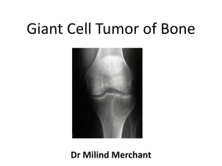
  • 1. Giant Cell Tumor of Bone Dr Milind Merchant
  • 2. Overview – Definition – Epidemiology – location – Presentation – Imaging – DDx – Tx
  • 3. Definition • 10 bone neoplasm • Generally benign • First described Cooper 1818 • Nelton microscopic description 1923 • Potential for : – Recurrence – Pulmonary metastasis – Frank malignancy
  • 4. Epidemiology • 5% of all bone tumors • 30 % of all bone tumours in India • 21% benign bone tumors • F : M 1.5 : 1 • 70-80% age 20-40 • Epiphyseometaphyseal • Rare skeletally immature • Monostotic
  • 5. Incidence • Ends of long bones • >50% about knee • Distal femur most common • 2nd MC is proximal tibia • 3rd MC is distal end radius • High local recurrence rate • Malignant GCT <1% • Rare polyostotic form <1%
  • 6. Gross • Involves epiphysiometaphyseal region of long bones • Tumour extends upto adjuvant articular cartilage which remains intact and rarely when neglected may involve diaphysis. • Usually eccentric to long axis of bone • The overlying cortex undergoes resorption and the contour of bone is expanded by the tumour which is covered by thin shell of subperiosteal new bone. • Tumour is grey to reddish brown in colour with soft vascular friable tissue. • Firm grey yellow areas of fibrosis and collagenisation and osteoid formation.
  • 7. Histology • Fibrohistiocytic origin • Osteoclast like multinucleated giant cells (40 to 60 nuclei) • Mononuclear stroma with single large nucleus, Round / ovoid / spindle shape with Indistinct cell membrane • Mitoses and intravascular extension of tumour may not indicate malignancy.
  • 8. Presentation • 3rd to 4th decade with 1.5 times female preponderance • Pain x wks. – mths • Mass x wks. – mths • Pathologic # at presentation in 5 to 10% • Neuro deficit (spine / sacrum) • Less common sites are sacrum,distal tibia,prox humerus,prox femur,prox fibula • Small bones of hands feet,ribs and spine are rare
  • 9. • Lytic lesion Radiology • Epiphysiometaphyseal location • Eccentric or central • Bulges beyond confines of cortex which has undergone varying degrees of resorption. • Occ. Cortical breakthrough • Thin shell of subperiosteal new bone • No periosteal rxns unless pathological # • Margins generally well defined or sometimes ill defined. • No sclerotic margin • Peripheral bony ridges give appearance of trabaculations • Soap bubble appearance on x ray
  • 10. Other modalities CT – Integrity cortical rim – Interosseous ext – Subchondral relation MRI • Low intensity on T1 and high on T2 • Intramedulary tumour best seen on T1W and extraosseous portion best seen on T2W • Best to see extraosseous extent and joint involvment Bone Scan – Suspect multicentric loci
  • 11. DDx – Chondroblastoma – Brown tumor – ABC – Chondromyxoid fibroma (rare) – Fibrogenic/Telangiectatic Osteosarcoma – Multiple fibrous histiocytoma
  • 12. Enneking Staging Stage 1 (Latent) Stage 2 (Active) Stage 3(local aggeressive) Pt % 10-15% ~70% 10-15% Symptoms asymp pain pain Radiograp h sclerotic rim expanded cortex cortical perforation Histology benign benign benign
  • 13. Campanacci grading • Based on radiological appearance. • Grade I: well marginated border of thin rim of mature bone, cortex intact thinned but not deformed. • Grade II: weel defined margins,no radioopaque rim,combined cortex and rim of reactive bone is thin and moderately expanded but still present. • Grade III: Fuzzy borders sugg/o rapid growth, bulges into soft tissues
  • 14. Biopsy • Necessary for Dx • Histologic grade not helpful • R/O 10 malignant GCT • Occ assoc. – ABC – Pagets
  • 15. Tx • Local control w/o sacrificing joint function • Traditionally: – Intralesional curettage / resection & bone graft – Recurrence 60% • En Bloc resection – Recurrence ~10% – Multiple complications • Adjuvant
  • 16. Curretage Adequate curretage? • Adequate exposure of lesion • Wide cortical window to access tumour to avoid having to curette under overhanging shelves or ridges of bones (windowing) • Head lamp or dental mirror use to see small pockets • high speed burr to break bony ridges • Pulatile jet lavage to expose raw cancellous bone and wash out tumour cells. Phenol in 5 to 80 pc concentration to decrease recurrence rates H2O2 may also be used as an adjuvant
  • 18. Adjuvant Tx • PMMA (polymethylmethacrylate), Liquid N2, Phenol, CO2 laser, Electrocautery – Local extension of margin – Kill residual foci • Radiation – for in-operable lesions of spine or pelvis ~10% risk of sarcomatous degeneration
  • 19. Cementation using methylmethacrylate • Fill tumor cavity • Exothermic reaction or chemical cytotoxic effect associated with polymerization • Heat kill the tumor cells • Cytotoxic agents like adriamycin and methotrexate added in cement • 8-26% recurrence • Easy recurrence detection
  • 20. Advantages of bone graft • Undergoes remodelling along stress lines • Once incorpoated,reconstruction is permanent Drawbacks of bone graft • Autograft qty is limited • Donor site morbidity • Allograft is expensive, requires bone bank • Recurrance relatively difficult to spot
  • 21. Advantages of cementing • PMMA monomer is cytotoxic • Thermal effect- hyperthermia extend the boundary of tumour kill • Xray detection of recurrance is easier • Immediate structural support and rapid weight bearing ambulation Drawbacks of cementing • Relatively weak in shear forces so when used in head and neck of femur,high chances of # thro cement • Chances of long term degen of articular cartilage in subchondral lesions an wt bearing areas.
  • 22. To decrease the chances of late articular degeneration in subarticular lesions where residual suchondral bone after extended curretage is <1cm • Multilayer reconstruction technique used • Mixture of morsellised autograft packed adjacent to subarticular surface. • Layer of gelfoam layered over this. • Remaining cavity is packed with cement. • If recurrance occurs danger of damage to articular cartilage during cement removal is reduced.
  • 24. Cryotherapy • 3 freeze thaw cycles • Irrigate cavity with liq nitrogen • Circumferential necrosis • Complications – Soft tissue injury – Late fractures – Delayed wound healing
  • 25. Excision • For expendable bones like lower end ulna and upper and fibula
  • 26. Marginal / wide local excision Reconstruction done using • Megaprosthetic joint replacement-prone to loosening,wear or breakage and revisions • Biologic reconstructions: 1. Autograft arthrodesis with internal fixation (wrist,knee, shoulder) 2. Live microvascular fibula reconstructions 3. Illizarov method 4. Osteo articular allografts
  • 28. Local recurrence in GCT • Characterised clinically by pain and radiologically by lysis of bone graft or adjacent cancellous bone. • Following curretage and cementation an osteolytic zone caused by thermal injury of 2mm surrounds the cement. This is bordered by thin outer sclerotic rim for 6 mths.lysis or failed development of this sclerotic rim may suggest recurrence. • Soft itssue recurrence seen on xray coz of peripheral calcification. • Total serum acid phosphatase used as a serum marker for monitoring response to t/t • Majority of recurrences occur in 1st 2 yrs. • High recurrence in grade 3 lesions • t/t like primary lesions
  • 29. Phenol • Wash cavity • Alcohol rinse • 10-20% recurrence Embolization Biphosphonates act by targeting osteoclast like giant cells inducing apoptosis and limiting tumour progression
  • 30. Thank you Have a nice day