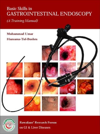
Gastrointestinal endoscopy training manual
•
71 gefällt mir•23,050 views

Gastrointestinal endoscopy training manual

Empfohlen
Weitere ähnliche Inhalte
Was ist angesagt?
Was ist angesagt? (20)
Andere mochten auch
Andere mochten auch (20)
Ähnlich wie Gastrointestinal endoscopy training manual
Ähnlich wie Gastrointestinal endoscopy training manual (20)
Mehr von Farhad Safi
Kürzlich hochgeladen
Kürzlich hochgeladen (20)
Gastrointestinal endoscopy training manual
- 1. Basic Skills in GASTROINTESTINAL ENDOSCOPY (A Training Manual) Muhammad Umar Hamama-Tul-Bushra RAWALIANSRESEARCHFORUM ON G.I & LIVER DISEASES Rawalians' Research Forum on GI & Liver Diseases
- 2. Basic Skills in GASTROINTESTINAL ENDOSCOPY (A Training Manual) Muhammad Umar MBBS, MCPS, FCPS, FRCP (Glasg), FRCP (L), FACG, AGAF Chairman & Professor of Medicine Rawalpindi Medical College Rawalpindi Chief Gastroenterology & Hepatology Division Holy Family Hospital Rawalpindi Hamama-tul-Bushra BSc, MBBS, FCPS, FRCP (Glasg), FACG Professor of Medicine Rawalpindi Medical College Rawalpindi Consultant Gastroenterology & Hepatology Division Holy Family Hospital Rawalpindi
- 3. Basic Skills in GASTROINTESTINAL ENDOSCOPY (A Training Manual) Basic Skills in GASTROINTESTINAL ENDOSCOPY - A Training Manual © 2007 Rawalians' Research Forum on GI & Liver Diseases. Library of Rawalians' Research Forum on GI & Liver Diseases - RS/ICT/437 All material in this manual is copyrighted by the author(s). No part of this publication may be reproduced, stored in a retrieval system, or transmitted, in any form or by means, electronic, mechanical, photocopying, recording or otherwise, without prior written permission of the author(s). Requests to reprint or reproduce material from this publication may be sent to Professor Muhammad Umar, Al-Shifa Clinic, 965-B Saidpur Road, Satellite Town, Rawalpindi, Pakistan. Although every effort has been made to ascertain the authenticity of the material provided by the contributors and to trace the copyright holders for borrowed material. If any omissions have occurred, the author(s), the editor and the publisher will be pleased to make the necessary arrangements at the first priority. ISBN: 978 969 9200 01 4 Edited by: Amir Rizwan,MBBS, DCPS-HCSM Designed & Composed by Jahanzeb Khan Printed & Published in the Islamic Republic of Pakistan in 2007 Rawalian’s Reasrch Forum Saidpur Road, Satellite Town, Rawalpindi - 46000 Tel: +92 51 4414174 / 4427614 / 9290422 / 9290321-7 Fax: +92 51 5591281 Gastroenterology & Hepatology Division Department of Medicine Holy Family Hospital Rawalpindi Tel: +92 51 4414174 / 4427614 / 9290422 Fax: +92 51 5591281 Email: drumarpk@yahoo.com
- 4. Basic Skills in GASTROINTESTINAL ENDOSCOPY (A Training Manual) Foreword It is nature of a human being to discover the undiscovered that's why he started attempting to look into human body orifices and body cavities date back from antiquity. Most of these efforts were fruitless because of lack of light source. In 1806, Philip Bozzini presented the theoretical idea of Lichtleiter (Light Conductor). This basic idea of light conduction was exclusively used in future developments in gastrointestinal, urological, gynecological endoscopies and eventually the laparoscopy. While technological advances have made endoscopy one of the most common procedures for examining the gastrointestinal tract, learning how to maneuver the instruments and interpret the images can be frustrating for those without experience. This manual is specifically designed for beginners in endoscopy training. It has very illustrative and user friendly format, which comprehensively covers all aspects of basic skills in endoscopy. ·Clear descriptions and images of the instruments and how and when they are used ·Step-by-step instructions for handling the endoscope, such as insertion, air insufflation, irrigation, and more ·Useful checklists and tables that lay out the procedures from beginning to end, including preparations, necessary medication and anesthesia, required staff and supplemental equipment, potential risks and complications, etc. The manual also encompasses a complete full-color atlas that illustrates the entire spectrum of both normal and pathological findings. In addition to detailed explanations of each finding, the authors provide ·The endoscopic criteria and the most important differential diagnoses for each disorder ·Series of images showing common variants, as well as comparison photographs of differential diagnoses ·Useful guidelines for proper documentation Amir Rizwan, MBBS, DCPS-HCSM Research Coordinator Gastroenterology & Hepatology Division Holy Family Hospital Rawalpindi – Pakistan
- 5. Basic Skills in GASTROINTESTINAL ENDOSCOPY (A Training Manual) About the Authors Muhammad Umar MBBS, MCPS, FCPS, FRCP (L), FRCP (Glasg), FACG, AGAF Graduated in 1981, with presidential gold medal from President of Pakistan from Rawalpindi Medical College Rawalpindi Pakistan, started his professional career as physician after obtaining membership (MCPS) and then fellowship (FCPS) of College of Physicians & Surgeons Pakistan in 1984 and 1985. He was awarded fellowships from American College of Gastroenterology (FACG) in 2003, Royal College of Physicians London (FRCP), Royal College of Physicians Glasgow (FRCP) in 2006, and American Gastroenterological Association (AGAF) in 2007. He was appointed Assistant Professor, then Associate Professor and currently the Professor & Chair of Medicine and Chief of Gastroenterology & Hepatology Division. He had keen zest for gastroenterology and specifically hepatology so he started pursuing his career in it. He started practicing gastroenterology by performing upper and lower GI procedures in general medical setting after formal training in endoscopy from CMH Rawalpindi and basic skills courses in the United States and the United Kingdom. He established GI & Liver Clinic, GI & Liver Learning Resource Centre, and Liver Research Clinic at Holy Family Hospital Rawalpindi. He has contributed to gastroenterology by publishing more than 70 review and original research papers in various national and international journals, two books on hepatology; Evidenced Based Approach to Hepatitis C Management and Hepatitis C in Pakistan. Moreover, he published National Hepatitis Practice Guidelines. He was awarded SJZ Research Award from Pakistan Society of Gastroenterology & GI Endoscopy in 2004. Currently, he is the President of Pakistan Society of Gastroenterology & GI Endoscopy, General Secretary of Pakistan Society of Hepatology, and President of Rawalians' Research Forum on GI & Liver Diseases. Hamama-tul-Bushra BSc, MBBS, FCPS, FRCP (Glasg), FACG She graduated from Rawalpindi Medical College Pakistan with distinction in 1981. She obtained fellowship from College of Physicians & Surgeons Pakistan (FCPS) in 1985 and started her career. She was honored with fellowships from Royal College of Physicians Glasgow (FRCP) and American College of Gastroenterology (FACG) in 2007. She was appointed Assistant Professor, then Associate Professor and currently the Professor of Medicine at Rawalpindi Medical College and Holy Family Hospital Rawalpindi Pakistan. She had very strong interest in gastroenterology and specifically hepatology so she started pursuing her career in it. She started practicing gastroenterology and developed GI Wing and Endoscopy Suit at District Headquarters Hospital Rawalpindi. She has a long list of publications including original and review papers, books, guidelines, and manuals. She has published about 80 research papers, two books on hepatology; Evidenced Based Approach to Hepatitis C Management and Hepatitis C in Pakistan. Moreover, she published National Hepatitis Practice Guidelines.
- 6. Basic Skills in GASTROINTESTINAL ENDOSCOPY (A Training Manual) Contributors Masood Ahmed MBBS, FCPS Assistant Professor Holy Family Hospital Rawalpindi – Pakistan Muhammd Khurram MBBS, FCPS Assistant Professor Holy Family Hospital Rawalpindi – Pakistan Zahid M. Minhas MBBS, DCPS, PhD Senior Registrar Medicine Holy Family Hospital Rawalpindi – Pakistan Saima Ambreen MBBS, FCPS Senior Registrar Holy Family Hospital Rawalpindi – Pakistan Abdul Naeem MBBS Registrar Holy Family Hospital Rawalpindi – Pakistan Atif Nazir MBBS Registrar Holy Family Hospital Rawalpindi – Pakistan Omar Hayder MBBS House Physician Holy Family Hospital Rawalpindi – Pakistan Salman Abdul Qayyum MBBS Registrar Holy Family Hospital Rawalpindi - Pakistan
- 8. 1 Endoscopic Equipment 2 The Endoscopy Suite, Facilities and Staff 3 Disinfection of Endoscopes 4 Quality Assurance in Reprocessing 5 Antibiotic Prophylaxis for Gastrointestinal Endoscopy 6 Safety and Sedation during Endoscopy 7 Indications, Contraindications, Complications of Upper Gastrointestinal Endoscopy 8 Upper Gastrointestinal Endoscopy 9 Lower Gastrointestinal Endoscopy 10 Colonoscopy / Sigmoidoscopy Technique Normal Endoscopic Findings 11 Basic Skills in GASTROINTESTINAL ENDOSCOPY (A Training Manual) Contents 1 Introduction 1 3 9 15 23 27 35 45 53 59 63 71 12 79 Lesion Recognition
- 10. Basic Skills in GASTROINTESTINAL ENDOSCOPY (A Training Manual) Introduction The word endoscopy is derived from the Greek word 'skopein' meaning 'to view' combining from prefix 'endo' meaning 'inside'. So Endoscopy means viewing inside the living body. th The endoscopes, which started as a simple, short, rigid, straight metallic tubes in 18 century went through a lot of innovation in last two centuries and now we have the long flexible fiberscope to video endoscopes. As the science and art of endoscopy evolved its role as purely a diagnostic tool has changed to a therapeutic armament. Endoscopy has become an important investigation in the management of dyspepsia and upper duodenal pain; similarly it has an important diagnostic and therapeutic role in upper GI bleed. As the incidence and prevalence of liver disease and especially de-compensated liver disease increases day by day, the demand of endoscopy is proportionally increasing. Gastrointestinal Endoscopy is a complex skill, requiring a period of formal training and experience with close supervision. Only a skilled professional can provide the high standards of patient care, accuracy in diagnosis, and appropriate therapeutic interventions with avoidance of complications. It has been further recommended that doctors performing endoscopy should have a professional commitment to two or more endoscopy sessions weekly. So to acquire and maintain proficiency, close liaison with other experienced endoscopists ensures that training is a continuous process. Multidisciplinary interactions between endoscopists, radiologists, pathologists, physicians and surgeons contribute to effective training and skills. Endoscopy Nurses 1. The main support to the endoscopist is given by nursing staff, although other properly trained health care workers can fit support roles. The specific role of the nurse in endoscopy is the care of patient before, during and after the procedure. 2. Training in patient monitoring and resuscitation techniques is essential for the nurse to cope with the unforeseen reactions and cardiorespiratory complications that may occur. 3. Endoscopes and their accessories are expensive and relatively delicate. Their incorrect maintenance and careless handling can damage them. Training in instrument handling and disinfection form an integral part of training of nurses and other endoscopy assistants and it is essential that all staff involved in endoscopy are kept up to date with ever-changing technology. 1
- 12. 1 Endoscopic Equipment Flexible endoscopes are complex. Basically they consist of control head and flexible shaft with a maneuverable tip. The head is connected to a light source via an umbilical cord, through which pass other tubes transmitting air, water and suction. The suction channel is used for the passage of diagnostic tools e.g. biopsy and therapeutic devices. Fig 1.1 Fibreoptic endoscope system Fiber optic instruments These are based on optical viewing bundles, well described as a highly flexible piece of illuminated spaghetti. The viewing bundle of standard fiber endoscope is 2-3mm in diameter and contains 20,000-40,000 fine glass fibers, each close to 10μm in diameter. However, fiber optic bundles are extremely flexible and an image can be transmitted even when tied in a knot. The distal lens which focuses the image on to the bundle is fixed. The image reconstructed at the top of the bundle is transmitted to the eye via focusing lens, adjusted to compensate for individual difference in refraction. Video Endoscope These are mechanically similar to fiber endoscopes, with a charged couple device (CCD) chip and supporting electronics mounted at the tip, to and from wiring replacing the optical bundle and further 3
- 13. Basic Skills in GASTROINTESTINAL ENDOSCOPY (A Training Manual) electronics and switches occupying the site of the ocular lens on the upper part of the control head. Removing any need to hold the instrument close to the endoscopist's eye has hygienic advantages. The CCD chip is an array of 33,000-100,000 individual photon cells known as pixels. The number of pixels determines the resolution of the image. The screen image quality of present video endoscopes equals that of present fiberscopes in both colour and resolution. Components of the endoscope The endoscope consists of the supply plug, umbilical cord, control head, insertion tube (shaft), and bending section. A fiber optic endoscope has an eyepiece, while a video endoscope has remote control buttons for the video control unit. Endoscopic Equipment Fig 1.2 Components of the Endoscope Angulation control wheels Eyepiece Suction button Air/water button Biopsy port Umbilical cord Insertion tube Connection for water bottle Air vent Air tube Connection for suction pump Light guide Lens Air / water jet Light guide Instrument channel 4
- 14. Basic Skills in GASTROINTESTINAL ENDOSCOPY Supply plug and umbilical cord (A Training Manual) The supply plug at the end of the umbilical cord has distal connectors for the light guide and air supply, side connectors for the water bottle and suction, and an air vent, which is not functional during endoscopy. The umbilical cord connects the supply plug to the control head. Control head and insertion tube Between the umbilical cord and insertion tube is the control head, which has control for air insufflation, irrigation, suction and for the bending section at the distal end of the scopes. At the intersection of the insertion tube and control head, there is a biopsy port for passing instruments down the endoscope shaft. The insertion tube has a distal bending section, whose tip carries the illuminating end of the light guide, air and water jets, the distal opening of the biopsy channel, and the lens of video chip. Fiber Optic vs. Video Endoscopy In a fiber optic endoscope, light is conducted from the distal lens to the eyepiece by bundles of optical glass fibers. In a video scope the image is captured with a video chip at the distal end of the endoscope, transmitted electronically and displayed on a monitor. Advantages and Disadvantages Video endoscopy offers several advantages over fiber optic endoscopy: high resolution, the convenience of a monitor display, which permits others in the room to view the image, and easier handling of the endoscope during the procedure. In therapeutic procedures, the assistant can coordinate in a better way by viewing the procedure. The main disadvantage of video endoscopy is its high cost. Illumination This is provided from an external high intensity source through one or Endoscopic Equipment Fig 1.3 Basic Design - control head and bending section 5
- 15. Basic Skills in GASTROINTESTINAL ENDOSCOPY (A Training Manual) more light carrying bundles. Because the light intensity is reduced at any optical interface, light bundles run uninterrupted from the tip of the instrument through its connecting umbilical cord directly to the point of focus of the lamp. These may be Xenon arc (300 w) or halogen-filled tungsten filament lamps (150 w). Instrument Tip Control of the instrument tip depends upon pull wires attached at the tip, just beneath its outer protective shaft, and passing back through the length of the instrument shaft to the angling controls in the control head. The two angling wheels/knobs (for up/down and right/left movement) incorporate a friction braking system, so that the tip can be fixed temporarily in any directed position. The instrument shaft is torque stable so that rotatory corkscrewing moments applied to the head are transmitted to the tip if the shaft is relatively straight at the time. Instrument Channels An operating channel (usually 2-4mm in diameter) allows the passage of fine flexible accessories (e.g. biopsy forceps, cytology brushes, sclerotherapy needles, diathermy snares) from a port on the endoscope control head through the shaft and into the field of view. Endoscopic Equipment Fig 1.4 The tip of a forward viewing endoscope. Fig 1.5 A side-viewer with cannula protruding from the elevator 6
- 16. Basic Skills in GASTROINTESTINAL ENDOSCOPY (A Training Manual) In some instruments (especially those with lateral viewing) the tip of the channel incorporates a small deflectable elevator or bridge which permits some directional control of the forceps and other accessories independent of the instrument tip, this elevator or bridge is controlled by a further thumb lever. The operating channel is also used for aspirations; an external suction pump is connected to the umbilical cord of the instrument near the light source and suction is diverted into the instrument channel by pressing the suction valve which has two positions, a neutral position and a suction position, the size varies with the instrument's purpose. Fig 1.6 Air insufflation and irrigation An ancillary small channel transmits air to distend the organ being examined. The air is supplied from a pump in the light source and is controlled by another valve. The air system also pressurizes the water Endoscopic Equipment Fig 1.7 Suction and instrumentation 7
- 17. bottle so that a jet of water can be squirted across the distal lens to clean it. In colonoscopies, there is a separate proximal opening for the water channel, to allow high pressure flushing with a syringe. The air/water valve has three positions (a) Neutral Position: no air insufflations, no water jet (b) Air insufflations (c) Irrigation (water jet). Fig 1.8 Carry endoscopes carefully to avoid knocks to the optics, control head and tip. Handling the Endoscope The control head of the endoscope is held in the left hand. The index and middle finger activate the suction and air/water valves. Many examiners operate angulations control wheels with the right hand, but an endoscopist with large hands can also manage these controls with the left hand. This leaves the right hand free to manipulate the insertion tube. This is advantageous in certain situations. 8
- 18. 2 The Endoscopy Suite, Facilities and Staff Setting up and running an endoscopy unit is a complex topic, of particular interest to directors and nurse managers. Staff and management Endoscopy units nowadays are complex organizations with many people of different back grounds and training. Procedures are performed by many different types of doctors including gastroenterologists, surgeons, internists and radiologists. There are also many different types of supporting staff. Most of the endoscopy assistants are nurses, whose primary role is to care for the patient's comfort and safety before, during and after the procedures. Nurses, nursing aides and technicians are responsible for cleaning, disinfection and maintenance of endoscopy equipment and accessories; other staff handles reception, documentation and billing duties. All of the staff needs to be specially trained and fully oriented. The unit must have a designated medical director responsible for overall policies. The director is assisted by an endoscopy nurse manager (Head Nurse) who helps to formulate policies and is responsible for implementing them on day to day bases. In the endoscopy room, a minimum of one qualified nurse and trained assistant will be required, with further nurse/endoscopy technician being responsible for cleaning and disinfection. The recovery area should have at least one qualified nurse and trained assistant to manage patients who are returning from the endoscopy room. Fig 2.1 Procedure room design Receptor Receptionist Endoscopy Room Nurses / Trained Assistant Recovery Room Nurses / Trained Assistant Reporting Room Doctor Cleaning and Disinfection Trained Nurse / Endoscopy Technician Medical Director Endoscopy Head Nurse 9
- 19. Basic Skills in GASTROINTESTINAL ENDOSCOPY (A Training Manual) The Endoscopy Unit The endoscopy unit should have a smart public face and a more functional back hall. It certainly requires dedicated space, which should be suitable for out patients and inpatients, and be easily accessible both day and night. Procedure Rooms The number of endoscopy rooms required, depend on the volume and spectrum of procedures, sedation practices, quality of technical support and the presence or absence of trainees. How many procedures a room can handle? Most endscopists say that they can complete simple upper GI procedures within 20 min, colons 45min and ERCPs in under an hour. The bulk of the work load is the 'standard' procedures (i.e. upper GI and colons). So, a routine procedure room can hold 1200 procedures per year. Six procedures can take a whole day, especially when teaching is involved. Procedure Room Design Endoscopy procedures vary in complexity, so do the procedure room requirements. However, all procedure rooms share many common design features. Rectangular rooms often work better than square ones. They must be wide enough to enable the trolley (Stretcher) to be spun around its axis. Functional planning of the endoscopy room with avoidance of cross-traffic is crucial to efficient work. Geographical sphere of activity should be defined for doctors, nurses and trainees, with their relative equipment in the appropriate sector. Fig 2.2 Procedure room design The Endoscopy Suite, Facilities and Staff 10
- 20. Basic Skills in GASTROINTESTINAL ENDOSCOPY (A Training Manual) The room should contain large sinks to accommodate contaminated accessories, adequate work surfaces, storage cupboards and power points. The floor should be washable and smooth. Any windows should have blackout curtains and blinds. Central room light should be dimmable with spot light over the work tops. The endscopist and nurses should have an easy access to the alarm systems. Door opening should be 4ft wide to facilitate moving patients' stretchers and large pieces of equipment. Procedure room should have a continuous supply of piped oxygen, always available suction (two outlets) and an alarm system. Fixed examination tables are unnecessary. Patient trolleys (stretchers) are convenient for examination and recovery. It should be possible to tilt the top into a head down position and it is convenient if the height of the trolley can be adjusted. There must be arrangements for maintaining a reasonable temperature and for extracting odors, both clinical and chemical. Efficiency of utilization would be improved if all procedure rooms could be used for all types of procedures. There is no need to separate upper GI examinations from colonoscopies, since the room requirements are similar. However some procedures require sophisticated, heavy and expensive equipment which cannot be moved or duplicated (i.e. X-ray machines and lasers); thus it is usual to have several standard rooms and one high-tech room. A floor area of 15-20 square meters (50-65 square feet) is adequate for a standard procedure room. Specialized procedure rooms Specialized rooms are usually large because of extra equipment for radiology, endoscopic ultrasound or laser. Fluoroscopic screenings must be available for many endoscopic procedures, especially dilatations. X-ray procedure rooms are larger than routine rooms. Correct siting of the table is essential. It is convenient for the radiologist and the endscopist to work on the same side of the table. It is essential to mount the video and fluoroscopy monitors side by side, across from the endoscopist and radiology staff. Rooms containing X-ray equipment need to be lead-lined with standard safety features. Rooms in which lasers are used must meet appropriate local safety standards for eye protection. The Endoscopy Suite, Facilities and Staff 11
- 21. Basic Skills in GASTROINTESTINAL ENDOSCOPY (A Training Manual) Fig 2.3 ERCP room layout. setup the fluoromonitor (F) and video scope monitor (V) side across the X-ray table from the endoscopist Preparation and recovery areas Procedure rooms are the heart of an endoscopy unit but patients and their relatives spend move time in waiting, preparation and recovery areas. The quality of the facilities and good attitude of the staff involved provide the main impression for the patients. Preparation consists of the preliminary interviews, teaching and consent process, undressing and setting up an intravenous line (best done with the patient lying down). After a sedated examination, the patient lies on the trolley on which procedure was done for period of 20-60 minutes. After dressing, the patient should be joined by relatives, have consultation with endoscopist or the nurse and then discharged. Physical arrangement one requires are a waiting area for relatives, interview and undressing and preparation rooms. Lockers or a place should be available for valuables and clothes of the patient or such can be carried in a basket which can be stored underneath the examination stretcher. Storage and cleaning space Large areas are required for cleaning, disinfection and storage of endoscopes and their accessories. Instrument disinfection should be done in a restricted area with strong ventilation, because toxic chemicals are used (e.g. glutaraldehyde). A 'sequential' design is practical. Used instrument are brought through one door into a 'dirty zone' by passing through a series of cleaning / disinfection process, to end up hanging in the final clean zone, accessible through a second door. The Endoscopy Suite, Facilities and Staff 12
- 22. Basic Skills in GASTROINTESTINAL ENDOSCOPY (A Training Manual) Fig 2.4 Cleaning area 'sequential' plan. A large work top is needed for packaging accessories. Reporting areas Reports are written, dictated or entered into computers immediately after procedures. A central reporting area should have arrangements for image management, video editing and record keeping. The Endoscopy Suite, Facilities and Staff 13
- 24. 3 Disinfection of Endoscopes ·Flexible endoscopes are complex, versatile instruments that require proper and specialized decontamination and disinfections. In addition to the external surface of endoscopes their internal channels for air, water, aspiration and accessories are all exposed to body fluids. Flexible endoscopes are heat labile and cannot be autoclaved. ·A guiding principle for decontamination is that of universal precautions. Any patient must be considered a potential infectious risk, and each endoscope and device must be reprocessed with the same rigor following every endoscopic procedure. ·Endoscopy induced infection is usually due to procedural errors in decontamination. Other potential risk factors for transmission of infection include the use of older endoscopes with associated surface and working channel irregularities and the use of contaminated water bottles or irrigation solutions using substandard disinfectant or inadequate drying or storage of endoscope. Endoscopy related infections Infections can spread from patient to patient or patient to healthcare worker or staff members if equipment is reprocessed inadequately. Bacterial infections have been acquired during endoscopy, such as salmonella and pseudomonas, viral disease, such as hepatitis B, and Hepatitis C have all been transmitted during endoscopy. There is generally no risk of transmission of helminths, nematodes, platyhelminths, ascaris or liver flukes. However, concerns have been raised with respect to the risk of transmission of Giardia lambliasis, cryptosporydium species and amoebas. Patients with immune deficiency syndrome or severe neutropenia and those receiving immunosuppressive chemotherapy or those who have artificial cardiac valves have an increased risk of infection. Diseases may be transmitted from patient to endoscopic personnel. Studies have suggested that endoscopes are potential vectors for the transmission of Helicobacter pylori. Another example is acquisition of 15
- 25. Basic Skills in GASTROINTESTINAL ENDOSCOPY (A Training Manual) herpes simplex ophthalmitis following esophageal biopsy. Healthcare workers are also at potential risk of infection with blood-borne viruses transmitted via sharps, such as spiked biopsy forceps. So, protection from direct contact with the endoscopes and accessories is essential. Gloves and aprons should be worn and protective masks and eye protection goggles should be available to avoid exposure to blood or body fluids. Traditionally, patients harboring potentially infectious microorganisms are scheduled for the end of endoscopy list in order to minimize cross infection. However, if universal endoscope decontamination regime is followed, which premises that all patients are potentially infectious, there is no need to place the patients with known infections last on the list. Nonetheless prevailing infection control policies often schedule patients with Methicillin Resistant Staphylococcus Aureus (MRSA) at the end of list. Staff known to be disease carriers should avoid duties that could transmit infection to patients. It is recommended that all staff be offered vaccination against hepatitis B. Decontaminant of Endoscopes Sterilization is defined as the complete destruction of all microorganism including bacterial spores. Sterilization is required for devices that are normally used in sterile areas of the body. Flexible endoscopes do not penetrate into the sterile areas of the body, but only have contact with mucosal membranes, so they only need a high level of disinfection rather than sterilization in order to kill bacteria, viruses, mycobacteria and some spores. Reprocessing of Endoscopes Before commencing with reprocessing of endoscopes and endoscopic accessories, protective clothing must be put on. Manual Cleaning a. As soon as the endoscope is removed from the patient, the air/water channel must be flushed for 10-15 seconds to eject refluxed blood and mucus. b. Detergent solution should be sucked/aspirated through the suction/biopsy channel to remove secretion and debris. c. The insertion tube should be wiped with discardable sponges or swabs to remove any blood, mucus or feces after conducting the leak testing. Disinfection of Endoscopes 16
- 26. Basic Skills in GASTROINTESTINAL ENDOSCOPY (A Training Manual) d. The endoscope should be immersed in water and detergent, and cleaned externally. The distal end is brushed with a soft toothbrush especially targeting the air/water outlet nozzle. All valves are removed and washed. The biopsy channel opening and the suction part should be cleaned with a cotton bud. e. Brushing through the suction/instrument channel and all accessible channels must be done using a cleaning brush. The brush must be passed through the channel several times until clean, and the brush itself must be cleaned with detergent using a soft toothbrush each time it emerges. Introduce the cleaning brush via the biopsy port through the shaft, until it emerges from the distal end at least three times. Pass the cleaning brush through the suction channel opening after removing the suction button and down the shaft until it emerges from the distal end at least three times. Pass the cleaning brush from the suction channel opening in the other direction through the umbilical cord, until it emerges from the suction connector at least three times. After the channels have been thoroughly brushed, either put the endoscope into the reprocessor to complete disinfection or follow the manual method. Rinsing Rinse the endoscope and valves under running tap water of drinking quality. Immerse the endoscope and irrigate all channels by flushing with water followed by air to expel as much air as possible prior to disinfection. Disinfection Manual disinfection:- Disinfection must be carried out in a separate room with proper ventilation. Protective gloves, eye protection and aprons must be used and splashing must be avoided. The instrument should be fully immersed in 2% glutaraldehyde or other chemical disinfectant of equally potency. All channels must be filled with disinfectant and soaked for not less than 10 minutes. Rinsing of the instruments with water must be undertaken after disinfection, internally and externally to remove all traces of disinfectant. The water must be of drinking quality or alternatively, filtered water may be used for rinsing. Disinfection of Endoscopes 17
- 27. Basic Skills in GASTROINTESTINAL ENDOSCOPY (A Training Manual) Dry the endoscope externally and flush each channel with air, wipe the eyepiece and light guide connector as well as the plugs before connecting the endoscope to the light source. Fit the air/water and suction valves and activate the air/water and suction channels. The endoscope is now ready for use again. Washer Disinfector: After manual cleaning, the endoscope is placed in an automatic disinfector and all channel connectors attached. This ensures exposure of all internal and external surfaces to the disinfectant. If automatic reprocessing cycle is interrupted disinfection cannot be assumed and entire process should be repeated. The advantages of automatic reprocessing are as follows. a. The likelihood of an essential step being omitted is reduced. b. All channels are properly irrigated. c. Exposure of skin, eyes and respiratory tract to disinfectants is reduced. d. Atmospheric pollution by the disinfectant is reduced. Drying and Storage of Endoscope Drying of endoscope prior to prolonged storage decreases bacterial colonization. Forced air can be used for drying. a. Storage should be done by hanging the scopes in a vertical position to facilitate drying. Remove all caps, valves and other detachable components. b. Uncoil insertion tube and umbilical cord. c. Endoscopes should be placed in well-ventilated rooms or cabinets. d. Clearly mark which endoscopes have been reprocessed. e. It is important to avoid the contamination of disinfected endoscopes by contact with environment. Endoscopic Accessories The following steps are important for disinfection/sterilization of all reusable accessories. i. Immediate washing in detergent solution. ii. Dismantle as far as possible iii. Brush with cleaning brush or tooth brush hollow iv. Flushing of detergent through lumen of hollow components v. Use an ultrasonic cleaner for all accessories vi. Rinse thoroughly in water of drinking quality Disinfection of Endoscopes 18
- 28. Basic Skills in GASTROINTESTINAL ENDOSCOPY (A Training Manual) vii. Sterilization can be achieved by either auto claving where recommended by the manufacturer, ethylene oxide gas can be used, and though it is time-consuming and expensive. viii. Non-sterlizable accessories should be disinfected prior to use. ix. Avoid the use of defective or damaged accessories because such instrument may not operate properly and may also damage the endoscope instrument channel Disinfectants The ideal disinfection is effective against wider range of organisms including viruses and prion proteins. It should be compatible with endoscopes and accessories and be non-irritant and safe for users. Aldehyde based disinfectants Glutaraldehyde (GA) and its derivative kill most bacteria and viruses, like HIV and HBV in less than 5 minutes. Mycobacterium is more resistant to 2% glutaraldehyde. So the endoscopes are immersed for more than 20 minutes at room temperature. The standard method of disinfection is immersing for 20 minutes in a 2% glutaraldehyde solution. The maximum shelf life is 14 days. The advantages of glutaraldehyde are that it is effective, relatively inexpensive and does not damage endoscopes and accessories. However it has irritant and sensitizing properties. So can lend to allergic problems and can cause dermatitis, conjunctivitis, nasal irritation and asthma in endoscopy staff. In patients residues of glutaraldehyde after insufficient rinsing can cause colitis, abdominal cramps and bloody diarrhea. Glutaraldehyde can fix proteins and allows for biofilm creation. Orthophthaldehyde (OPA) It is a high level disinfectant. OPA completely destroys all viable common bacteria is 5 minutes of exposure. It is non irritant. High level disinfection is achieved in 12 minutes life span is 2 weeks. Peracetic acid (PAA) When compared peracetic acid has similar or better biocidal activity. A contact time of 5 minutes is recommended for the destruction of vegetative bacteria and viruses. The sporicidal activity requires immersion for 10 minutes (0.35 PAA), a contact time at 10-15minutes and a concentrations >0.09%. Disinfection of Endoscopes 19
- 29. Basic Skills in GASTROINTESTINAL ENDOSCOPY (A Training Manual) PAA can remove hardened material in biopsy channels resulting from use of GA, Development of microorganism resistant has not been reported. PAA is less stable than GA. Once prepared it requires replacement every 24 hours. PAA has corrosive abilities and has vinegar like order. Damage of flexible scope has been reported after some brands of PAA. Personnel Protection during Endoscope Decontami-nation 1. Wear long-sleeved waterproof gowns. These should be changed between patients. 2. Use sterile gloves which are long enough to protect the forearms from splashes. 3. Goggles prevent conjunctival irritation and protect the wearer from splashes. 4. Disposable charcoal-impregnated face masks may reduce inhalation of vapour from disinfectants, but experience with them is not yet widespread. 5. An HSE-approved vapour respirator should be available in case of spillage or other emergencies. It should be stored away from disinfectants as the charcoal adsorbs fumes and respirators should be regularly replaced. Steps in Endoscope Decontamination As soon as possible after use: 1 Wipe down insertion tube 2 Flush air/water channels 3 Aspirate water through biopsy/suction channel 4 Dismantle detachable parts (e.g. valves) 5 Manually clean with enzymatic detergent followed by rinsing 6 Disinfect and rinse in an automatic reprocessor 7 Dry 8 Store appropriately Disinfection of Endoscopes 20
- 30. Basic Skills in GASTROINTESTINAL ENDOSCOPY (A Training Manual) Microbicidal Activity Inactivation Disinfectant Spores Mycobacteria Bacteria Viruses Stable by organic Disinfection of Endoscopes matter Corrosive/ damaging Irritant (I) Env. Non Env. Glutaraldehyde 2% Moderate 3 hours Good 20 mins Good <5 mins Good <5 mins Good <5 mins Moderate (e.g. 14-28 days) No (?xative) No I/S Ortho-phthalaldehyde (0.55%) Poor >6 hours Good <5 mins Good <5 mins Good <5 mins Good <5 mins Moderate (30 days) No (?xative) No (staining) I Peracetic acid 0.2 - 0.35%* Varies 10 – 20 mins Varies 5 – 20 mins Good <5 mins Good <5 mins Good <5 mins No (1-3 days) No Slight I Alcohol (usually 70%) None Good <5 mins Good <5 mins Good <5 mins Moderate Variable Yes Yes (?xative) Slight (lens cements) No Chlorine dioxide Good 10 mins Good <5 mins Good <5 mins Good <5 mins Good <5 mins No (1-5 days) Yes Yes I Superoxidised water Good 10 mins Good <5 mins Good <5 mins Good <5 mins Good <5 mins No (< 1 day) Yes Yes No * activity varies with concentration of product Instrument Disinfectants: Properties Table 3.1 21
- 31. Basic Skills in GASTROINTESTINAL ENDOSCOPY (A Training Manual) Endoscope Disinfection Process (Step – 1) Pre-cleaning ****** (Step – 2) *** * (Step – 3) *** * (Step – 4) * *** (Step – 5) * * (Step – 6) ** (Step – 7) *** Clear gross debris by sucking water through the working channel (250 ml/min). Expel any blood, mucus or other debris. Flush the air/water channel and wipe down the insertion shaft. Check for bite marks or other surface irregularities. Detach the endoscope from the light source/videoprocessor. Transport in a closed container to the reprocessing room. Cleaning Conduct leak testing and block testing. Immerse the endoscope in detergent or a soap solution. Clean all surfaces, brush channels and valves with a clean dedicated brush and a clean swab or tissue. Follow the same procedures for all accessories as for endoscope processing Rinsing Rinse the endoscope and valves under running tap water (must be drinking water quality). Immerse the endoscope and irrigate all channels. Discard the rinse water after each use to avoid concentration of the detergent and the risk of reduced efficacy of the disinfectant solution. Clean and rinse the container before the next procedure. Disinfection Immerse the endoscope and valves in a disinfectant solution of proven efficacy (GA, PAA, OPA etc). Irrigate all channels with a syringe until air is eliminated to avoid dead spaces. Contact time with the solution should be according to the manufacturer's recommendation Disinfection solution should be removed by flushing air before rinsing. Final Rinsing Rinse the endoscope and valves in drinking-quality or boiled water by immersing the endoscope and irrigating all channels. Discard the rinse water after each use to avoid concentration of the disinfectant and thus damage to mucosa. Drying Ensure correct final drying before storage. Dry with compressed air or if not available inject air with clean syringe. Storage Disassemble the endoscope. Store in well ventilated storage cupboard. Ensure the valves are dry and lubricate if necessary. v Store separately or store the endoscope in a clean closed box with the valves. Disinfection of Endoscopes 22
- 32. 4 Quality Assurance in Reprocessing Microbiological surveillance is an important means of evaluating the outcome quality of reprocessing procedures and is an instrument of regular quality control in gastrointestinal endoscopy, whether endoscopic procedures are performed in hospital, in private clinics or doctors offices. It is an instrument for detecting and redressing procedure, and for preventing the transmission of infections agents through endoscopy. Table. 4.1 Weaknesses and deficiencies in endoscope reprocessing (modified from reference[12]) a. Inadequate reprocessing of endoscopes and accessories - Inadequate cleaning (e. g. inadequate manual cleaning and brushing of endoscope channels) - Contaminated cleaning accessories (e. g. cleaning brushes) - Use of unsuitable or incompatible detergents and disinfectants - Inadequate concentrations and contact time of agents - Contaminated or time-expired solutions - Contaminated rinsing water - Fixed organic material (biofilm) in endoscopes, water pipes , containers, or washer-disinfectors - Use of nonsterile accessories in invasive diagnosis and treatment (e. g. nonsterile biopsy forceps, polypectomy snares) - Inadequate reprocessing of water bottles (e. g. no sterilization) - Use of tap water in water bottles b. Inadequate transport and storage of endoscopes - Insufficient drying before storage (e. g. Pseudomonas spp.) - Inappropriate storage conditions c. Contaminated or defective washer-disinfector - Contaminated pipes, containers, etc. - Contaminated final rinsing water - Mechanical/electronic defects of washer-disinfector - Incorrect use of washer-disinfector (e. g. wrong connections) - Lack of regular maintenance of washer-disinfector according to manufacturer’s recommendations d. Design limitations and damaged endoscopes - Small lumina, branched channels, not accessible to cleaning brushes - Damage to the surfaces (internal and external) of the endoscope, providing potential for contamination e. Contaminated water in the endoscopy unit - Contaminated main water pipes/supply - Contaminated or inadequate water supply systems (filtration etc.) 23
- 33. Basic Skills in GASTROINTESTINAL ENDOSCOPY (A Training Manual) Quality Assurance in Reprocessing Sample for routine tests Endoscopes microbiological testing of endoscopes should cover 1. Channels 2. Outer surfaces 3. The connected water bottle. Liquid samples from endoscope channels Flash the appropriate channels with 20ml sterile saline and collect the liquid in a sterile container; the sample may be collected from all channels separately swabs from the outer surfaces of the endoscope saline swabs from the moistured distal end, valve parts and bridge elevators are taken and put in containers containing culture both. Swabs from the outer surfaces of the endoscope Saline moistened swabs from the distal end, valve ports, and bridge elevators are taken and put in containers containing culture broth. Organisms identified in microbiological tests Indication of origin Troubleshooting Escherichia coli, entero-cocci and Enterobacte-riaceae A: Insufficient cleaning and/or disinfection procedures, e. g.: - No brushing - Inadequate concentrations or exposure times of process chemicals B: Mechanical or electronic defects of washer-disinfector,e. g.: - Incorrect amounts and/or concentration of processing chemicals - Design flaws of washer-disinfector, with dead volumes A: Review whole reprocessing cycle with special emphasis on manual cleaning B: Initiate full maintenance of washer-disinfector Pseudomonas aerugino-sa and other gram-neg-ative nonfermenters A: - Insufficient final rinsing - Contamination of final rinsing water - Contamination of washer-disinfector due to mechanical or electronic defects - Contamination of filter systems - Design flaws of washer-disinfector with dead volumes B: Insufficient drying of endoscopes before storage A: Review water supply systems and procedures: - Water quality - Manual and/or washer-disinfector rinsing - Initiate full maintenance of washer-disinfector and filtration systems - Initiate autodisinfection cycle according to manufac-turer’s instructions (thermal disinfection is preferred) B: Review drying procedures before storage, and ventilation of storage facilities Staphyloccus aureus, Staphylococcus epider-midis Recontamination of endoscopes due to: - Inadequate storage and transport - Inadequate hand hygiene Review of hygiene arrangement for storage, transport and of manual handling Contamination from sampling Repeat sampling Atypical mycobacteria Legionella organisms Contamination of washer-disinfector and water system Review water supply systems and procedures: - Manual and/or washer-disinfector rinsing - Initiate autodisinfection cycle according to manufacturer’s instruction (thermal disinfection is preferred) - Initiate full maintenance of washer-disinfector and filtration systems Table. 4.2 24
- 34. Basic Skills in GASTROINTESTINAL ENDOSCOPY (A Training Manual) Quality Assurance in Reprocessing Liquid sample from water bottles Two samples of 100ml are taken from the water bottle; ready for use should be tested. Adequate connectors should be used in order to take liquid samples from water bottles via the connection tube of the water bottle. Final rinse water of washer-disinfectors Two sample of 100ml water collected using a sterile syringe in sterile containers. Water supply Water sample should be taken from the top water. The samples should be sent to microbiologists for culture. Interpretation of results and corrective measure in case of contamination Total microbiological count The maximum total count in liquid samples from endoscope channels should be < 20cful channel. Indicator organism should not be found at any time. Cultures taken from swabs should be focused on the growth of indicator organisms. The maximum count from water samples should be < 10/100 efu/ml. Indicator organism should not be found at any time. 25
- 36. 5 Antibiotic Prophylaxis for Gastrointestinal Endoscopy Bacteraemia occurring in a patient with a susceptible cardiac lesion may lead to infective endocarditis. Gastrointestinal endoscopy can infrequently give rise to transient bacteraemia but there is little evidence to suggest that endoscopic procedures have led to cases of endocarditis. Nevertheless, endoscopic procedures with a higher than average risk of bacteraemia in a patient with a susceptible cardiac lesion may carry a significant danger, and in these it is prudent to use antibiotic prophylaxis. Apart from cardiac infections there are a number of other infectious complications that may be attributable to gastrointestinal endoscopic procedures. These include pancreato-biliary sepsis following endoscopic retrograde cholangiopancreaticography (ERCP), infection of orthopaedic and other non-cardiac prosthesis and wound infections secondary to percutaneous endoscopic gastrostomy. The rate of bacteraemia after endoscopic procedures 1. Diagnostic upper gastrointestinal endoscopy and colonoscopy The bacteraemia rate is low, up to 4%. The risk of bacteraemia does not seem to increase with biopsy or polypectomy. Bacteria most often cultured after upper GI endoscopy are contaminants. 2. Esophageal dilatation and insertion of prosthesis This causes a significant bacteraemia reaching approximately 45%. Although mouth commensals are found most often following esophageal dilatation, oral decontamination with Clindamycin was not an effective prophylaxis. Disinfection of bougie dilators did however reduce post-procedural bacteraemia. The risk after balloon dilatation is probably small as this kind of dilator traverses the endoscope channel and has no contact with the oral cavity. However, no data is available to confirm or refuse these findings. Antibiotic Prophylaxis For Gastrointestinal Endoscopy 27
- 37. Basic Skills in GASTROINTESTINAL ENDOSCOPY (A Training Manual) Antibiotic Prophylaxis For Gastrointestinal Endoscopy 3. Injection sclerosis of esophageal varices This is the second most important cause of bacteraemia associated with GI endoscopic procedures. Bacteraemia has been reported in up to 50% of patients under-going endoscopic sclerotherapy, but it has also been detected in up to 13% prior to diagnostic endoscopy. Bacteraemia as a result of contamination of the injection needle catheter that passes through the suction channel of the endoscope probably cannot be eliminated altogether. The technique of submucosal injection is increasingly being used for polypectomy or resection of flat mucosal lesions. As the suction channel contaminants are those of the digestive tract, contamination during submucosal injection cannot be avoided by using disinfected endoscope, sterile needles and injection fluids. Using an aseptic injection needle protected from contamination by a covered tip may reduce bacteraemia. 4. Endoscopic band ligation of esophageal varices This is considered a safe technique with a low risk of bacteraemia, ranging from 3-6%. 5. Laser therapy This may cause significant bacteraemia depending upon the nature of the procedure. In the upper GI tract the rate of bacteraemia following laser therapy is 31-34%. In the lower GI tract the likelihood of bacteraemia is less, approximately 19%. Laser therapy is believed to mechanically irritate the tumor tissue or mucosa and promote invasion of bacteria. 6. Percutaneous endoscopic gastrostomy (PEG) The most common infection is peristomal, which occurs in 30-34% of patients. Bacteria originating from two sources are involved depending on the techniques; esophageal in pull method and cutaneous in both push and pull method. 28
- 38. 7. ERCP Basic Skills in GASTROINTESTINAL ENDOSCOPY (A Training Manual) Cholangitis and sepsis are the commonest causes of death following ERCP. The major risk factor is biliary obstruction, which increases the risk of bacteraemia to 11-16%. There are two potential pathways for developing bacteraemia; infection of the pancreatic biliary system by instrumentation, or dissemination of already existing organism in an obstructed biliary tree. 8. The risk of infectious complications The majority of bacteraemia associated with gastrointestinal procedures are asymptomatic. As transient bacteraemia is unlikely to harm a normal individual, antibiotic prophylaxis is unnecessary unless there is susceptibility for endocarditis. There are only few endoscopic procedures, where the potential risk of infectious complications is significant. Table. 5.1 Endoscopic procedures associated with higher risk of infectious complications. · Oesophageal stricture dilation · Endoscopic sclerotherapy for oesophageal varices · Laser therapy in upper gastrointestinal tract · Endoscopic placement of percutaneous feeding tube Endoscopic retrograde cholangiopancreatography for known biliary obstruction or pancreatic pseudocyst Sepsis is unlikely to follow upper gastrointestinal endoscopy. Endocarditis attributable to colonoscopy is infrequently reported. Incidence of post dilatation bacteraemia is 21%, entirely caused by Streptococcus viridans. Reported cases of bacterial endocarditis attributable to dilatation of esophageal stricture occurred in patients with mitral insufficiency and mitral valve prolapse. These data support the use of antibiotic prophylaxis prior to endoscopic dilation of esophageal stricture. Bacteraemia associated with endoscopic Antibiotic Prophylaxis For Gastrointestinal Endoscopy 29
- 39. Basic Skills in GASTROINTESTINAL ENDOSCOPY (A Training Manual) Antibiotic Prophylaxis For Gastrointestinal Endoscopy sclerotherapy for esophageal varices is well recognized and a number of septic complications such as septicemia, cerebral abscess, perinephric abscess and endocarditis have been reported. It has been shown that intravenous cefotaxime significantly reduces the frequency of bacteraemia after endoscopic sclerotherapy. Antibiotic prophylaxis should be instituted for individuals with a higher risk of endocarditis. Infectious complications after submucosal injection for polypectomy are exceedingly rare post polypectomy leucocytosis and fever associated with pain one usually caused by burning syndrome or inflammatory response due to hot biopsy forceps. Bacteraemia following laser therapy of the upper gastrointestinal tract is common and septic complications involving organisms commonly associated with endocarditis do occur. The risk therefore seems considerable and so antibiotic prophylaxis is recommended for high-risk patients. As the risk of bacteraemia following laser therapy in the colon is lower, the need for antibiotic prophylaxis is less clear. The wound infection is quite common after percutaneous endoscopic gastrostomy and antibiotic prophylaxis with piperacillin, cefotaxime and amoxycillin/clavulanic acid has been shown to be effective in reducing the rate of early local infection. Biliary sepsis is one of the major complications of ERCP, and although it occurs in only 0.4- 30
- 40. Basic Skills in GASTROINTESTINAL ENDOSCOPY (A Training Manual) Antibiotic Prophylaxis For Gastrointestinal Endoscopy 0.8%, it is associated with 8-20% mortality rate. Antibiotic prophylaxis is recommended for patients who are likely to undergo therapeutic ERCP if there has been previous biliary sepsis, bile duct obstruction or pancreatic pseudocyst. 9. Identification of high risk patients The risk of endocarditis depends, largely on the nature of the cardiac condition. Cardiac and other clinical conditions have been divided into three groups according to their potential for the development of infectious complication. Table. 5.2 Conditions associated with a risk of endocarditis or symptomatic bacteraemia. ·High risk: §Prosthetic heart valve. §Previous endocarditis. §Surgically constructed systemic-pulmonary shunt or conduit. §Synthetic vascular graft less than 1 year old. 3 §Severe neutropenia (neutrophils<1 x 10 /l) ·The moderate, low or theoretical risk with: §Mitral valve prolapse with insufficiency. §Rheumatic valvular or congenital cardiac lesion. §Hypertrophic cardiomyopathy. §Ventriculo-peritoneal shunt. §Heart transplant. 3 §Moderate neutropenia (neutrophils 1 x 10 /l) § ·And no increased risk with: §Mitral valve prolapse without insufficiency. §Uncomplicated secundum atrial septal defect. §Cardiac pacemaker. §Coronary artery bypass graft. §Implanted defibrillator. All other patients. The risk of developing endocarditis is also 31
- 41. Basic Skills in GASTROINTESTINAL ENDOSCOPY (A Training Manual) Antibiotic Prophylaxis For Gastrointestinal Endoscopy dependent upon bacteraemia. Although bacteraemia is common following many invasive endoscopic procedures, only certain bacteria commonly cause endocarditis. Haemolytic streptococci and staphylococci are the most likely, comprising respectively 55% and 25% cases associated with native valve and 30% and 45% of vascular prosthesis infections. Streptococcus viridians is the commonest cause of endocarditis following esophageal high risk therapeutic procedures. Antibiotic prophylaxis for bacterial endocarditis should be specifically directed against these organisms. It should be stressed that endocarditis hardly ever develops as a consequence of an endoscopic procedure. 10. Patients related risk factors Infection of synthetic vascular graft has a serious and potentially fatal consequence. Complete endothelialization of the graft does not occur for a period of one year and bacteraemia is potentially dangerous. Antibiotic prophylaxis is therefore recommended over this period. Existing evidence is insufficient to recommend antibiotic prophylaxis concerning the possibility of infection of orthopaedic, neurosurgical and other prosthesis. Antibiotic prophylaxis for transplant recipients on immunosuppression is not recommended, as little data on the incidence of infectious complication is available. Nevertheless neutropenia appears to increase the risk of post endoscopic symptomatic bacteraemia. Table Recommendations of the antibiotic prophylaxis according to the procedure. 32
- 42. Basic Skills in GASTROINTESTINAL ENDOSCOPY (A Training Manual) Antibiotic Prophylaxis For Gastrointestinal Endoscopy Table. 5.3 Recommendations of the antibiotic prophylaxis according to the procedure. Procedure Patients risk group Antibiotic prophylaxis high risk procedures: high risk patients regimen A or B oesophageal dilation severe neutropenia regimen A or B plus C variceal sclerosis moderate risk patients not necessary laser therapy in upper GI regimen A or B may be considered low or average risk patients not recommended low risk procedures: high risk patients not necessary regimen A or B may be considered moderate or low risk patients not recommended ERCP · all patients with: biliary occlusion pancreatic pseudocyst previous cholangitis or · therapeutic ERCP regimen C PEG all patients regimen D Table. 5.4 Recommended antibiotic regimens A. Patients not allergic to penicillin. Adults: 1g amoxycyllin intramuscularly in 2.5ml 1% lignocaine hydrochloride plus 120 mg gentamicin intramuscularly just before start of the procedure, followed by 500 mg amoxycillin orally 6 hours later. Children under 10 years: 500 mg amoxycillin intramuscularly in 2.5ml 1% lignocaine hydrochloride plus 2 mg/kg body weight gentamicin intramuscularly, followed by 250 mg (children 5 -9 years) or 125 mg (children 0-4 years) amoxycillin orally 6 hours later. B. Patients allergic to penicillin or who have had penicillin more than once in the previous month. Adults: 1g vancomycin in slow intraven ous infusion over 100 minutes followed by 120 mg gentamicin intravenously 15 minutes before the procedure or 400 mg teicoplanin intravenously followed by 120 mg gentamicin 15 minutes before the procedure Children under 10 years: 20 mg/kg vancomycin by slow intravenous infusion followed by 2 mg/kg gentamicin intravenously or 6 mg/kg teicoplanin intravenously followed by 2 mg/kg gentamicin intravenously C. Prior to biliary procedures. 750 mg ciprofloxacin orally 60-90 minutes before the procedure or 120 mg gentamicin intravenously just before the procedure or a parenteral quinolon, cephalosporin or ureidopenicillin just before the procedure. D. Prior to percutaneous endoscopic gastrostomy 2 g cefotaxime (or equivalent) parenterally 30 minutes before th e procedure or 4 g piperacillin/0.5 g tazobactam parenterally or 1 g amoxycillin/clavulanic acid intravenously E. Patients with severe neutropenia Adults: Add 7.5 mg/kg metronidazole intravenously to any of the above regimens Children: Add 7.5 mg/kg metronidazole intravenously to any of the above regimens 33
- 44. 6 Safety and Sedation during Endoscopy Preprocedure Assessment Assessment of patients through a medical history and evaluation of their physical condition should be done before administration of intravenous sedation. Essential elements of medical history include the following: i. Significant cardiac or pulmonary disease ii. Neurological or seizure disorder iii. Strider, snoring or sleep apnea iv. History of adverse reaction to sedation or anesthesia v. Current medications, drugs and allergies vi. Alcohol or drug abuse vii. Time of last oral intake In addition to above assessments, a patient's overall disease risk should be classified according to the American Society for Anesthesiology (ASA) physical status classification. Table 6.1 ASA Classification Class Description I The patient is normal and healthy II The patient has mild systemic disease that does not limit their activities (eg, controlled hypertension or controlled diabetes without systemic sequelae) III The patient has moderate or severe systemic disease, which does limit their activities (eg, stable angina or diabetes with systemic sequelae) IV The patient has severe systemic disease that is a constant potential threat to life (eg, severe congestive heart failure, end-stage renal failure) V The patient is morbid and is at substantial risk of death within 24 hours (with or without a procedure) E Emergency status: in addition to indicating underlying ASA status (1–5), any patient undergoing an emergency procedure is indicated by the suffix ”E” ASA Class I-III patients are appropriate candidates for administration of sedation by an endoscopist while administration of sedation in classes IV-V patients should be done by anesthesia specialists. There are a variety of recommendations for fasting before procedural sedation. This reflects that there are no studies showing a direct relationship between fasting time and the risk of pulmonary aspiration. 35
- 45. Basic Skills in GASTROINTESTINAL ENDOSCOPY (A Training Manual) Safety and Sedation during Endoscopy The ASA guidelines indicate that patients should fast a minimum of 2 hours for clear liquids, and 6 hours for light meals, before sedation. In contrast the American College of Emergency Physicians does not endorse recent food intake as a contraindication for administering procedural sedation and analgesia. The focused physical examination should include: a. Vital signs and weight b. Auscultation of heart and lungs c. Level of consciousness d. Assessment of airway Airway Assessment/Evaluation This is required to identify patients with anatomy that may make endotracheal intubations or positive pressure ventilation more difficult. This includes patients with obesity, short, thick neck, cervical spine disease, decreased hyoid-mental distance and structural abnormalities of the mouth, jaw and oral cavity. Examination of oral cavity can identify individuals with anatomy that is associated with more difficult intubations. Mallampati score is used to predict difficult intubations. Patient is asked to open the mouth maximally, if the faucial pillars and uvula cannot be adequately visualized (Mallampati III and IV), laryngoscopy and visualization of the glottis is likely to be more difficult. Grade - II Grade - I Grade - III Grade - IV Fig 6.1 Mallampati score used to predict difficult intubation. This system emphasizes the importance of the base of the tongue in predicting difficulty with laryngoscopy. If the faucial pillars and uvula cannot be adequately visualized on maximal mouth opening (Mallampati III and IV), visualization of the glottis is likely to be more difficult. 36
- 46. Basic Skills in GASTROINTESTINAL ENDOSCOPY (A Training Manual) Safety and Sedation during Endoscopy Pharmacology of Sedation The goal of endoscopic sedation is to maximize patient comfort while minimizing the risk of drug related side effects. The drugs most widely used for endoscopic sedation are benzodiazepines and opioids. Opioids: The principal effects of the opioids are analgesia and sedation. In clinical practice, opioids are usually combined with a benzodiazepine for endoscopic sedation. The pharmacological effects of benzodiazepines include anxiolysis, sedation, amnesia, anticonvulsant effects, muscle relaxation and anaesthesia. The amnestic effect may persist after sedation has worn off. Diazepam The initial induction dose for endoscopic procedure is 5-10mg intravenously over 1 minute. If required, additional doses may be administered at 5 minutes interval. Dose reduction is required in debilitated or elderly patients. In general, 10mg is sufficient for most endoscopic procedures, although up to 20mg may be necessary if a narcotic is not being co-administered. The major side effect of diazepam is respiratory depression. This is dose dependent and results from depression of the central ventilatory response to hypoxia and hypercapnea. Respiratory depression is more likely to occur in patients with underlying respiratory disease or those receiving combination of a benzodiazepine and an opioid. Midazolam Midazolam is a water soluble, short acting benzodiazepine. It is 1.5-3.5 times more potent than diazepam. Midazolam is distinguished from the other benzodiazepines by its more rapid onset of action and shorter duration of effect, the onset of effect for midazolam is 1-2 minutes and the peak effect occurs in 15-80 minutes. Midazolam clearance is reduced in the elderly, obese and those with hepatic and renal impairment. The bioavailibility of midazolam is increased by approximately 30% in patients using a hisamine (H2) receptor antagonist. The initial intravenous dose in healthy adults younger than 60 years of age is l mg (or no more than 0.03mg/kg injected over 1-2 minutes. 37
- 47. Basic Skills in GASTROINTESTINAL ENDOSCOPY (A Training Manual) Safety and Sedation during Endoscopy Additional doses of 1mg may be admistered at 2-minutes intervals until adequate sedation is achieved. When midazolam is used with an opioid, a synergistic interaction occurs and reduction in the dose of midazolam may be indicated. Patients older than 60 years and those with ASA physical states III or greater require a dose reduction of 20% or more. A total intravenous dose greater than 6 mg is not usually required for routine endoscopic procedures. The major side effect of midazolam is respiratory depression. In some cases apnea may last as long as 30 minutes after administration of the last dose of midazolam. Midazolam induced respiratory depression may be an administration related phenomenon, with more rapid administration resulting in greater number of apneic episodes. Cardiac dysrhythmia has been reported rarely after administration of midazolam. Flumazenil Flumazenil is a benzodiazepine specific antagonist. Flumazenil competitively antagonizes the central effects of benzodiazepines, reversing sedation, psychomotor impairment, memory loss and respiratory depression. Flumazenil is water-soluble and can be administered by either continuous or bolus infusion. The half life of Flumazenil after intravenous administration is 0.7-1.3 hours and average duration of antagonism is 1 hour. Flumazenil has also been shown to reverse ventilatory depression in patients sedated with a combination of benzodiazepines and opioids. Midazolam induced reversal of respiratory depression occurs approximately 120 seconds after the intravenous administration of Flumazenil. Propofol Propofol is a hypnotic with minimal analgesic effect. At sub-hypnotic doses, propofol produces sedation and amnesia. Its duration of effect is 4-8 minutes. The presence of cirrhosis or renal failure does not significantly affect its pharmacokinetic profile. Co-administration of other central nervous system medications such as opioids potentates the sedative effect of propofol. The cardiovascular effects of propofol include a decrease in cardiac output, systemic vascular resistance and arterial pressure. These effects respond rapidly to a dose reduction or interruption of drug infusion. 38
- 48. Basic Skills in GASTROINTESTINAL ENDOSCOPY (A Training Manual) Safety and Sedation during Endoscopy Combination Propofol By combining small doses of several drugs that possess desirable pharmacologic actions it is possible to maximize the therapeutic actions of each while minimizing the likelihood of a dose related adverse reaction. This pharmacologic principle can be applied to procedural sedation. So, when propofol is combined with small doses of an opioid analgesic and a benzodiazepine, analgesia and amnesia can be achieved with sub-hypnotic doses of propofol, eliminating the need for deep sedation. Furthermore, precise dose titration is possible with smaller bolus doses of propofol (5-15mg) and the potential for reversibility is retained using Naloxone or Flumazenil. The protocols for combination propofol include a pre-induction dose of either an opioid or a benzodiazepine or both. An induction dose of propofol is then administered (5-15mg), followed by additional boluses of (5-15mg). The average cumulative doses of propofol in a combination regimen are 65-100mg during colonoscopy and 35-70mg during upper GI endoscopy. Ketamine Ketamine possesses both analgesic and sedative properties. After intravenous administration ketamine has a rapid onset of action (<1minute) and short duration of effect (10-15min). Ketamine is easy to administer and in contrast to benzodiazepine/narcotic regimens, does not depress airway or cardiovascular reflexes. The dose of ketamine for endoscopic sedation has been studied predominantly in the pediatric settings. Pharyngeal Anesthesia Pharyngeal Anesthesia is often used to suppress the gag-reflex during procedures involving the upper gastrointestinal tract. Commonly used topical anesthetics include benzocaine and lidocaine. They are administered by aerosol spray or gargling. The effect lasts for up to an hour. It probably provides little benefit for most patients receiving intravenous sedation. It may be acceptable to use topical anesthesia for some patients, particularly if light or no conscious sedation is administered. Patient Monitoring Patient monitoring is an essential element of endoscopic sedation. This includes both visual assessment as well as the use of devices to measure physiological parameters. Before sedating the patient, all monitoring and resuscitation equipment required for the intended level of sedation should be present and functioning. 39
- 49. Basic Skills in GASTROINTESTINAL ENDOSCOPY (A Training Manual) Safety and Sedation during Endoscopy Staffing When sedation is used for gastrointestinal endoscopic procedures, there must be a minimum of three appropriate staff present for endoscopy, the endoscopist, the assistant to the endoscopist and the person providing sedation and monitoring of the patient. For all sedation for gastrointestinal endoscopic procedures, a medical practitioner with specific training and experience in airway management and resuscitation must be involved in the sedation or procedure itself. From the time of initiation of sedation a person with appropriate training must be present whose sole responsibility will be to monitor the patient's level of consciousness and cardio-respiratory status during the procedure and to assist in resuscitation if required. If major risk factors are identified or difficulties are anticipated, involvement of an anesthetist in administering sedation is recommended. Facilities and Equipment The procedure must be performed in a location, which is adequate in size and staffing and is equipped to deal with a cardiopulmonary emergency. A list of emergency resuscitation equipment and drugs is given in table. Table 6.2 Emergency Resuscitative Equipment vAssorted syringes, tourniquets, adhesive tape vIntravenous access equipment including fluids vBasic airway management equipment vOxygen supply vSuction machine and catheter vNasal cannulae and face-masksa vBag-mask ventilation device vOral and nasal airways (all sizes) vAdvanced airway management equipment vLaryngoscope handles and bladesa vEndotracheal tubes and styletsa vLaryngeal mask airwaya vCardiac equipment vPulse oximeter vCardiac defibrillator vEmergency medications vAtropine vDiphenhydramine vEpinephrine vEphedrine vFlumazenil vGlucose, 50% vHydrocortisone vLidocaine vNaloxone vSodium bicarbonate All appropriate sizes should be available. 40
- 50. Basic Skills in GASTROINTESTINAL ENDOSCOPY (A Training Manual) Safety and Sedation during Endoscopy The equipment stocked on an emergency cart should be tailored to the practice environment and the training of the sedation team. It is strongly recommended that a cardiac defibrillator be available on site. Equipment for providing positive-pressure ventilation must be immediately available. This includes oral and nasal airways and an Ambu-bag. Advanced airway management equipment including laryngoscopes and endotracheal tubes, or laryngeal mask airways should be available on the emergency cart. Appropriate drugs for cardiopulmonary resuscitation and drugs for reversal of sedation due to benzodiazepines and opioids should be readily available. A pulse oximeter and cardiac monitor recording pulse, blood pressure and ECG should be attached to the patient. Technique and Monitoring All patients should have reliable venous access for endoscopic procedures requiring sedation. Dosage of benzodiazepines and opiates should be kept to a minimum to achieve sedation. Opioids should, whenever possible, be given before benzodiazepines and their effect observed before proceeding. Most endoscopic practices recommend that 5mg of midazolam should usually be the maximum dose given and that elderly patients are given 1- 2mg initially with a sensible pause to observe effect. Doses of pethidine in excess of 50mg or Fentanyl 100mcg are seldom required and elderly patients will receive dose reduction (usually below 50%) when these drugs are used. Oxygen should be given to all sedated patient throughout the procedure and recovery. Clinical Monitoring Pulse oximetry should always be used during endoscopy. Oxygen de-saturation can occur during the endoscopic procedure when sedation is used, especially when the patient has been over sedated. A reading below 90% SpO2 is dangerous and requires immediate intervention. Preoxygenation with oxygen enriched air (2 liters per minute) is safe even in chronic obstructive pulmonary disease and greatly diminishes or prevents hypoxemia. This should be used routinely in all at-risk patients. In the event of de-saturation, encouraging the patient to breathe deeply with supplemental oxygen, termination of the procedure, assisted ventilation and pharmacological reversal of either the benzodiazepine and/or the opioid may be required. Homodynamic parameters also provide important feed back during an endoscopic procedure. Tachycardia and hypertension may indicate that the patient is sedated inadequately, whereas bradycardia and or hypotension may be 41
- 51. Basic Skills in GASTROINTESTINAL ENDOSCOPY (A Training Manual) Safety and Sedation during Endoscopy an early indication of over-sedation. A base line blood pressure should be noted before the administration of sedation, and then at 3 – 5 minutes interval throughout the procedure. The use of continuous electrocardiography during routine endoscopic procedure in low risk patients is not required. High-risk patients (including those with a history of dysrythmias) receiving sedation should be considered for cardiac monitoring. Continuum Sedation Sedation and /or analgesic agents produce alteration in a patient's level of consciousness. These changes in consciousness occur along a continuum that ranges from anxiolysis to general anesthesia. Four stages of sedation have been defined by a stimulus response relationship. Table. 6.3 Levels of Sedation and Analgesia Minimal sedation (anxiolysis) Moderate sedation/analgesia (conscious sedation) Deep sedation/analgesia General anesthesia Responsiveness Normal response to verbal stimulation Purposefula response to verbal or tactile stimulation Purposefula response following repeated or painful stimulation Unarousable, even with painful stimulus Airway Spontaneous ventilation No intervention required Adequate Intervention may be required May be inadequate Intervention often required Frequently inadequate Cardiovascular function Unaffected Unaffected Unaffected Usually maintained Usually maintained May be impaired A patient level of consciousness should be assessed and documented, beginning before the initiation of Sedation and continuing through out the procedure and recovery period, until the patient is suitable for discharge. Recovery And Discharge After the completion of endoscopy, patients receiving intravenous sedation require observation and monitoring. Standardized discharge criteria should be used to assess recovering from sedation. Several recovery scales have been developed. One such system is Aldrete scoring system, which evaluates 5 physiological parameters: respiration, oxygen saturation, blood pressure, consciousness and activity. 42
- 52. Basic Skills in GASTROINTESTINAL ENDOSCOPY (A Training Manual) Safety and Sedation during Endoscopy Table 6.4 Aldrete Scoring Systems Respiration Able to take deep breath and cough 2 Dyspnea/shallow breathing 1 Apnea 0 Oxygen saturation SaO2 95% on room air 2 SaO2 90%–95% on room air 1 SaO2 90% even with supplemental O 2 0 Consciousness Fully awake 2 Arousable on calling 1 Not responding 0 Circulation BP 20 mm Hg baseline 2 BP 20–50 mm Hg baseline 1 BP 50 mm Hg baseline 0 Activity Able to move 4 extremities 2 Able to move 2 extremities 1 Able to move 0 extremities 0 NOTE. Monitoring may be discontinued and patient discharged to home or appropriate unit when Aldrete score is 9 or greater. Monitoring may be discontinued and patient discharged to home when Aldrete score is 9 or greater. In addition to a physiological assessment, suitability for discharge includes an ability to dress and walk independently. All patients should receive verbal and written instructions for diet, activity, medications and follow up evaluation on discharge. A responsible individual should accompany the patient home. Informed Consent The philosophy behind the law of informed consent is that a patient has the right to make informed decisions about what happens to his or her body. Informed consent for endoscopic sedation requires that the patient understand the nature of the proposed sedations, the risks and benefits associated with it, the options (including no sedation) and the risks and benefits associated with the options. A direct face-to-face informed consent discussion also provides a good opportunity for the endoscopist to assess the patient's clinical sedation risks such as history of sedation reaction or difficult intubations and abnormal head and neck anatomy as well as the details and risks of the endoscopy procedure. All this should be documented in the form of a hand written note or signed pre-printed form. 43
- 54. 7 Indications, Contraindications, Complications of Upper Gastrointestinal Endoscopy Upper gastrointestinal endoscopy, also known as esophago gastroduodenoscopy (EGD) is the method of choice for examining the esophagus, stomach and duodenum. In one, sitting it permits the gross visual inspection of the upper gastrointestinal tract, the collection of tissue and fluid sample, as well as emergency and elective therapeutic interventions. It can be performed quickly and safely with good patient tolerance and without extensive preparation. Indications 1. Dysphagia 2. Upper abdominal pain, heart burn, reflux 3. Nausea, vomiting, anorexia, fullness, weight loss, anemia, haematemesis 4. Diarrhea (tropical or non tropical sprue) 5. Portal hypertension 6. Unexplained aspiration, chronic cough Contraindications 1. Corrosive ingestion 2. Perforation 3. Full stomach 4. Advance heart failure, acute myocardial infarction 5. Advanced respiratory failure Risks and Complications The rate of serious complication in upper GI endoscopy is small and is measured in tenths of a percent. Reports based on larger reviews show that mortality rate is less then 0.01%. Table 7.1 Complication rates in upper GI endoscopy Complication Complication rate Percentage of all complication Cardiac 1: 2000 60% Pulmonary 1: 4000 30% Perforation, bleeding 1: 15000 9% Infection 1: 50000 1% 45
- 55. Basic Skills in GASTROINTESTINAL ENDOSCOPY (A Training Manual) Risk factors and high risk patient 1. Advanced age 2. NYHA class III-IV heat failure 3. Severe aortic stenosis 4. Severe pulmonary disease 5. Bleeding tendency, platelet count < 50,000 and markedly prolonged prothrombin time 6. Anaemia (hemoglobin < 8g/dl) 7. Emergency Procedures Local Anesthesia Anesthetic throat sprays have the potential to incite an allergic reaction, produce cardiac side effects and promote aspiration. The overall risk of complication from pharyngeal anesthesia is approximately once every 10,000 procedures. Sedation and Analgesia The principal risks are a fall in blood pressure and hypoxemia induced cardiac arrhythmia. Respiratory complications can range from hypoventilation to apnea. The most common problem is aspiration. Cardiac Complications Approximately 50% of the complications that occur in upper GI endoscopy are cardiac in nature. The most common arrhythmias are tachycardia and extra systoles, which have no clinical significance and are spontaneously reversible. Bradycardia is observed in less than 5%, occurring predominately in patients with coronary heart disease. This reflects silent myocardial ischemia, caused by arterial hypoxia due to increased cardiac workload. Respiratory complications Respiratory complications consist of hypoventilation, apnea and aspiration. Their overall incidence is low. The mortality is less than 1:50,000. Gastrointestinal Complications Perforation and Bleeding Although perforation and bleeding from gastroscopy are the complications that patients fear the most, they account for less than 10% of all complications. The most common sites of perforation in descending order of frequency are the esophagus, hypopharynx, duodenum and stomach. Predisposing factors Indications, Contraindications, Complications of Upper Gastrointestinal Endoscopy 46
- 56. Basic Skills in GASTROINTESTINAL ENDOSCOPY are diverticula, severe cervical spondylosis, and endoscopic interventions such as dilatation, prosthesis insertion and laser therapy. Severe post biopsy bleeding is quite rare. Infection The risk of clinically overt infection is extremely small, but does exist. The direct transmission of microorganisms from patient to patient by contaminated endoscopes has been described for Salmonellae, Mycobacteria, Helicobacter pylori, hepatitis B virus and other pathogens. Potential sources of infection are contaminated water bottles and the endoscope channel, which are more difficult to access and clean. Meticulous cleaning and disinfections after each endoscopy and before the first endoscopy of the day are essential elements of risk management. Nature of the procedure It is clear that procedures which inflict mucosal injuries are associated with a higher infection risk, such as dilatation, sclerotherapy, laser therapy etc. Patient associated risk Patient having cardiac anomalies, prosthetic valves and immunosuppression have increased risk of infection after upper GI endoscopy Diagnosis and Treatment of Complications Complications during examination Restlessness, agitation, pain, coughing, gagging. Causes Reasons for stopping the examination and withdrawing the endoscope ØNormal response to the examination ØFaculty examination technique (especially, poor intubation technique) ØNot enough verbal reassurance, inadequate preparation for the procedure ØMisdirected intubation, retroflexion in the pharynx ØPain due to unfavorable patient position ØParadoxical reaction to sedation ØRespiratory distress, pain, angina pectoris ØPreexisting illness: - Bronchial asthma, chronic obstructive lung disease - Alcohol abuse - Previous unpleasant endoscopy Brief assessment ØEndoscope position (retroflexed? Misdirected?) ØVerbal responsiveness of patient, cooperativeness ØHeart rate ØSerious preexisting illness ØElderly patient ØTachycardia > 140bpm, arrhythmia ØO2 saturation < 90%, declining ØUncooperative patient ØAngina pectoris Reasons for pausing but leaving the instrument in place ØNo preexisting illness ØYoung patient ØO2 saturation > 90% ØPatient responsive to verbal commands ØPatient and endoscopist agree to continue Treatment ØAccording to cause (A Training Manual) Indications, Contraindications, Complications of Upper Gastrointestinal Endoscopy Table 7.2 47
- 57. Basic Skills in GASTROINTESTINAL ENDOSCOPY (A Training Manual) Indications, Contraindications, Complications of Upper Gastrointestinal Endoscopy Dyspnea Causes General treatment ØPsychogenic ØObstructed nasal breathing ØMisdirected intubation, aspiration ØCardiac decompensation Brief assessment ØRespiratory rate, breath sound, cyanosis ØO2 saturation ØPulse Reasons for stopping the examination and withdrawing the endoscope ØElderly patient ØPre-existing cardiopulmonary disease ØO2 saturation < 90% declining ØTachycardia > 140bpm Reasons for pausing but leaving the instrument in place ØNo preexisting illness ØYoung patient ØAdequate O2 saturation (>95%) Table 7.3 ØFor significant dyspnea, treat according to the general rules for cardiopulmonary resuscitation: A Clear the airway and keep it Open (suction of mucus, vomitus, etc). B Ventilate (O2 by mask or nasal catheter): intubate if response is poor. C Maintain and stabilize the circulation, if necessary using external cardiac massage. D Place an intravenous line, give specific pharmacotherapy Specific treatments ØFor bronchoconstricion - Theophylline, 200mg i.v. - Prednisolone, 100mg i.v. - Beta-2 mimetics by aerosol - Beta-2 mimetics s.c. (e.g, Bricanyl 0.5-1.0mgsc.) ØFor cardiac decompensation - Nitroglycerine sublingual - Furosemide, 40-80mg i.v - With concomitant bronchospasm: theophylline, 200mg i.v. - Tratment of cardiac arrhythmias Bleeding Causes Course of action ØInstrument tip contact (pharynx, upper esophageal sphincter, duodenal bulb) ØVarices (ulcerations in the esophagus, stomach, and duodenum) ØMallory-Weiss syndrome ØLess common causes (see p. 155) ØCoagulation disorders, thrombocytopenia Table 7.4 ØDo not with draw the endoscope Specific Treatment ØTreat for upper gastrointestinal bleeding (see p. 146) 48
- 58. Basic Skills in GASTROINTESTINAL ENDOSCOPY Apnea Causes Specific treatment ØPharmacological: sedation, analgesia ØCardiac: asystole, ventricular fibrillation, bradycardia ØAnaphylactic shock Course of action ØDiscontinue examination, withdraw endoscope Brief Assessment ØRespiratory excursions, O2 saturation ØCarotid pulse General treatment ØFollow the general rules for cardiopulmonary resuscitation: A Clear the airway and keep it open (suction out mucus, vomitus, etc.). B Ventilate (O2 by mask or nasal catheter): intubate if response is poor. C Maintain and stabilize the circulation, if necessary using external cardiac massage. D Place an intravenous line, give specific pharmacotherapy. ØFor antagonizing the effect of benzodiazepines - Flumazenil (Anexate). 0.5-1mg repeat after three minutes if necessary - Caution: half-life of flumazenil < half-life of midazolam <<half-life of diazepam ØFor antagonizing the effect of opiates - Naloxone (Narcanti), 0.4- 2mg, repeat after three minutes if necessary ØFor a cardiac cause - Bradycardia: 0.5-1mg atropine i.v., may require temporary pacemaker - Asystole: precordial thump, 0.5-1mg epinephrine in 10mL NaCI i.v., temporary pacemaker - Ventricular fibrillation: cardioversion (200, 300, 360J), repeat after 1mg epinephrine in 10mL NaCI i.v. Ø For anaphylaxis - Epinephrine, 0.25-0.5mg in 10mL NaCI i.v. - Prednisolone, 250mg i.v. - Antihistamine (e.g., 2mg clemastine i.v.) - Volume: 500 -1000mL (e.g., crystalline or colloidal plasma substitute) (A Training Manual) Indications, Contraindications, Complications of Upper Gastrointestinal Endoscopy Loss of Consciousness Causes General Treatment ØSedation or analgesia ØHypoxia ØAsystole or ventricular fibrillation ØAnaphylaxis Course of action ØDiscontinue examination, withdraw instrument Brief Assessment ØVerbal responsiveness, response to painful stimuli. ØRespiratory excursions, O2 saturation ØCarotid pulse ØFollow the general rules for cardiopulmonary resuscitation: A Clear the airway and keep it open (suction out mucus, vomitus. etc.). B Ventilate (O2 by mask or nasal catheter): intubate it response is poor C Maintain and stabilize the circulation, if necessary using external cardiac massage. D Place an intravenous line, give specific pharmacotherapy. Specific Treatment ØAntagonize effects of benzodiazepines (see Apnea. P. 13). ØAntagonize effects of opiates (see Apnea, P.13). ØTreat for anaphylaxis (see Apnea, P.13.). Table 7.5 Table 7.6 49
- 59. Basic Skills in GASTROINTESTINAL ENDOSCOPY (A Training Manual) Indications, Contraindications, Complications of Upper Gastrointestinal Endoscopy Complication immediately after the examination or after a complaint free interval Pain Causes General Treatment ØNeck: pharyngeal injury, esophageal perforation ØChest: esophageal perforation, angina pectorisØ Abdomen: overdistention, perforation of the stomach or duodenum Diagnosis ØClinical findings - Pharyngeal inspection, cutaneous emphysema, abdominal findings. Table 7.7 ØRadiographic findings - Plain film of the neck and chest (cutaneous emphysema? air in mediastinum?) - Standing abdominal plain film (free air?) - Abdominal plain film in LLD (free air?) ØECG if a cardiac cause is suspected Treatment ØNPOØFurther treatment according to cause Esophageal Perforation Risk Factor Diagnosis ØUncooperative patient ØDiverticula (especially Zenker diverticula). Strictures, ulcers. ØOsteophytes ØInterventions ØBougie or balloon dilation, sclerotherapy of verices Symptoms ØPain (neck, chest, back) ØCutaneous emphysema ØDysphagia Table 7.8 ØRadiography findings - Plain film of chest and neck (free air?) - Contrast examination of the esophagus (with water-soluble medium) Treatment ØSurgical treatment ØConservative treatment only in selected cases ØNPOØ Antibiotic, proton pump inhibitor (PPI) i.v. 50
- 60. Basic Skills in GASTROINTESTINAL ENDOSCOPY (A Training Manual) Indications, Contraindications, Complications of Upper Gastrointestinal Endoscopy Perforation of the Stomach or Duodenum Risk Factor Diagnosis ØDuodenal diverticula ØInterventions (polypectomy, laser therapy) Symptoms ØPain (epigastric, diffuse). Peritonism ØDiminished bowel sounds ØFall in blood pressure, tachycardia, sweating ØFever ØRadiographic findings - Standing abdominal pain film (free air?) - Abdominal plain film in LLD (free air?) Treatment ØSurgical treatment ØConservative treatment only in selected cases ØNPO nasogastric tube ØAntibiotic, PPI Dyspnea Causes Diagnosis ØAspiration ØCardiac decompensation Symptoms ØTachypnea? Cyanosis? Tachycardia? ØAuscultation ØArterial blood gas analysis ØChest radiograph ØECG Treatment ØAccording to cause Aspiration or Aspiration Pneumonia Risk Factor Treatment ØSedation, pharyngeal anesthesia ØUncooperative patients, elderly patients ØPatients with swallowing difficulties, with multiple morbidity ØEmergency endoscopy Symptoms ØDyspnea, cyanosis ØTachycardia, fever Diagnosis ØAuscultation - Fine of coarse rales ØRadiographs - May be negative (1), diffuse patchy infiltration ØAntibiotics (e.g., cefoxitin, tobramycin) ØPrednisolone, 250mg i.v ØO2 by nasal catheter if no response, intubate and ventilate Complications ØGlobal respiratory failure ØAdult respiratory distress syndrome (ARDS) ØAbscess Table 7.9 Table 7.10 51
- 61. Basic Skills in GASTROINTESTINAL ENDOSCOPY (A Training Manual) Indications, Contraindications, Complications of Upper Gastrointestinal Endoscopy Cardiac Decompenation Risk Factor Diagnosis ØElderly patients ØPreexisting cardiovascular disease ØAnemia Symptoms ØDyspnea, tachycardia, frothy sputum ØAcrocyanosis ØCold sweats ØAuscultation Table 7.11 - Sometimes normal (1): moist rales, bronchospasm ØRadiography findings - Symmetrical hillar shadow, cardiac enlargement. Kerley B lines ØECG - Ischemia? Arrhythmia? Treatment ØO2 by nasal catheter ØNitrates, furosemide ØTreatment of arrhythmia ØAntianginal therapy as required Hepatemesis, Malena, Hypovolemia Risk Factor Diagnosis ØVarices ØGastric and duodenal ulcer ØThrombopenia, coagulation disorders, anticoagulant therapy ØInterventions (polypectomy, mucosectomy) Symptoms ØHematemesis ØMelena ØSymptoms of hemorrhagic shock ØEndoscopy Treatment Table 7.12 ØSee relevant chapters. 52
- 62. 8 Upper Gastrointestinal Endoscopy Examination Technique Instrument Handling The patient lies in left lateral position; the chin is tucked against the chest. The endoscopist should stand comfortably and the endoscope has to be held correctly. Its head should be placed in the palm of the left hand and gripped between the fourth and little fingers and base of the thumb, with the tip of the thumb resting on the up/down control. This grip leaves the first finger free to activate the air/water and suction button. The middle finger assists the thumb during major movements of the up/down control; with practice the left/right control can also be managed with the left thumb. The right hand thus remains free to push, pull and torque the instrument and also to control accessories such as the biopsy forceps, injection needle and diathermy snares etc. The right hand may be used intermittently to manage the left/right tip control and the brakes. The basic left hand grip should be maintained throughout the examination. Acute rotation of the instrument should be effected by rotating the hand not by rotating the instrument in the hand. The thumb can reach across to the left/right control. The thumb rests on the up / down control wheel, the forefinger on the air/water button and the middle finger can also assist. Fig 8.1 Instrument handling 53
- 63. Basic Skills in GASTROINTESTINAL ENDOSCOPY (A Training Manual) Upper Gastrointestinal Endoscopy Orientation When referring to tip deflection it is convenient to use up/down and right/left in relation to the instrument view and the neutral position of the instrument head i.e. buttons up rather than to ceiling or floor, thus turning the up /down control anticlockwise as seen from the right (pushing the button of the wheel away from the endoscopist with the thumb) always moves the tip up relative to the field of view. This applies whatever the shaft rotation, if the hand and straight scope is rotated so that the buttons face the floor, 'up' deflection of the tip now points it towards the floor. Fig 8.2 Inversion of the endoscope and the 'up / down' convention Inserting the Endoscope Pre-check the endoscope for proper functioning and lubricate the distal 20cms of the shaft just before inserting the endoscope through the mouth guard/bite guard, pre-rehearse up/down moments of the controls to ensure that the tip moves in the correct longitudinal axis to follow the pharynx, then pass the tip into the mouth guard. You will see a rough pale surface of the tongue in the upper part of the view. While advancing over the curve of tongue take care to stay in the mid line. The uvula is often seen projected upwards in the lower part of the view then as the tip advances the epiglottis and finally, the cricoarytenoid cartilage with the false vocal cords above it are visible in the upper part of the view. Due to the normal tonic contraction of the cricopharyngeal sphincter, the pharyngeal part of the esophagus is poorly seen except transiently during swallowing. At this point there will often be a 'red out' as the tip impacts into the cricopharyngeal sphincter; insufflate air, maintain gentle inward pressure and the instrument should slip into the esophagus. If necessary, ask the patient to swallow at this stage. Fig 8.3 Inversion of the endoscope and the 'up / down' convention a. Follow the centre of the tongue... b. ..... past the uvula..... c. ..... and the epiglottis..... d. ..... to pass below the cricoarytenoid on either side. 54