SlideShare ist ein Scribd-Unternehmen logo
1 von 46
COMMON INJURIES TO THE UPPER EXTREMITY
Rotator Cuff Problems
The rotator cuff is a group of four muscles that surround the humeral head (ball of joint).
The muscles are referred to as the "SITS" muscles-Supraspinatus, Infraspinatus, Teres
minor and Subcapularis. The muscles function to provide rotation and elevate the arm
and give stability to the shoulder joint (glenohumeral joint). The supraspinatus is most
frequently involved in degenerative tears of the rotator cuff. More than one tendon can be
involved. There is a bursa (sac) between the rotator cuff and acromion that allows the
muscles to glide freely when moving. When rotator cuff tendons are injured or damaged,
this bursa often becomes inflamed and painful.
Rotator cuff tendinitis
Swimmer's shoulder; Pitcher's shoulder; Shoulder impingement syndrome;
Tennis shoulder
Definition
Rotator cuff tendinitis is an inflammation (irritation and swelling) of any of the 4
tendons of the rotator cuff muscles of the shoulder. The most common ones
injured are supraspinatus and infraspinatus. Often the patient can develop what
is known as “impingement syndrome”.
What causes impingement syndrome?
Repeated movement of the arm overhead can cause the rotator cuff to contact
the outer end of the shoulder blade where the collarbone is attached, called the
acromion. When this happens, the rotator cuff becomes inflamed and swollen, a
condition called tendonitis. The swollen rotator cuff can get trapped and pinched
under the acromion. All these conditions can inflame the bursa in the shoulder
area. A bursa is a fluid-filled sac that provides a cushion between a bone and
tissues such as skin, ligaments, tendons, and muscles. An inflammation of the
bursa is called bursitis
Neer developed a system of categorizing rotator cuff tendonitis/impingement
syndrome. Stage I is when there is edema and swelling but is completely
reversible. Stage II is when there is thickening of the tendon; there may be spurs
on the undersurface of the acromion and the condition is irreversible. Stage III
has all the characteristics of Type II but there is now a tear of some sort in the
rotator cuff tendon.
Causes, incidence, and risk factors
The shoulder joint is a ball and socket type joint where the top part of the arm
bone (humerus) forms a joint with the shoulder blade (scapula). The rotator cuff
holds the head of the humerus into the scapula.
Inflammation of the tendons of the shoulder muscles can occur in sports
requiring the arm to be moved over the head repeatedly as in tennis, baseball
(particularly pitching), swimming, and lifting weights over the head. Chronic
inflammation or injury can cause the tendons of the rotator cuff to tear.
The risk factors are being over age 40 and participation in sports or exercise that
involves repetitive arm motion over the head (such as baseball).
Symptoms
• Pain associated with arm movement; mainly overhead activities
• Pain in the shoulder at night, especially when lying on the affected
shoulder
• Weakness with raising the arm above the head, or pain with overhead
activities (brushing hair, reaching for objects on shelves, etc.)
• If they go into late stage II or stage III they may develop loss of range of
motion
Signs and tests
A physical examination will reveal tenderness over the shoulder. Pain may occur
when the shoulder is raised overhead. There is usually weakness of the shoulder
when it is resisted in certain positions. The Neer’s test for impingement is usually
positive. Another commmon impingement test is the Hawkins Kennedy test.
Both tests place the arm in such a position that it minimizes the subacromial
space and if the tissues are enlarged, they will be pinched in these positions
causing an increase in pain.
X-rays may show a bone spur, while MRI may demonstrate inflammation in the
rotator cuff. If a tear in the rotator cuff is present, this can usually be identified on
MRI.
Treatment
The injured shoulder should be rested from the activities that caused the problem
and from activities that cause pain. Ice packs applied to the shoulder and non-
steroidal anti-inflammatory drugs will help reduce inflammation and pain.
Physical therapy to strengthen the muscles of the rotator cuff and the scapula
should be started; attention to any causative factors such as poor posture should
also be addressed. If the pain persists or if therapy is not possible because of
severe pain, a steroid injection may reduce pain and inflammation enough to
allow effective therapy.
If the rotator cuff has sustained a complete tear, or if the symptoms persist
despite conservative therapy, surgery may be necessary. Arthroscopic surgery
can remove bone spurs and inflamed tissue around the shoulder. The most
common surgery is called a subacromial decompression where they remove
the coracoacromial ligament; remove about a third of the width of the acromian or
in some cases, remove the distal end of the acromion.
Small tears can be treated with arthroscopic surgery. Newer techniques allow
even large tears to be repaired arthroscopically, although some large tears
require open surgery to repair the torn tendon.
Rotator Cuff Tears
Rotator cuff tears are a common source of shoulder pain. . The incidence of
rotator cuff damage increases with age and is most frequently due to
degeneration of the tendon, rather than injury from sports or trauma. While the
information that follows can be used as a guide for all types of rotator cuff tears, it
is intended specifically for complete degenerative tears of the rotator cuff.
Treatment recommendations vary from rehabilitation to surgical repair of the torn
tendon(s). The best method of treatment is different for every patient. The
decision on how to treat rotator cuff tears is based on the patient's severity of
symptoms and functional requirements, and presence of other illnesses that may
complicate treatment. In consultation with an orthopaedic surgeon, the
information that follows is intended to assist patients in deciding on the best
management of their rotator cuff tear with the understanding that all patients are
unique.
Rotator cuff tears increase in frequency with age, are more common in the
dominant arm, and can be present in the opposite shoulder even if there is no
pain1,7
. The true incidence of rotator cuff tears in the general population is hard to
determine because 5 percent to 40 percent of people without shoulder pain may
have a torn rotator cuff. This was determined by studies using MRI and
ultrasound to image the shoulders of patients with no symptoms. One study6
revealed a 34 percent overall incidence of rotator cuff tears. The highest
incidence occurred in patients who were more than 60 years old. This study
supported the concept that rotator cuff damage has a degenerative component
and, importantly, that a tear of the rotator cuff is compatible with a painless,
normal functioning shoulder.
There are intrinsic and extrinsic causes of rotator cuff tears. An example of an
intrinsic factor is tendon blood supply. The blood supply to the rotator cuff
diminishes with age and transiently with certain motions and activities. The
diminished blood supply may contribute to tendon degeneration and complete
tearing3,4,5
. The substance of the tendon itself degenerates over time. Due to an
age related decrease in tendon blood supply, the body's ability to repair tendon
damage is decreased with age; this can ultimately lead to a full-thickness tear of
the rotator cuff.
An extrinsic cause would be damage to the rotator cuff from bones spurs
underneath the acromion. The spurs rub on the tendon when the arm is elevated;
this is often referred to as impingement syndrome. Bone spurs are another result
of the aging process. The rubbing of the tendon on the bone spur can lead to
attrition (weakening) of the tendon. Combining this with a diminished blood
supply, the tendons have a limited ability to heal themselves. These factors are
at least partly responsible for the age-related increase in rotator cuff disease and
the higher frequency in the dominant arm.
Natural History
What will happen if a torn rotator cuff is not treated with surgery? Will I lose the
use of my arm? Will the tear get larger over time? These are common concerns
patients have, and the answers are not always clear. In one study, 40 percent of
patients with a rotator cuff tear showed enlargement of the tear over a five-year
period; however, 20 percent of those patients had no symptoms. Therefore, less
than half of patients with a rotator cuff tear will have tear enlargement, but 80
percent of patients whose tear enlarges will develop symptoms7
. This data is
based on a small group of patients; it is important to realize that once symptoms
develop, progression may have already progressed and enlarged.
Surgical and Non-Surgical Options
Treatment options include:
1. Non-operative (conservative) treatment
2. Operative - Rotator cuff repair
a. Open
b. Mini-open
c. All-arthroscopic
Non-Operative Treatment Benefits and Limits
Non-surgical treatment typically involves:
1. Injections
2. Activity modification (avoidance of activities that cause symptoms)
Non-operative management of a rotator cuff tear can provide relief in approximately 50
percent of patients.
Hawkins &
Dunlop8
Itoi &
Tabata9
Bokor et
al.10
Bartolozzi et
al.11
Percentage of patients who
were satisfied
54 percent 82 percent 56 percent 25 percent
Percentage of patients with
pain relief
45 percent 55 percent 77 percent Not reported
These studies show that about half (50 percent) of patients have decreased pain
and improved motion, and are satisfied with the outcome of nonsurgical
treatment. Surgeons may recommend nonsurgical treatment for patients who are
most bothered by pain, rather than weakness, because strength did not tend to
improve without surgery. There are a few predictors of poor outcome from
nonsurgical treatment:
1. Long duration of symptoms (more than 6-12 months)
2. Large tears (more than 3 centimeters)
Nonsurgical treatment has both advantages and disadvantages.
Advantages:
• Patient avoids surgery and its inherent risks:
a. Infection
b. Permanent stiffness
c. Anesthesia complications
• Patient has no "down time"
Disadvantages:
• Strength does not improve
• Tears may increase in size over time
• Patient may need to decrease activity level
Surgical Intervention and Considerations
Surgical management is indicated for a rotator cuff tear that does not respond to
non-operative management and is associated with weakness, loss of function
and limited motion. Because there is no evidence of better results in early versus
delayed repairs, many surgeons consider a trial of non-operative management to
be appropriate1
. Tears that are associated with profound weakness, are caused
by acute trauma, and/or are very large (greater than 3cm) on initial evaluation
may also be considered for early operative repair. Operative treatment of a torn
rotator is designed to repair the tendon back to the humeral head (ball of joint)
from where it is torn. This can be accomplished in a number of ways. Each of the
methods available has its own pros and cons; all have the same goal--getting the
tendon to heal to the bone. The choice of surgical technique depends upon
several factors including the surgeon's experience and familiarity with a particular
procedure, the size of the tear, patient anatomy, quality of the tendon tissue and
bone, and the patient's needs. Regardless of the repair method used, studies
show similar levels of pain relief, strength improvement, and patient satisfaction.
The three commonly employed surgical techniques for rotator cuff repair are:
1. Open repair
2. Mini-open repair
3. All-arthroscopic repair
An individual surgeon's ability to repair a torn rotator cuff and achieve a
satisfactory result varies by technique. Variation is based on experience and
familiarity with the technique. While one surgeon may be capable of achieving a
quality repair through all-arthroscopic means, another may have better results
with mini-open repair. Prior to surgery, patients should discuss the options
available to them with their surgeon. The surgeon can share results of using
different techniques so that the most appropriate treatment plan can be
designed.
Operative Procedure
Open repair
Open repair is performed without arthroscopy. The surgeon makes an incision
over the shoulder and detaches the deltoid muscle to gain access to and improve
visualization of the torn rotator cuff. The surgeon will usually perform an
acromioplasty (removal of bone spurs from the undersurface of the acromion) as
well. The incision is typically several centimeters long. Open repair was the first
technique used to repair a torn rotator cuff; over the years, the introduction of
new technology and surgeon experience has led to development of less invasive
measures. Although a less invasive procedure may be attractive to many
patients, open repair does restore function, reduce pain and is durable in terms
of long-term relief of symptoms12, 13
.
Mini-open repair
As the name implies, mini-open repair is a smaller version of the open technique.
The incision is typically 3 cm to 5 cm in length. This technique also incorporates
an arthroscopy to visualize the tear, assess and treat damage to other structures
within the joint (i.e., labrum and remove the spurs under the acromion.
Arthroscopic removal of spurs (acromioplasty) avoids the need to detach the
deltoid muscle. Once the arthroscopic portion of the procedure is completed, the
surgeon proceeds to the mini-open incision to repair the rotator cuff. Mini-open
repair can be performed on an outpatient basis. Currently, this is one of the most
commonly used methods of treating a torn rotator cuff; results have been equal
to the open repair. The mini-open repair has also proven to be durable over the
long-term14
.
All-arthroscopic repair
This technique uses multiple small incisions (portals) and arthroscopic
technology to visualize and repair the rotator cuff. All-arthroscopic repair is
usually an outpatient procedure. The technique is very challenging and the
learning curve for surgeons is steep. It appears that the results are comparable
to the mini-open and open repairs15
.
Below is a picture of a open repair.
Results
After rotator cuff repair, 80 percent to 95 percent of patients achieve a
satisfactory result, defined as adequate pain relief, restoration or improvement of
function, improvement in range of motion, and patient satisfaction with the
procedure. Certain factors decrease the likelihood of a satisfactory result16
:
1. Poor tissue quality
2. Large or massive tears
3. Poor compliance with post-operative rehabilitation and restrictions
4. Patient age (older than 65 years)
5. Worker's Compensation claims
Surgical techniques for rotator cuff repair have progressed to more minimally
invasive procedures. With each advance in technique, surgeons must undergo a
learning curve. Initially, some tears were considered too large to be treated with
less invasive techniques. As surgeons become more experienced in using the
technique, they are better able to treat most tears with less invasive means. The
most recent development is the all-arthroscopic technique. As more surgeons
gain experience with this technique, more tears will become amenable to an all-
arthroscopic repair.
Each step toward less invasive surgery has benefited the patient by:
1. Decreasing pain from surgery
2. Decreasing post-operative stiffness
3. Decreasing operative blood loss
4. Decreasing length of stay in the hospital
Each technique has similar results in terms of satisfactory relief of pain,
improvement in function and patient satisfaction. Less invasive surgery results in
an easier rehabilitation process and less postoperative pain.
Open repair results:
Author Satisfied Patients Range of Motion
Improved
Strength
Improved
Ellman18
98 percent Yes Yes
Hawkins et al.17
94 percent Yes Yes
Cofield19
77 percent Yes Yes
Mini-open repair results:
Author Satisfied Patients Range of Motion
Improved
Strength
Improved
Levy et al.20
96 percent Yes Yes
Paulos et al.21
94 percent Yes Yes
Blevins et al.22
89 percent Yes Yes
All-arthroscopic repair results:
Author Satisfied Patients Range of Motion
Improved
Strength
Improved
Tauro23
92 percent Yes Yes
Gartsman24
90 percent Yes Yes
Wilson25
88 percent Yes Yes
The above studies represent on a few of many papers on this topic. A large
review of all published material relating to outcomes from rotator cuff repair
surgery was presented in 200330
. This paper demonstrated:
1. Results were equal between open, mini-open, and arthroscopic
techniques measured by:
a. Patient satisfaction
b. Pain relief
c. Strength
2. Surgeon expertise is more important in achieving satisfactory results than
the choice of technique
Improvement in pain, function, and strength typically occurs over a 4-6 month
period following the procedure.
Source:
http://orthoinfo.aaos.org/indepth/thr_report.cfm?
Thread_ID=2&topcategory=Shoulder
SHOULDER INSTABILITY
What is traumatic shoulder instability?
Traumatic shoulder instability begins with a first dislocation that injures the supporting
ligaments of the shoulder. The glenoid (the socket of the shoulder) is a relatively flat
surface that is deepened slightly by the labrum, a cartilage cup that surrounds part of the
head of the humerus. The labrum acts as a bumper to keep the humeral head firmly in
place in the glenoid. More importantly, the labrum is the attachment point for ligaments
stabilizing the shoulder. When the labrum is torn from the glenoid, the support of these
ligaments is lost. The development of recurrent instability depends upon the type and
amount of damage that is done to the labrum and the supporting ligaments.
The most common dislocation that leads to traumatic instability is in the anterior
(forward) and inferior (downward) direction. A fall on an outstretched arm that is forced
overhead, a direct blow on the shoulder, or a forced external rotation of the arm are
frequent causes of this type of dislocation. Much less common is a posterior (backward)
dislocation, which is usually related to a seizure disorder or electrocution, events in
which the muscular forces of the shoulder cause the dislocation.
What are the signs and symptoms of a dislocation?
If the shoulder is dislocated, it is usually very apparent:
• The shoulder is quite painful.
• Motion is severely restricted.
• The shoulder appears to hang down and forward, with a large dimple
evident under the acromion (in the area of the collar bone).
• The humeral head may be visible as a bump on the front of the shoulder,
or in the armpit.
To return the dislocated arm to its socket (called a reduction) usually requires a
visit to the emergency department, where expert assistance can be found. Some
individuals with recurrent dislocations eventually become experienced at
reducing the arm themselves.
How is a dislocation and traumatic shoulder instability diagnosed?
As a rule, a sudden dislocation is quite evident. The patient usually holds the arm
against the side, since any attempts at motion cause pain. A large crease under
the acromion and a bulge in the armpit are clues to the direction of the
dislocation. However, when the shoulder spontaneously relocates into its proper
position, the diagnosis can be more difficult. Patients may only report the feeling
of having the shoulder "slip" before the spontaneous reduction occurred.
A qualified individual usually can relocate the humerus at the site of the injury
occurrence. Once the reduction is performed, there is immediate pain relief.
Without medications, some patients may be unable to relax the shoulder muscles
enough to allow the reduction to take place. Often, these patients must go to the
emergency department to get the reduction accomplished.
• X-rays are usually taken to confirm the dislocation, its direction, and to
check for a related fracture. After the reduction, follow up X-rays will
confirm proper positioning and assess any other injuries. X-rays may
reveal a "bony Bankart", which is a fracture of the anterior-inferior
glenoid (front, lower portion of the glenoid). The presence of this
fracture indicates that the labrum and ligaments in the front part of
the shoulder are no longer attached to the glenoid.
• If X-rays do not reveal such a fracture, an MRI or arthrogram may be
ordered. In this diagnostic test, the status of the labrum and ligaments can
be assessed. A Bankart lesion (detachment of the anterior-inferior
portion of the labrum from the glenoid) is the most common cause of
recurrent instability after an injury.
How is a dislocation and traumatic shoulder instability
treated?
The initial reduction of a dislocation can be quite difficult.
Contractions of the shoulder muscles can trap the humeral
head against the glenoid. Gentle traction, and at times,
medication may be needed to accomplish the reduction. Once
the shoulder is reduced, a sling is used for a few days to
protect it, and relieve discomfort. Physical therapy may help
the patient regain motion in the joint.
Non-Operative Treatment
Initial treatment for recurrent instability of the shoulder centers
on physical therapy. Strengthening the rotator cuff muscles
and periscapular muscles (those around the scapula) gives
stability to the joint. The goal of physical therapy is to help the
muscles provide stability to the shoulder that the torn
ligaments can no longer supply. The therapy for recurrent instability should be
carefully designed for each patient since this condition often causes
apprehension about certain arm positions or exercise maneuvers. Very often,
physical therapy can help regain lost motion, reduce apprehension, and restore
shoulder function.
Operative Treatment
Surgery is usually recommended if recurrent instability cannot be controlled with
physical therapy and activity modification. The goal of surgery is to return stability
to the shoulder with the least loss of motion. All shoulder procedures designed
to stabilize the shoulder involve some loss of motion. The current
procedures for anterior shoulder instability attempt to restore the normal anatomy
without over tightening the ligaments. In certain instances, such as in young
persons who have a higher risk of re-dislocation and in contact athletes who plan
on continuing to participate in sports that put their shoulders at risk, surgery may
be performed after the first dislocation.
Open Labral Repair
Currently, the preferred procedure for anterior instability is an open labral repair
with an anterior capsular shift. This procedure is performed through a two to
three inch incision on the front of the shoulder. The torn labrum is repaired and
the stretched-out anterior shoulder capsule is imbricated (overlapped) to make it
smaller. This procedure is successful approximately 95% of the time in
eliminating recurrent dislocations.
Arthroscopic Techniques
Recently, arthroscopic procedures such as Bankart repair have been used to
repair the torn labrum and reduce capsular laxity. Arthroscopic techniques are
approximately 80% successful. These procedures are performed with
visualization through a small fiberoptic scope. Instruments are inserted into the
joint through two or three small incisions to repair the labrum. The surgical
technique is similar to the one used in an open repair. A loose capsule is more
difficult to address arthroscopically. Procedures using thermal energy to shrink
the loose capsule have been developed, and are still being evaluated.
What types of complications may occur?
The major complications of anterior stabilization techniques are recurrent
instability and/or loss of motion. The rate of recurrent instability depends largely
on the technique used for the repair. The loss of motion can be severe, and is a
function of over tightening the anterior capsule. In general, the operative
shoulder should lose no more than ten degrees of external rotation. Other
small risks (less than 1%) include infection, post-operative stiffness, nerve
damage, or blood vessel injury.
Non-Operative Recovery
• Patients who have a first dislocation, and do not develop recurrent
instability, will often regain full motion from a four to six week course of
physical therapy.
• Patients who do develop recurrent instability have a longer rehabilitation
course and should concentrate on strengthening the shoulder muscles.
Daily exercises in a home program may be recommended to help prevent
instability events.
Operative Recovery
Following either arthroscopic or open operative repair and stabilization:
• The patient will usually wear a sling for the first four to six weeks. This
immobilization protects the repaired labrum while it heals to the glenoid.
Until the ligaments heal, the repair must depend on the sutures used to
secure the labrum.
• During this immobilization period, elbow and wrist motion are maintained
with gentle range of motion exercises.
• Once the initial healing process is complete, physical therapy may begin.
Exercises stressing range of motion are done for approximately eight
weeks after surgery, or until full strength is regained.
• Overhead sports, such as baseball or tennis, may resume about three
months after surgery.
• Contact sports are restricted for six months.
http://www.steadman-hawkins.com/shoulder3/overview.asp
Thoracic Outlet Syndrome
Thoracic outlet syndrome is actually a collection of syndromes brought about by
abnormal compression of the neurovascular bundle by bony, ligamentous or
muscular obstacles between the cervical spine and the lower border of the axilla.
What does that mean?
First of all a syndrome is defined as a group of signs and symptoms that
collectively characterize or indicate a particular disease or abnormal condition.
• The neurovascular bundle which can suffer compression consists of the
brachial plexus plus the C8 and Tl nerve roots and the subclavian artery
and vein.
• The brachial plexus is the network of motor and sensory nerves which
innervate the arm, the hand, and the region of the shoulder girdle.
• The vascular component of the bundle, the subclavian artery and vein
transport blood to and from the arm, the hand, the shoulder girdle and the
regions of the neck and head.
What are the signs and symptoms of thoracic outlet syndrome?
It is important to understand that presenting with the symptoms listed below in no way
indicates a definitive diagnosis for thoracic outlet syndrome. Professionals understand the
importance of coupling diagnostic testing skills with the patient?s report of what hurts
and what doesn?t seem to be working properly. Don?t self diagnose! Neurologic and
vascular symptoms can be indicative of more serious conditions.
The bony, ligamentous, and muscular obstacles all define the cervicoaxillary
canal or the thoracic outlet and its course from the base of the neck to the axilla
or arm pit. Look at the scheme of this region and it all becomes more easily
understood.
What causes the
neurovascular
compression?
Compression occurs when the
size and shape of the thoracic
outlet is altered. The outlet can
be altered by exercise, trauma,
pregnancy, a congenital
anomaly, an exostosis, postural
weakness or changes.
Below is a list of the component
syndromes which comprise
Vascular symptoms include: Neurologic symptoms include:
1. Swelling or puffiness in the arm
or hand
2. Bluish discoloration of the hand
3. Feeling of heaviness in the arm
or hand
4. Pulsating lump above the
clavicle
5. Deep, boring toothache-like pain
in the neck and shoulder region
which seems to increase at night
6. Easily fatigued arms and hands
7. Superficial vein distention in the
hand
1. Parasthesia along the inside forearm and
the palm (C8, T1 dermatome)
2. Muscle weakness and atrophy of the
gripping muscles (long finger flexors)
and small muscles of the hand (thenar
and intrinsics)
3. Difficulty with fine motor tasks of the
hand
4. Cramps of the muscles on the inner
forearm (long finger flexors)
5. Pain in the arm and hand
6. Tingling and numbness in the neck,
shoulder region, arm and hand
thoracic outlet syndrome along with a brief description of each. Refer to the
scheme for questions about the gross anatomy of the region.
Anterior scalene tightness
Compression of the interscalene space between the anterior and
middle scalene muscles-probably from nerve root irritation,
spondylosis or facet joint inflammation leading to muscle spasm.
Costoclavicular approximation
Compression in the space between the clavicle, the first rib and the
muscular and ligamentous structures in the area-probably from
postural deficiencies or carrying heavy objects.
Pectoralis minor tightness
Compression beneath the tendon of the pectoralis minor under the
coracoid process-may result from repetitive movements of the arms
above the head (shoulder elevation and hyperabduction).
What sort of activities can cause these compression syndromes?
Thoracic outlet syndrome has been described as occurring in a diverse
population. It is most often the result of poor or strenuous posture but can also
result from trauma or constant muscle tension in the shoulder girdle.
Static postures such as those sustained by assembly line workers, cash register
operators, students of, for example, those who do needle work often result in a
drooping shoulder and forward head posture. This position of the shoulders and
head is also indicative of poor upper body posture. Middle aged and elderly
women who suffer from osteoporosis often display this type of posture as a result
of increased thoracic spinal kyphosis.
Carrying heavy loads, briefcases and shoulder bags can also lead to
neurovascular compression. Humans are not well adapted as beasts of burden
and heavy loads hung form the shoulders and arms can stress the supporting
structures of the shoulder girdle which is basically suspended by the clavicle and
all of the component ligaments and muscles.
Occupations which require repetitive over head arm movements can also
produce symptoms of compression . Electricians, painters and plasterers may
develop hyperabduction syndrome. Compression of the neurovascular structures
also occurs in athletes who repetitively hyperabduct their arms. Swimmers,
volleyball players, tennis players and baseball pitchers may suffer compression
of the neurovascular structures as well. However, compression of these
structures may be caused by stretching or microtrauma (small tears in muscle
tissue) to the muscles which support the scapula.
Are there other causes of thoracic outlet syndrome?
Some people are born with an extra rib right above the first rib. Since this
intersection of nerves, vessels, muscles, bones and ligaments is already quite
involved one can imagine what the presence of an extra rib in the region might
do. A fibrous band extends from this cervical rib to the first rib causing an extra
bend in the lower part of the brachial plexus which may produce a compression
in this region.
How is thoracic outlet syndrome treated?
The first step to beginning any treatment begins with a trip to the doctor. Make a
list of all of the symptoms which seem to be present even if the sensations are
vague. Make a note of what activities and positions produce or alleviate the
symptoms and the time of day when symptoms are worst. Also, note when the
symptoms first appeared. This list is important and should also include any
questions one may have.
Due to overlapping in terms of symptoms it’s difficult to make a definitive
diagnosis; this is why a list is so important. Certain diagnostic tests have been
designed which are very useful for examination. These tests involve maneuvers
of the arms and head and can help the practitioner by providing information as to
the cause of the symptoms and help in designing an approach to treatment.
These tests, accompanied by a thorough history help in ruling out other causes
which may produce similar symptoms. These include Pancoast tumor,
neurofibromas, cervical spondylosis, cervical disk herniation, carpal tunnel
syndrome and cubital tunnel syndrome. Don?t forget to ask your practitioner
about these conditions as well.
Here are a few more commonly applied provocation tests used in the diagnosis
of thoracic outlet syndrome. These tests may or may not momentarily reproduce
symptoms but as was mentioned earlier are important in ruling out other causes
which may produce similar symptoms
EAST Test or "Hands-up" Test
The patient brings their arms up as shown with elbows slightly behind the
head. The patient then opens and closes their hands slowly for 3 minutes.
A positive test is indicated by pain, heaviness or profound arm weakness
or numbness and tingling of the hand.
Adson or Scalene Maneuver
The examiner locates the radial pulse. The patient rotates their head
toward the tested arm and lets the head tilt backwards (extends the neck)
while the examiner extends the arm. A positive test is indicated by a
disappearance of the pulse.
Costoclavicular Maneuver
The examiner locates the radial pulse and draws the patient?s shoulder
down and back as the patient lifts their chest in an exaggerated "at
attention" posture. A positive test is indicated by an absence of a pulse.
This test is particularly effective in patients who complain of symptoms
while wearing a back-pack or a heavy jacket.
Allen Test
The examiner flexes the patient?s elbow to 90 degrees while the shoulder
is extended horizontally and rotated laterally. The patient is asked to turn
their head away from the tested arm. The radial pulse is palpated and if it
disappears as the patient?s head is rotated the test is considered positive.
Provocative Elevation Test
This test is used on patients who already present with symptoms. The
patient sits and the examiner grasps the patient's arms as shown. The
patient is passive as the shoulders are elevated forward and into full
elevation. The position is held for 30 seconds or more. This activity is
evidenced by increased pulse, skin color change (more pink) and
increased hand temperature. Neurological signs go from numbness to
pins and needles or tingling as well as some pain as blood flow to the
nerve returns. Similar to what is felt after an arm "falls asleep" and
circulation returns.
Once a diagnosis is decided, every effort is made for a conservative treatment
approach. That means it should hurt less as treatment progresses. Should
symptoms persist over 3 or 4 months or if there is intractable pain, vascular loss
or neuralgic loss then surgery should be considered. Surgery is consistent in
relieving pain but muscle weakness and atrophy do not usually improve
significantly.
Conservative treatment usually includes mobilization or manipulation to
contributory hypomobile cervical or thoracic segments or ribs; mobilization of
hypomobile neural elements and a program which address postural retraining,
shoulder strengthening and stretching exercises. The practitioner should create a
treatment program specific to the presenting symptoms of each individual patient.
http://www.nismat.org/ptcor/thoracic_outlet/index.html
ELBOW INJURIES
Lateral Epicondylitis or Tennis Elbow
Injury to the lateral aspect of the elbow is the most common upper extremity
tennis injury. Tennis elbow is generally caused by overuse of the extensor
tendons of the forearm, particularly the extensor carpi radialis brevis. Commonly
experienced by the amateur player, this injury is often a result of (1) a one-
handed backhand with poor technique (the ball is hit with the front of the shoulder
up and power generated from the forearm muscles), (2) a late forehand swing
preparation with resulting wrist snap to bring the racquet head perpendicular to
the ball, or (3) while serving, the ball is hit with full power and speed with wrist
pronation (palm turned downward) and wrist snap which increases the stress on
the already taught extensor tendons.
Causes
One theory on the cause of lateral epicondylitis is that small tears in the tendon
occur through overuse. They begin to heal but when reinjured by continued use, the
tendons seem to finally give up on trying to heal and a condition called
angiofibroblastic degeneration begins to take over. Think of this as scar tissue
that never reaches maturity and remains weak and painful. Others feel that the
tendon changes are primarily a result of decreased blood flow in the area, a sort of
heart attack of the tendon. The end result is still the formation of the
angiofibroblastic tendinosis tissue. The same events can happen with repeated
strains like hammering a nail, picking up a heavy bucket, or pruning shrubs.
Signs and Symptoms
General
• difficulty holding onto, pinching, or gripping objects
• pain, stiffness, or insufficient elbow and hand movement
• forearm muscle tightness
• insufficient forearm functional strength
• point tenderness at or near the insertion sites of the muscles of the lateral or
medial elbow
Specific
Lateral Epicondylitis
painful resisted wrist extension
painful resisted radial deviation
(bending wrist toward pinky)
palpation tenderness of the lateral epicondyle
Treatment
1. Initial treatment focus’ on R.I.C.E if acute
2. If chronic, focus of treatment is on restoring blood supply to the area and
promoting the formation of healthy collagen tissue in the tendon.
3. Treatment therefore will consist of TFM, many reps of submaximal
exercises with a focus on eccentric work
4. Regaining full ROM of the extensor musculature (Mills position)
5. Eventually re-strengthening the entire forearm and hand through specific
exercises and ultimately functional re-training
6. In some cases, counterforce brace assists recovery
7. Physicians often inject with cortisone….be careful!
8. Very chronic cases may have surgery.
In general, the surgical procedures are designed to remove the diseased and
degenerated tissue around the outside of the elbow and stimulate the
improvement of the blood supply to the involved area. Release of a portion or all
of the origin of the affected extensor muscles may also be part of the operation.
Medial Elbow Pain
Causes in Throwers:
• avulsion fractures of the medial epicondyle
• ulnar collateral ligament (UCL) sprains or tears
• Little league elbow (LLE) is a valgus overload or overstress injury to the
medial elbow
o During the throwing motion, valgus stress is placed on the elbow.
This valgus stress results in tension on the medial structures (ie,
medial epicondyle, medial epicondylar apophysis, medial collateral
ligament complex) and compression of the lateral structures (ie,
radial head, capitellum).
o Repeated stress results in overuse injury when tissue breakdown
exceeds tissue repair.
o Recurrent microtrauma of the elbow joint can lead to LLE, a
syndrome that encompasses
 delayed or accelerated growth of the medial epicondyle
(medial epicondylar apophysitis),
 traction apophysitis (medial epicondylar fragmentation),
 medial epicondylitis.
Ulnar Collaterol Ligament injuries can manifest as acute ligament tears
following a single valgus stress or as overuse sprains following repetitive
valgus overloads. The clinical presentation is similar to LLE; however, the
typical age range of the athlete is the older teenager who is skeletally
mature. Suspected UCL injuries should be referred for further evaluation
by a sports medicine specialist. Athletes with UCL injuries should not be
allowed to pitch until they have been evaluated.
Although uncommon in children, neurological injuries such as C8-T1
radiculopathy and ulnar neuritis can manifest as medial elbow pain and
should be included in the differential diagnosis.
Lateral compression of the elbow most frequently results in injuries to the
capitellum and radial head. Osteochondrosis of the capitellum (known
as Panner disease) generally occurs in children aged 7-12 years and
manifests as dull, achy activity-related lateral elbow pain. Swelling,
clicking, and decreased range of motion are uncommon associated
symptoms. Panner disease tends to be a benign self-limited condition that
does well over time and is treated with complete rest from inciting
activities such as throwing and weightbearing on the elbow.
Osteochondral injuries can also be observed in the radial head.
Osteochondritis dissecans (OCD) of the capitellum occurs in
adolescents aged 13-17 years. This is a localized injury to subchondral
bone that results from repetitive lateral compression of the elbow during
overhead motions. These patients report a general dull elbow pain that
worsens with activity, often have a flexion contracture of 15° or greater,
and may have mechanical symptoms of clicking or popping. Loose body
formation, residual capitellum deformity, and elbow degenerative joint
disease are potential sequelae. Different treatment options are used
based on the age and skeletal maturity of the patient and the type of
lesion present. OCD lesions can be separated into type I, which has no
displacement and no articular cartilage fracture; type II, which has
evidence of articular cartilage fracture or partial displacement; and type III,
which is completely displaced with loose bodies in the joint.
http://www.emedicine.com/SPORTS/byname/Little-League-Elbow-Syndrome.htm
Medial Epicondylitis (Golfer's Elbow)
Introduction
Medial epicondylitis is commonly known
as golfer's elbow. This does not mean
that only golfers have this condition. But
the golf swing is a common cause of
medial epicondylitis. Many other repetitive
activities can also lead to golfer's elbow--
throwing, chopping wood with an ax,
running a chain saw, and using many
types of hand tools. Any activities that
stress the same forearm muscles can
cause symptoms of golfer's elbow.
Commonly involves the wrist and finger flexors –common flexor tendon. Same
issues as in lateral epicondylitis with repetitive tendon injuries leading to
angiofibroblastic degeneration or tendinosis. Treatment is the same as lateral
epicondylitis.
http://www.handuniversity.com/topics.asp?Topic_ID=4
Cubital Tunnel Syndrome
Cubital tunnel syndrome is the common term used for uInar compressive
neuropathies at the elbow (from the midarm to the midforearm). UInar
entrapment neuropathy develops because of the predisposing anatomy of the
elbow region and the biomechanics of the ulnar nerve at the elbow; it is based on
compressive, traction, and frictional forces, with the possible association of a
nerve at risk. Systemic diseases such as diabetes, chronic alcoholism, renal
failure, and malnutrition may predispose the patient to compressive neuropathy
(i.e., a nerve at risk).' Ultimately, the cumulative effect on the nerve is to cause a
region of ischemia and inflammation resulting in ulnar nerve dysfunction or
cubital tunnel syndrome.
Compression of the ulnar nerve at the elbow may be idiopathic, but often there is
a component of extrinsic compression aiding in the entrapment. Moreover, the
compression of the nerve may be dynamic or static. The dynamic anatomy and
biomechanics of the cubital tunnel dramatically affect the uInar nerve, resulting in
relative regional ischernia to the nerve . Dynamic impingement tends to occur
early in the clinical course of the disease (i.e., position-dependent, intermittent).
Although initially reversible, fixed structural changes occur over time, leading to
static compression. Static compression also can develop from structural
abnormalities of osseous architecture or space occupying lesions .
With flexion of the elbow, the aponeurosis covering the cubital tunnel stretches,
changing the cross-sectional geometry of the cubital tunnel from smooth and
round to flattened and triangular. This both decreases the volume of the tunnel
by 55%, and significantly increases the intraneural pressure, therefore putting the
nerve at risk of ischernia. Intraneural pressure can be increased up to 600% with
shoulder abduction, elbow flexion, and wrist extension. Moreover, contraction of
the FCU muscle may increase the pressure on the ulnar nerve. Normally, the
uInar nerve at the cubital tunnel is known to elongate 4.7 mm during elbow
flexion. Should the nerve be tethered by perineural fibrosis (e.g., postoperative,
posttrauma), it can no longer elongate and may experience up to doubled
intraneural pressures.
The ulnar nerve has five common sites of compression in the elbow region. The
most common sites are at the level of the elbow. As the nerve passes by the
medial epicondyle (or epicondylar groove) and into the cubital tunnel (passing
deep to the aponeurosis linking the two heads of the FCU), it is at its greatest risk
of compression. These are the primary sites of idiopathic disease. The other
three tend to be sites of secondary or iatrogenic compression (often because of
inadequate release). The most proximal site is the arcade of Struthers, a fascial
band 8 cm proximal to the medial epicondyle, extending from the medial head of
the triceps to the medial intermuscular septum. (Note: This is different from the
ligament of Struthers, which extends from a supracondylar process to the medial
humeral epicondyle and is involved primarily with proximal median nerve
compression.)
Table 1. CAUSES OF CUBITAL TUNNEL SYNDROME
Idiopathic
Trauma
Acute compression or direct injury
Entrapment in distal humeral fracture or elbow dislocation
Compression secondary to deformity--cubitus vaIgus, cubitus varus, malunion, nonunion
Heterotopic ossification-posttraurnatic, secondary to burns,80 secondary to head injury8l
Aberrant or abnormal musculature
anconeus epitrochlearis3O.45 reverse flexor carpi uInaris,69 triceps brachii (snapping)
Cumulative trauma disorders-keyboard operator, baseball pitcher
Arthritides
Osteoarthritis-secondary to osteophytes, loose bodies, synovial cysts37
Inflammatory arthritis-synovitis
Synovial chondromatosis
Vascular bands--often branches of uInar artery
latrogenic-postanesthetic palsy 61
Space-occupying lesions of the cubital tunnel and epicondyle region--e.g., lipomata
CLINICAL PRESENTATION OF
CUBITAL TUNNEL SYNDROME
Patients with cubital tunnel syndrome tend to present with a pain, often aching or
lancinating, primarily in the region of the elbow. It may radiate proximally or
distally. The most frequent complaint, however, is of paresthesias in the ring and
small fingers, initially intermittent and position-related, often waking them at night.
Objective sensory loss usually occurs later, with progression of the disease
(Table 2). A clue to the location of proximal uInar nerve entrapment is the
presence of dorsal sensory loss. There is usually no sensory loss along the
medial forearm because it is supplied by the medial antebrachial cutaneous
nerve, a branch of the medial cord of the brachial plexus. Most importantly,
examining for Tinel's sign helps localize the entrapment.
Patients often note they have "weak or clumsy hands," often dropping objects or
being unable to open jars. Muscle weakness or atrophy often occurs and, in
many patients, in the absence of any objective sensory change. The fascicles to
the intrinsic ulnarly supplied hand muscles are more susceptible because of their
superficial topographic location within the ulnar nerve at the cubital tunnel (FCU
and FDP often are spared). Muscle weakness may be demonstrated by clawing
of the hand, Wartenberg's sign or Froment's sign although these indications are
not helpful in localizing the lesion. Because of the common anomalous
connections between the median and uInar nerves (17% incidence of Martin-
Gruber anastomosis) that primarily affect the intrinsic innervation, however,
weakness from significant cubital tunnel syndrome may be absent.
Table 2.
STAGING OF ULNAR NERVE COMPRESSION AT THE ELBOW
Mild
Sensory, Intermittent paresthesias; vibratory perception increased
Motor, Subjective weakness, clumsiness, or loss of coordination
Tests, Elbow flexion test or Tinel's sign may be positive
Moderate
Sensory, Intermittent paresthesias; vibratory perception normal or decreased
Motor, Measurable weakness in pinch or grip strength
Tests, Elbow flexion test or Tinel's sign is positive; finger crossing may be abnormal
Severe
Sensory, Persistent paresthesias; vibratory perception decreased; abnormal two-point
discrimination
(static >6 mm, moving >4 mm)
Motor, Measurable weakness in pinch and grip plus muscle atrophy
Tests, Positive elbow flexion test or positive Tinel's sign may be present; finger crossing usually
abnormal
TREATMENT OF CUBITAL TUNNEL
SYNDROME
Progress in the management of ulnar nerve syndromes has been affected by: (1)
delayed diagnosis and (2) treatment that is often based on personal bias rather
than scientific results.13
The principles of treatment of uInar nerve compression
and chronic arm pain are:
1.identification of the presenting signs and symptoms
2.reaching the correct diagnosis and ruling out other common differential
diagnoses
3.treating the patient appropriately to optimize recovery of neural function and
lower the risk of postoperative complications (neuroma, neuritis, inadequate
release)
Nonoperative Treatment
As discussed, idiopathic uInar neuropathy often initially manifests as intermittent
or mild symptoms. At the early, mild stage, the process may be reversible if
treated prior to the onset of chronic neural changes.78
The goals of conservative
treatment are to reduce the inflammatory state of the perineural tissues, to
enhance vascular perfusion of the nerve, and, indirectly, to restore normal axonal
transport. This could include using splints to prevent elbow flexion and physical
therapy consisting of neural mobilization therapy.
Urbaniak believes patients with the following traits should undergo a trial of
conservative treatment:
1.early symptoms, intermittent episodes
2.mild paresthesias without significant
pain
3. minimal physical findings (slight numbness), with normal motor examination
Operative Treatment
Can consist of decompression by medial epicondylectomy and nerve
transposition
http://www.simmonsortho.com/literature/cubitaltunnelsyndrome/cubitaltunnelsynd
rome.html
Posterior Interosseous Syndrome: mimics tennis elbow
1. Inciting causes:
a. radiocapitellar joint ganglions and synovitis
b. congenital tightness of ligamentous arcade of Frohse
c. include poorly placed screws for fracture fixation
i. PIN is vulnerable during ORIF of proximal radius
ii. In 25% of pts, PIN lies in direct contact w/ periosteum of
radius just dorsal to the biciptial tuberosity
iii. fixation device applied to radius, which might have its proximal
screw at level of the bicipital tuberosity, could cause PIN
syndrome by entrapment beneath the plate
d. idiopathic compression syndrome:
i. sites of compression
1. fibrous bands anterior to the radial head at the entrance
of radial tunnel (uncommon cause of compression)
2. radial recurrent vessels (leash of Henry)
3. tendinous origin of ECRB: the ECRL is more superficial
and is not a source of compression
4. arcade of Froshe
a. this is the most common location of nerve
compression in radial tunnel syndrome
b. lies deep to the extensor carpi radialis brevis
c. tendinous proximal border of supinator (arcade of
Frohse)
5. distal edge of the supinator at exit: this is the least
common source of compression
2. Differential Diagnosis:
a. C7 radiculopathy: unlike PIN, there will be weakness of triceps and
wrist flexors
b. lateral epicondylitis (ECRB)
1. It is often misdiagnosed as resistant tennis elbow or PIN
Syndrome
2. unlike tennis elbow, there is tenderness about 4 cm distal to
the lateral humeral epicondyle;
c. Distal PIN syndrome:Pts w/ distal posterior interosseous nerve
syndrome have pain with repetitive dorsiflexion & tenderness
centered over the 4th extensor compartment
3. Exam
a. pts commonly have tenderness over lateral epicondyle & almost
always have tenderness more distally over the arcade of Froshe
b. pain is almost always experienced w/ resisted supination of the
forearm and frequently w/ resisted pronation
c. full pronation of forearm produces pressure on PIN by sharp
tendinous edge of the origin of ECRB muscle
d. active supination from a pronated position (tightening supinator)
along w/ wrist flexion (which tighens the ECRB) may reproduce the
patient's symptoms
e. most will have pain w/ resisted extension of extension of middle
finger
f. pt will unable to extend thumb or other digits at MCP joints;
w/ complete palsy, pts will continue to have wrist extension (ECU)
but they are unable to extend wrist at neutral or in ulnar deviation;
they can extend the digits at the interphalangeal joints, but not at
MP joints
g. pain is relieved by blocking the posterior interosseous nerve 3 cm
proximal to the wrist joint; performed by injecting approx 1 cm ulnar
to Lister's tubercle
1. Injection Test:
• lidocaine injection 4 finger breadths distal to the lateral
epicondyle will result in temporary PIN palsy and, in the
case of PIN syndrome, will result in temporary relief of
pain
• with lateral epicondylitis, the patient should note pain
relief following injection at the origin of the ECRB tendon
(which is usually more proximal than the site of injection
for PIN compression)
4. Treatment
As with cubital tunnel syndrome, can try physical therapy in mild cases;
otherwise needs surgical decompression
http://www.wheelessonline.com/ortho/posterior_interosseous_nerve_compressio
n_syndrome
Anterior Interosseous Nerve Syndrome
Anterior interosseous nerve (AIN) compression is also known as Kiloh Nevin
syndrome. The AIN is the branch of the median nerve arising approximately 6 cm
below the elbow and supplys motor function to the FPL, pronator quadratus, and
FDP to the index finger, as well as the pronator quadratus.
Causative factors include tendinous bands, a deep head of the pronator teres,
accessory muscles (including the Gantzer muscle, which is the accessory head
of the FPL), aberrant radial artery branches, and injury may occur following
fractures to the arm causing fibrous bands to form or from impingement by
enlarged bursae or thrombosed veins.
Symptoms
These include vague pain in the proximal forearm and weakness of the FPL and
FDP to the index finger. Pain in the forearm is exacerbated by exertion.
Complete paralysis produces a 'pinch deformity'. Affected persons cannot form a
circle by pinching their thumb and index finger (ie, hyperextension of index distal
interphalangeal joint and thumb interphalangeal joint). Sensory involvement is
not described.
Treatment:
Nonsurgical treatment includes rest, anti-inflammatory medications, and splints.
Surgical treatment includes exploration of the median nerve through an approach
similar to that for pronator syndrome, release of the lacertus fibrosus and division
superficially more or less to the deep heads of the pronator teres, and ligation of
crossing vessels.
http://www.patient.co.uk/showdoc/40001151/
http://www.emedicine.com/plastic/topic300.htm
Carpal Tunnel Syndrome
Carpal tunnel syndrome develops when a large nerve — the median nerve — is
compressed inside the wrist. This nerve controls feeling in the thumb, index
finger and thumb side of the ring finger. The median nerve also controls the
muscles at the base of the thumb. The condition gets its name from the eight
carpal bones that surround the median nerve in the wrist, forming a tunnel to the
hand.
The tunnel is just big enough for the median nerve and several tendons to run
through it. Anything that makes the tunnel smaller (such as arthritis) or makes the
tendons larger (such as thickening of the tendon linings) can cut off the
circulation to the nerve. When this happens, you notice pain, numbness and
tingling in your fingers.
Causes of carpal tunnel syndrome
Pressure on the median nerve can be caused by:
 A wrist injury or a fracture of the wrist or the end of the forearm
 Frequent use of vibrating hand tools
 Any repetitive, forceful motion with the wrist bent, especially when done
for prolonged periods without rest
Carpal tunnel syndrome can also be caused by underlying medical conditions
including:
 Rheumatoid arthritis
 Osteoarthritis
 Hypothyroidism and, less often, hyperthyroidism
 Diabetes
 Pregnancy
 Amyloidosis
 Acromegaly
 Systemic lupus erythematosus
Symptoms of carpal tunnel syndrome usually improve or go away when these
underlying medical conditions are treated — or in the case of pregnancy, after
the baby is born.
In many cases, the syndrome has no specific cause — in spite of the widespread
belief that everyone in the work force is at risk. Jobs that require people to grip
something repeatedly and forcefully or hold their fingers or wrists in an abnormal
position may contribute to the condition in some people, but studies seeking to
find a cause-and-effect relationship are inconclusive.
One clear finding from these studies is that frequent computer use doesn't cause
carpal tunnel syndrome, though it may worsen the symptoms. Several studies
have found no relationship between computer use of up to seven hours a day
and risk of carpal tunnel syndrome.
What increases the risk?
 Being female and middle-aged. Women are three times as likely as
men to develop carpal tunnel syndrome. Doctors aren't sure why. Fluid
retention caused by hormone shifts may be a factor, as may having large
breasts. So might the fact that women's carpal tunnels are smaller than
men's.
 Obesity. Obesity increases the risk of carpal tunnel syndrome.
Numerous studies have found more than twice as many carpal tunnel
syndrome cases among the obese as among those with average weights.
 Genetics. Genetic makeup may increase the risk of carpal tunnel
syndrome. Researchers have found that in fraternal twins, the chance that
both twins will develop carpal tunnel syndrome is significantly lower than
that in identical twins, who share all the same genes. One specific — but
rare — hereditary nerve disorder seems to cause carpal tunnel syndrome in
a small number of people.
 Smoking. People who smoke cigarettes may experience worse
symptoms and slower recovery from carpal tunnel syndrome than
nonsmokers do.
Symptoms of carpal tunnel syndrome
Carpal tunnel syndrome usually develops gradually. Pressure on the median
nerve produces a specific pattern of numbness, tingling and pain that usually is
worse at night. As the condition worsens, the patient may lose some hand
strength and dexterity. The patient may experience any or all of these symptoms:
• Numbness, burning, tingling or pain in the hand or fingers — but not in
the little finger
• Increased discomfort at night or first thing in the morning
• Increased discomfort with prolonged grasping or flexing of the wrist
• Temporary symptom relief when "shaking out" the hands
• Hand weakness, especially loss of pinch strength or difficulty holding
and picking up objects
• Loss of feeling in the thumb, index and middle fingers, and the radial
side of the ring finger
• Atrophy or weakness of the muscle at the base of the thumb
• Inability to distinguish temperature changes
Non Surgical Treatment
Splinting
Splints (braces) are the most commonly used nonsurgical treatment for carpal
tunnel syndrome. Splints that immobilize the wrist in a neutral position are most
likely to relieve discomfort. An neutral wrist maximizes the size of the carpal
tunnel, which reduces pressure on the median nerve, relieving the symptoms.
The patient may need to wear a splint for a few days to a week or more before
noticing significant improvement. Some studies show that continuous splinting —
wearing a splint both day and night — is more effective than wearing it just at
night. Others, however, show little or no added benefit from 24-hour splinting,
particularly in terms of symptom relief. Also, many people find that a splint
restricts their hand and wrist movements too much to be worthwhile at work and
around the house, so night wear alone is a good option.
Splints are safe, relatively inexpensive and give many people with carpal tunnel
syndrome excellent short-term relief from their symptoms. Besides relieving
discomfort, splints can sometimes improve conduction of nerve impulses along
the median nerve, which can improve strength, dexterity and sensation.
Splints are most likely to work for if there are mild to moderate carpal tunnel
syndrome symptoms for one year or less. Splints may be a good choice in
pregnancy related carpal tunnel.
Sometimes, physical therapy or special hand exercises relieve mild to moderate
symptoms of carpal tunnel syndrome.
Gliding exercises. Some people who don't get adequate relief from splints and
activity modification do get more relief when these treatments are combined with
nerve and tendon gliding exercises. Gliding exercises alone may help, but
usually not as much as when they're combined with other treatments.
Drug therapy
Drug treatment of carpal tunnel syndrome attempts to reduce any inflammation in
the carpal tunnel that might put pressure on the median nerve. These
medications are commonly used:
NSAIDs. If tendinitis or another inflammatory condition accompanies carpal
tunnel syndrome, the patient can relieve some of the pain by taking nonsteroidal
anti-inflammatory drugs (NSAIDs). These drugs include ibuprofen (Advil, Motrin,
others), naproxen sodium (Anaprox, Aleve, others) and aspirin — either
prescription or over-the-counter.
NSAIDs are unlikely to relieve the symptoms if inflammation isn't causing the
discomfort — and most people don't have inflammation around their nerve.
NSAIDs do nothing to relieve other carpal tunnel syndrome signs and symptoms,
such as tingling, numbness or hand weakness. Even when an inflammatory
condition is present, NSAIDs may be most effective when used in combination
with splinting, activity modification and gliding exercises. When used in such
combination, it's difficult to know if the NSAIDs are adding to the known benefit of
those other treatments. Even nonprescription NSAIDs may cause side effects in
some people if taken for a long time.
Diuretics. Diuretics are high blood pressure medications sometimes prescribed
on the theory that fluid retention in the carpal tunnel can cause pressure on the
median nerve. There's no evidence this is true. Diuretics have undesirable side
effects and are rarely used.
Corticosteroids. If the patient doesn’t get adequate relief from splints or activity
modification, the doctor may inject the carpal tunnel with a corticosteroid, such as
cortisone. Corticosteroid injections can be quite effective in providing temporary
relief from carpal tunnel syndrome symptoms. The main effect of these drugs is
to reduce inflammation, whether it's from arthritis, asthma or some other
condition. Because surgery for carpal tunnel syndrome rarely reveals any
evidence of inflammation, though, the reasons for corticosteroids' effectiveness in
carpal tunnel syndrome are unknown. Corticosteroid pills don't work as well as
corticosteroid injections.
Corticosteroid injections will most likely give long-term relief from carpal tunnel
syndrome symptoms if the patient:
 Have mild to moderate symptoms for one year or less
 Have no loss of feeling or strength in your hand
Corticosteroid injections may provide significant short-term relief from pain,
tingling and numbness. Often, some relief is immediate. Relief then builds over
time, often peaking about one month after injection. The length of time an
injection is effective varies greatly from several weeks to more than a year. Relief
may last longer if the carpal tunnel syndrome symptoms are mild to moderate at
the time of injection.
The patient may get renewed relief from a second injection. However, some trials
have found that fewer than half of those who got good relief from the first
injection got adequate relief from the second.
Complications with corticosteroid injections are rare. However, the risk of
complications increases as the patient has more injections. These rare
complications include nerve injury and tendon rupture.
Surgical
Carpal tunnel surgery cuts the transverse carpal ligament. Cutting the ligament is
like cutting a rubber band wrapped over a rolled-up newspaper. It allows the
carpal tunnel to expand in size, relieving pressure on the median nerve and
thereby reducing or eliminating carpal tunnel syndrome symptoms.
Carpal tunnel surgery is usually done by an orthopedic surgeon, a plastic
surgeon, a hand surgeon or a neurosurgeon. Two types of carpal tunnel surgery
are widely available: open carpal tunnel release (OCTR) and endoscopic carpal
tunnel release (ECTR). Both surgeries are commonly performed in the United
States.
Open carpal tunnel release (OCTR) is performed under local anesthesia. The
anesthetic, which is injected into the palm, causes burning that many people
consider the most painful part of the operation. This burning can be significantly
reduced if the surgeon mixes some sodium bicarbonate with the anesthetic. The
doctor makes a small incision in the base of the palm and sometimes extending
into the wrist, measuring about 1 to 2 inches (about 2 to 5 centimeters) in length.
This opens the skin so that thesurgeon can see the ligament as it's cut with a
sterile surgical blade. The cut ligament springs open and immediately provides
more space for the median nerve to pass through the carpal tunnel. The skin
incision is then closed with stitches (sutures). Complications include nerve injury
and infection, but these rarely happen — infection occurs in about one in 100
people, and accidental nerve injury in fewer than one in 1000.
Endoscopic carpal tunnel release (ECTR) surgery also is an outpatient procedure
typically done in a hospital or surgical center operating room. The surgeon
makes one or two small incisions called portals. Many surgeons deem it safer to
make two incisions — one in the palm and one in the forearm just above the wrist
(double-portal ECTR). With two portals, it may be easier for the surgeon to see
that the carpal ligament has been completely cut. Each incision is about 1/2 inch
long.
Other surgeons make just one incision — either in the palm or in the forearm just
above the wrist (single-portal ECTR). A hollow, flexible tube is inserted through
an incision hole, passing beneath the transverse carpal tunnel ligament. The
wrist must be cocked back quite far to get the tube to fit in properly. This part of
the operation may be painful, unless the patient is given some sedation. An
attached video camera (endoscope) is inserted through the tube. The camera
lets the surgeon see the ligament so that it can be accurately cut with tiny knives
inserted through either the same or a different tube.
Pros and cons
Which type of carpal tunnel release surgery the patient has — ECTR or OCTR —
largely depends on the surgeon's experience and the patient’s preference. ECTR
and OCTR share similar outcomes. Both:
 Have excellent long-term success rates and low complication rates
 Restore muscle strength, manual dexterity and sensation in most cases
There are, however, some important distinctions between the two types of
surgery, as well as the different techniques.
ECTR
 Technique. Single-portal ECTR has the same overall success rate as
double-portal, though single-portal has a slightly higher complication rate.
 Who shouldn't have it. Patients who have abnormal nerve anatomy or
scarring from a previous surgery in the area — both of which are rare;
patients having repeat surgery
 Complications. Complications such as a nerve injury are rare from any
kind of carpal tunnel release, but are slightly higher for ECTR. The chances
of needing a second operation also are higher with ECTR than with OCTR
— about one in 50 compared to one in 300. Partly, the need for a second
operation arises from complications, but mostly, it's because with ECTR
there's a slightly higher risk of not completely cutting the carpal ligament.
 Recovery. ECTR usually has slightly less immediate post-surgical pain
and tenderness at the incision sites than does OCTR. ECTR requires mild
sedation. OCTR doesn't.
 Return to work. Getting back to work after ECTR and modified OCTR is
usually a few weeks earlier than for traditional OCTR. That's primarily
because the incisions are smaller.
 Cost. ECTR costs more than does OCTR for the actual surgery because
an endoscope and sedation are needed. Long-term costs for both surgeries
are similar and vary greatly depending primarily on whether complications
arise and how long the patient is off work.
OCTR
 Technique. OCTR uses a larger incision than does ECTR. This allows
the surgeon to better see the anatomy of the hand and thereby reduces the
risk of injuring a nerve or vessel. A larger incision makes initial recovery
slightly more painful, though the discomfort is easily controlled with oral
pain medication.
 Complications. Complications are rare and are slightly lower for OCTR
than for ECTR. It's extremely rare to need a second carpal tunnel operation
after OCTR.
 Cost. OCTR costs less than ECTR for the actual surgery because less
equipment and supplies are needed. Long-term cost is variable, depending
mainly on whether complications arise and how long the patient is off work.
Pros
For long-term symptom relief, surgery is more effective than splinting, injection or
any other nonsurgical treatment.
Even if some symptoms remain after surgery, they tend to be less severe than
those persisting or recurring after other types of therapies.
Despite the time it takes to heal, surgery works faster. Often, pain, tingling and
numbness are gone immediately after surgery.
Cons
Surgery isn't always a cure-all. Symptoms may lessen, but still persist. Hand
strength may be slow to return to normal, or not return to normal.
Surgery may offer partial or no relief if another medical condition, such as
obesity, arthritis or thyroid disease, is partly causing the carpal tunnel syndrome.
Possible surgical complications include nerve damage and infection, though both
are rare.
For as long as four to eight weeks, the may not be able to perform their job or do
other routine activities.
http://www.mayoclinic.com/health/carpal-tunnel-
syndrome/CP99999/PAGE=CP00017
Dupuytren's Contracture Presentation and Anatomy:
Thickening of the palmar fascia, which is usually painless and develops slowly
over time. If pronounced, it may prevent the hand from being able to fully open.
Examination:
• Obvious focal thickening on palmar aspect hand.
• On palpation, feels tough and thick, though non-tender and without signs
of inflammation.
• May interfere with ability to fully open hand
]
Dupuytren’s Contracture
4. Heberden's Nodes
Presentation and Anatomy: Bony excresences that cause deformity at the DIP
joints of the fingers. Occurs slowly over time and is associated with
Osteoarthritis. May affect many joints or only a few, though not usually
symmetric. Similar protrusions at the PIP joints are called Bouchard's nodes.
Examination:
o Obvious bony protrusions at DIP joints
o Non-tender on palpation with an absence of inflammation
o Some times interfere with joint movement and function
Heberden's Nodes
5. Trigger Finger
Presentation and Anatomy: Flexor tendons connect muscles proximal to the wrist
to the fingers. When the muscles shorten, they pull on the tendons, causing the
fingers to flex. Occasionally, nodules/irregularities develop along the tendons,
which then interfere with their smooth movement thru "pulleys" on the palm.
Patients note difficulty flexing and extending the affected finger and lack of
smooth movement. This is associated with a sensation of sudden freeing of the
tendon ("triggering") when the irregularity slips through the pulley.
Examination:
o The palm and fingers usually appear normal. The affected tendon is not
visible.
o Ask the patient to fully flex the affected finger. When they attempt to
extend and flex it, the movement will be impaired. It's worth noting that
sometimes the triggering does not occur with every movement.
o If you place one of your fingers over the affected tendon, you may feel the
"pop" when it finally pulls thru. There is usually no associated pain or
inflammation.
6. Tenosynovitis of the Thumb (DeQuervain's type)
Presentation and Anatomy: Repetitive abduction and adduction of the thumb can
irritate the tendons of the extensor policis brevis and abductor policis longus
muscles. When this occurs, any movement of the thumb (in particular, gripping)
may cause pain at its base.
Examination:
o The thumb usually appears normal. In cases of severe tendonitis, there
may be swelling overlying the tendons.
o Tenderness at the point where the tendons of the extensor pollicis brevis
and longus cross the radial styloid (distal end of the radius)
o Pain with passive stretching of the tendons (a.k.a. Finklestein Test):
a. Direct the patient to place the thumb in their palm.
b. Have them cover the thumb with the fingers of the same hand,
forming a fist.
c. Gently deviate the wrist towards the ulna. This stretches the
inflamed tendons over the radial styloid, reproducing the patient's
pain.
Finkelstein's Test
medicine.ucsd.edu/clinicalmed/Joints3.html
Boxer's Fracture
Presentation: When a closed fist strikes a solid surface, the force may cause a break in
the 5th metacarpal.
Examination:
o Pain and swelling over the 5th metacarpal
Ulnar Collateral Ligament Disruption (Gamekeeper's Thumb)
The ulnar collateral ligament (UCL) is a strong band of tissue that connects the
first phalanx of the thumb to the metacarpal bone along the ulnar side. Injury to
this structure was first described in Scottish Gamekeepers, who damaged the
ligament as a result of the manner in which they killed rabbits. The head of the
rabbit was grasped between thumb and first finger of one hand while they pulled
on the rabbit's hind quarters with their other. This force chronically stressed the
UCL, leading to weakening or frank rupture. After its initial description, it was
quickly recognized that the ligament could be torn by any strong force that
acutely abducts the extended thumb. Patient's are usually immediately aware
that something is wrong, developing swelling, pain and instability at the
metacarpal-phalangeal (MCP) joint . It has become a relatively common ski
injury, occurring when a person falls on a hand that has a ski pole gripped
between the thumb and forefinger.
Hand Injuries: See syllabus for different hand/finger deformities.

Weitere ähnliche Inhalte

Was ist angesagt?

Supracondylar fractures in children
Supracondylar fractures in childrenSupracondylar fractures in children
Supracondylar fractures in children
orthoprince
 
Tests for shoulder joint
Tests for shoulder jointTests for shoulder joint
Tests for shoulder joint
Aarti Sareen
 
Scapular dyskinesis
Scapular dyskinesisScapular dyskinesis
Scapular dyskinesis
Tony Tompos
 

Was ist angesagt? (20)

Common injuries of lower extremity
Common injuries of lower extremity Common injuries of lower extremity
Common injuries of lower extremity
 
Facet Joint Syndrome
Facet Joint SyndromeFacet Joint Syndrome
Facet Joint Syndrome
 
Deformities Of Human Body
Deformities Of  Human  BodyDeformities Of  Human  Body
Deformities Of Human Body
 
The distal radioulnar joint and tfcc
The distal radioulnar joint and tfccThe distal radioulnar joint and tfcc
The distal radioulnar joint and tfcc
 
Pes planus
Pes planusPes planus
Pes planus
 
Galeazzi fracture dislocation
Galeazzi fracture  dislocationGaleazzi fracture  dislocation
Galeazzi fracture dislocation
 
Congenital vertical talus BY DR.NAVEEN RATHOR
Congenital vertical talus BY DR.NAVEEN RATHORCongenital vertical talus BY DR.NAVEEN RATHOR
Congenital vertical talus BY DR.NAVEEN RATHOR
 
Rotator cuff injuries
Rotator cuff injuriesRotator cuff injuries
Rotator cuff injuries
 
The elbow fracture
The elbow fractureThe elbow fracture
The elbow fracture
 
Shoulder Instability
Shoulder InstabilityShoulder Instability
Shoulder Instability
 
Supracondylar fractures in children
Supracondylar fractures in childrenSupracondylar fractures in children
Supracondylar fractures in children
 
Flat foot
Flat footFlat foot
Flat foot
 
Tests for shoulder joint
Tests for shoulder jointTests for shoulder joint
Tests for shoulder joint
 
Scapular dyskinesis
Scapular dyskinesisScapular dyskinesis
Scapular dyskinesis
 
Pes planus
Pes planusPes planus
Pes planus
 
Acromio clavicular joint injury
Acromio clavicular joint injuryAcromio clavicular joint injury
Acromio clavicular joint injury
 
Hallux valgus UG lecture
Hallux valgus UG lectureHallux valgus UG lecture
Hallux valgus UG lecture
 
MCL,LCL & ALL injuries of the knee
MCL,LCL & ALL injuries of the knee MCL,LCL & ALL injuries of the knee
MCL,LCL & ALL injuries of the knee
 
Fracture of Distal End Humerus.
Fracture of Distal End Humerus.Fracture of Distal End Humerus.
Fracture of Distal End Humerus.
 
knee biomechanics
knee biomechanicsknee biomechanics
knee biomechanics
 

Ähnlich wie Common injuries to the upper extremity

Biomechanics Assignment
Biomechanics AssignmentBiomechanics Assignment
Biomechanics Assignment
Liz Sims
 
Common sports-relatedshoulder injuriesShoulder pain is.docx
Common sports-relatedshoulder injuriesShoulder pain is.docxCommon sports-relatedshoulder injuriesShoulder pain is.docx
Common sports-relatedshoulder injuriesShoulder pain is.docx
cargillfilberto
 
Common sports-relatedshoulder injuriesShoulder pain is.docx
Common sports-relatedshoulder injuriesShoulder pain is.docxCommon sports-relatedshoulder injuriesShoulder pain is.docx
Common sports-relatedshoulder injuriesShoulder pain is.docx
drandy1
 

Ähnlich wie Common injuries to the upper extremity (20)

Bicipital tendonitis & Rehabilitation
Bicipital tendonitis & RehabilitationBicipital tendonitis & Rehabilitation
Bicipital tendonitis & Rehabilitation
 
Bicipital tendonitis
Bicipital tendonitisBicipital tendonitis
Bicipital tendonitis
 
Periarthritis shoulder & painful arc
Periarthritis shoulder & painful arcPeriarthritis shoulder & painful arc
Periarthritis shoulder & painful arc
 
Shoulder Impingement Diagnosis And Rehabilitat
Shoulder Impingement Diagnosis And RehabilitatShoulder Impingement Diagnosis And Rehabilitat
Shoulder Impingement Diagnosis And Rehabilitat
 
SPORTS INJURIES OF UPPER EXTREMITIES
SPORTS INJURIES OF UPPER EXTREMITIESSPORTS INJURIES OF UPPER EXTREMITIES
SPORTS INJURIES OF UPPER EXTREMITIES
 
Rotator cuff injury
Rotator cuff injuryRotator cuff injury
Rotator cuff injury
 
Recent Advances in Arthroscopic Hip Treatment
Recent Advances in Arthroscopic Hip TreatmentRecent Advances in Arthroscopic Hip Treatment
Recent Advances in Arthroscopic Hip Treatment
 
Approach to common orthopaedic problems
Approach to common orthopaedic problemsApproach to common orthopaedic problems
Approach to common orthopaedic problems
 
Biomechanics Assignment
Biomechanics AssignmentBiomechanics Assignment
Biomechanics Assignment
 
fractured elbow
fractured elbow fractured elbow
fractured elbow
 
MSK L017 Upper 06 Joints of upper limb anatomy.pdf
MSK L017 Upper 06 Joints of upper limb anatomy.pdfMSK L017 Upper 06 Joints of upper limb anatomy.pdf
MSK L017 Upper 06 Joints of upper limb anatomy.pdf
 
Common sports-relatedshoulder injuriesShoulder pain is.docx
Common sports-relatedshoulder injuriesShoulder pain is.docxCommon sports-relatedshoulder injuriesShoulder pain is.docx
Common sports-relatedshoulder injuriesShoulder pain is.docx
 
Common sports-relatedshoulder injuriesShoulder pain is.docx
Common sports-relatedshoulder injuriesShoulder pain is.docxCommon sports-relatedshoulder injuriesShoulder pain is.docx
Common sports-relatedshoulder injuriesShoulder pain is.docx
 
Supraspinatus tear - medial information
Supraspinatus tear - medial information Supraspinatus tear - medial information
Supraspinatus tear - medial information
 
Hip & thigh injuries in sports
Hip & thigh injuries in sportsHip & thigh injuries in sports
Hip & thigh injuries in sports
 
Bursitis/Tendinitis
Bursitis/TendinitisBursitis/Tendinitis
Bursitis/Tendinitis
 
Medial Epicondylitis conditioning and rehab
Medial Epicondylitis conditioning and rehabMedial Epicondylitis conditioning and rehab
Medial Epicondylitis conditioning and rehab
 
ROTATOR CUFF INJURY.pptx
ROTATOR CUFF INJURY.pptxROTATOR CUFF INJURY.pptx
ROTATOR CUFF INJURY.pptx
 
Painful arch syndrome
Painful arch syndromePainful arch syndrome
Painful arch syndrome
 
the elbow for orthopedics
 the elbow for orthopedics  the elbow for orthopedics
the elbow for orthopedics
 

Mehr von Medvizz institute of medical education

Mehr von Medvizz institute of medical education (20)

arterial puncture procedure
arterial puncture procedure arterial puncture procedure
arterial puncture procedure
 
Brachial plexus detailed lecture
Brachial plexus detailed lectureBrachial plexus detailed lecture
Brachial plexus detailed lecture
 
MEMBRANE POTENTIALS
MEMBRANE POTENTIALS MEMBRANE POTENTIALS
MEMBRANE POTENTIALS
 
Cardiac cycle Detailed
Cardiac cycle DetailedCardiac cycle Detailed
Cardiac cycle Detailed
 
CARDIOLOGY - ECG MADE SIMPLE - HIGH YIELD USMLE
CARDIOLOGY - ECG MADE SIMPLE - HIGH YIELD USMLECARDIOLOGY - ECG MADE SIMPLE - HIGH YIELD USMLE
CARDIOLOGY - ECG MADE SIMPLE - HIGH YIELD USMLE
 
Biochemistry chart dr.g.bhanu prakash
Biochemistry chart dr.g.bhanu prakashBiochemistry chart dr.g.bhanu prakash
Biochemistry chart dr.g.bhanu prakash
 
Guillain barre syndrome
Guillain  barre syndromeGuillain  barre syndrome
Guillain barre syndrome
 
Ecg interpretation ; USMLE
Ecg interpretation ; USMLEEcg interpretation ; USMLE
Ecg interpretation ; USMLE
 
Atrial fibrillation
Atrial fibrillationAtrial fibrillation
Atrial fibrillation
 
Aortic stenosis
Aortic stenosisAortic stenosis
Aortic stenosis
 
Atrial flutter
Atrial flutterAtrial flutter
Atrial flutter
 
Acute coronary syndrome
Acute coronary syndromeAcute coronary syndrome
Acute coronary syndrome
 
Patent ductus arteriosus
Patent ductus arteriosusPatent ductus arteriosus
Patent ductus arteriosus
 
Motor lesions : Neuroanatomy
Motor lesions : NeuroanatomyMotor lesions : Neuroanatomy
Motor lesions : Neuroanatomy
 
paediatric gastroenteritis : emergency medicine
paediatric gastroenteritis : emergency medicinepaediatric gastroenteritis : emergency medicine
paediatric gastroenteritis : emergency medicine
 
Placental development
Placental developmentPlacental development
Placental development
 
Embryologic derivatives endoderm
Embryologic derivatives  endodermEmbryologic derivatives  endoderm
Embryologic derivatives endoderm
 
Long Case Template : Abdominal Pain and Vomiting 50 Year Old Male
Long Case Template   : Abdominal Pain and Vomiting 50 Year Old MaleLong Case Template   : Abdominal Pain and Vomiting 50 Year Old Male
Long Case Template : Abdominal Pain and Vomiting 50 Year Old Male
 
Biochemistry chart dr.g.bhanu prakash
Biochemistry chart dr.g.bhanu prakashBiochemistry chart dr.g.bhanu prakash
Biochemistry chart dr.g.bhanu prakash
 
Usmle complete gold collection
Usmle complete gold collectionUsmle complete gold collection
Usmle complete gold collection
 

Kürzlich hochgeladen

Spellings Wk 3 English CAPS CARES Please Practise
Spellings Wk 3 English CAPS CARES Please PractiseSpellings Wk 3 English CAPS CARES Please Practise
Spellings Wk 3 English CAPS CARES Please Practise
AnaAcapella
 

Kürzlich hochgeladen (20)

Spatium Project Simulation student brief
Spatium Project Simulation student briefSpatium Project Simulation student brief
Spatium Project Simulation student brief
 
General Principles of Intellectual Property: Concepts of Intellectual Proper...
General Principles of Intellectual Property: Concepts of Intellectual  Proper...General Principles of Intellectual Property: Concepts of Intellectual  Proper...
General Principles of Intellectual Property: Concepts of Intellectual Proper...
 
Key note speaker Neum_Admir Softic_ENG.pdf
Key note speaker Neum_Admir Softic_ENG.pdfKey note speaker Neum_Admir Softic_ENG.pdf
Key note speaker Neum_Admir Softic_ENG.pdf
 
HMCS Max Bernays Pre-Deployment Brief (May 2024).pptx
HMCS Max Bernays Pre-Deployment Brief (May 2024).pptxHMCS Max Bernays Pre-Deployment Brief (May 2024).pptx
HMCS Max Bernays Pre-Deployment Brief (May 2024).pptx
 
Mehran University Newsletter Vol-X, Issue-I, 2024
Mehran University Newsletter Vol-X, Issue-I, 2024Mehran University Newsletter Vol-X, Issue-I, 2024
Mehran University Newsletter Vol-X, Issue-I, 2024
 
Interdisciplinary_Insights_Data_Collection_Methods.pptx
Interdisciplinary_Insights_Data_Collection_Methods.pptxInterdisciplinary_Insights_Data_Collection_Methods.pptx
Interdisciplinary_Insights_Data_Collection_Methods.pptx
 
Making communications land - Are they received and understood as intended? we...
Making communications land - Are they received and understood as intended? we...Making communications land - Are they received and understood as intended? we...
Making communications land - Are they received and understood as intended? we...
 
UGC NET Paper 1 Mathematical Reasoning & Aptitude.pdf
UGC NET Paper 1 Mathematical Reasoning & Aptitude.pdfUGC NET Paper 1 Mathematical Reasoning & Aptitude.pdf
UGC NET Paper 1 Mathematical Reasoning & Aptitude.pdf
 
Wellbeing inclusion and digital dystopias.pptx
Wellbeing inclusion and digital dystopias.pptxWellbeing inclusion and digital dystopias.pptx
Wellbeing inclusion and digital dystopias.pptx
 
Spellings Wk 3 English CAPS CARES Please Practise
Spellings Wk 3 English CAPS CARES Please PractiseSpellings Wk 3 English CAPS CARES Please Practise
Spellings Wk 3 English CAPS CARES Please Practise
 
Accessible Digital Futures project (20/03/2024)
Accessible Digital Futures project (20/03/2024)Accessible Digital Futures project (20/03/2024)
Accessible Digital Futures project (20/03/2024)
 
Towards a code of practice for AI in AT.pptx
Towards a code of practice for AI in AT.pptxTowards a code of practice for AI in AT.pptx
Towards a code of practice for AI in AT.pptx
 
FSB Advising Checklist - Orientation 2024
FSB Advising Checklist - Orientation 2024FSB Advising Checklist - Orientation 2024
FSB Advising Checklist - Orientation 2024
 
HMCS Vancouver Pre-Deployment Brief - May 2024 (Web Version).pptx
HMCS Vancouver Pre-Deployment Brief - May 2024 (Web Version).pptxHMCS Vancouver Pre-Deployment Brief - May 2024 (Web Version).pptx
HMCS Vancouver Pre-Deployment Brief - May 2024 (Web Version).pptx
 
Understanding Accommodations and Modifications
Understanding  Accommodations and ModificationsUnderstanding  Accommodations and Modifications
Understanding Accommodations and Modifications
 
Unit 3 Emotional Intelligence and Spiritual Intelligence.pdf
Unit 3 Emotional Intelligence and Spiritual Intelligence.pdfUnit 3 Emotional Intelligence and Spiritual Intelligence.pdf
Unit 3 Emotional Intelligence and Spiritual Intelligence.pdf
 
Kodo Millet PPT made by Ghanshyam bairwa college of Agriculture kumher bhara...
Kodo Millet  PPT made by Ghanshyam bairwa college of Agriculture kumher bhara...Kodo Millet  PPT made by Ghanshyam bairwa college of Agriculture kumher bhara...
Kodo Millet PPT made by Ghanshyam bairwa college of Agriculture kumher bhara...
 
Micro-Scholarship, What it is, How can it help me.pdf
Micro-Scholarship, What it is, How can it help me.pdfMicro-Scholarship, What it is, How can it help me.pdf
Micro-Scholarship, What it is, How can it help me.pdf
 
Food safety_Challenges food safety laboratories_.pdf
Food safety_Challenges food safety laboratories_.pdfFood safety_Challenges food safety laboratories_.pdf
Food safety_Challenges food safety laboratories_.pdf
 
Unit-V; Pricing (Pharma Marketing Management).pptx
Unit-V; Pricing (Pharma Marketing Management).pptxUnit-V; Pricing (Pharma Marketing Management).pptx
Unit-V; Pricing (Pharma Marketing Management).pptx
 

Common injuries to the upper extremity

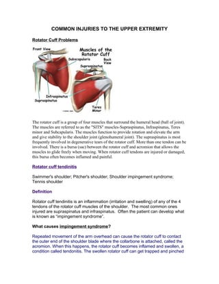
  • 1. COMMON INJURIES TO THE UPPER EXTREMITY Rotator Cuff Problems The rotator cuff is a group of four muscles that surround the humeral head (ball of joint). The muscles are referred to as the "SITS" muscles-Supraspinatus, Infraspinatus, Teres minor and Subcapularis. The muscles function to provide rotation and elevate the arm and give stability to the shoulder joint (glenohumeral joint). The supraspinatus is most frequently involved in degenerative tears of the rotator cuff. More than one tendon can be involved. There is a bursa (sac) between the rotator cuff and acromion that allows the muscles to glide freely when moving. When rotator cuff tendons are injured or damaged, this bursa often becomes inflamed and painful. Rotator cuff tendinitis Swimmer's shoulder; Pitcher's shoulder; Shoulder impingement syndrome; Tennis shoulder Definition Rotator cuff tendinitis is an inflammation (irritation and swelling) of any of the 4 tendons of the rotator cuff muscles of the shoulder. The most common ones injured are supraspinatus and infraspinatus. Often the patient can develop what is known as “impingement syndrome”. What causes impingement syndrome? Repeated movement of the arm overhead can cause the rotator cuff to contact the outer end of the shoulder blade where the collarbone is attached, called the acromion. When this happens, the rotator cuff becomes inflamed and swollen, a condition called tendonitis. The swollen rotator cuff can get trapped and pinched
  • 2. under the acromion. All these conditions can inflame the bursa in the shoulder area. A bursa is a fluid-filled sac that provides a cushion between a bone and tissues such as skin, ligaments, tendons, and muscles. An inflammation of the bursa is called bursitis Neer developed a system of categorizing rotator cuff tendonitis/impingement syndrome. Stage I is when there is edema and swelling but is completely reversible. Stage II is when there is thickening of the tendon; there may be spurs on the undersurface of the acromion and the condition is irreversible. Stage III has all the characteristics of Type II but there is now a tear of some sort in the rotator cuff tendon. Causes, incidence, and risk factors The shoulder joint is a ball and socket type joint where the top part of the arm bone (humerus) forms a joint with the shoulder blade (scapula). The rotator cuff holds the head of the humerus into the scapula. Inflammation of the tendons of the shoulder muscles can occur in sports requiring the arm to be moved over the head repeatedly as in tennis, baseball (particularly pitching), swimming, and lifting weights over the head. Chronic inflammation or injury can cause the tendons of the rotator cuff to tear. The risk factors are being over age 40 and participation in sports or exercise that involves repetitive arm motion over the head (such as baseball). Symptoms • Pain associated with arm movement; mainly overhead activities • Pain in the shoulder at night, especially when lying on the affected shoulder • Weakness with raising the arm above the head, or pain with overhead activities (brushing hair, reaching for objects on shelves, etc.) • If they go into late stage II or stage III they may develop loss of range of motion Signs and tests A physical examination will reveal tenderness over the shoulder. Pain may occur when the shoulder is raised overhead. There is usually weakness of the shoulder when it is resisted in certain positions. The Neer’s test for impingement is usually positive. Another commmon impingement test is the Hawkins Kennedy test. Both tests place the arm in such a position that it minimizes the subacromial
  • 3. space and if the tissues are enlarged, they will be pinched in these positions causing an increase in pain. X-rays may show a bone spur, while MRI may demonstrate inflammation in the rotator cuff. If a tear in the rotator cuff is present, this can usually be identified on MRI. Treatment The injured shoulder should be rested from the activities that caused the problem and from activities that cause pain. Ice packs applied to the shoulder and non- steroidal anti-inflammatory drugs will help reduce inflammation and pain. Physical therapy to strengthen the muscles of the rotator cuff and the scapula should be started; attention to any causative factors such as poor posture should also be addressed. If the pain persists or if therapy is not possible because of severe pain, a steroid injection may reduce pain and inflammation enough to allow effective therapy. If the rotator cuff has sustained a complete tear, or if the symptoms persist despite conservative therapy, surgery may be necessary. Arthroscopic surgery can remove bone spurs and inflamed tissue around the shoulder. The most common surgery is called a subacromial decompression where they remove the coracoacromial ligament; remove about a third of the width of the acromian or in some cases, remove the distal end of the acromion. Small tears can be treated with arthroscopic surgery. Newer techniques allow even large tears to be repaired arthroscopically, although some large tears require open surgery to repair the torn tendon.
  • 4. Rotator Cuff Tears Rotator cuff tears are a common source of shoulder pain. . The incidence of rotator cuff damage increases with age and is most frequently due to degeneration of the tendon, rather than injury from sports or trauma. While the information that follows can be used as a guide for all types of rotator cuff tears, it is intended specifically for complete degenerative tears of the rotator cuff. Treatment recommendations vary from rehabilitation to surgical repair of the torn tendon(s). The best method of treatment is different for every patient. The decision on how to treat rotator cuff tears is based on the patient's severity of symptoms and functional requirements, and presence of other illnesses that may complicate treatment. In consultation with an orthopaedic surgeon, the information that follows is intended to assist patients in deciding on the best management of their rotator cuff tear with the understanding that all patients are unique. Rotator cuff tears increase in frequency with age, are more common in the dominant arm, and can be present in the opposite shoulder even if there is no pain1,7 . The true incidence of rotator cuff tears in the general population is hard to determine because 5 percent to 40 percent of people without shoulder pain may have a torn rotator cuff. This was determined by studies using MRI and ultrasound to image the shoulders of patients with no symptoms. One study6 revealed a 34 percent overall incidence of rotator cuff tears. The highest incidence occurred in patients who were more than 60 years old. This study supported the concept that rotator cuff damage has a degenerative component
  • 5. and, importantly, that a tear of the rotator cuff is compatible with a painless, normal functioning shoulder. There are intrinsic and extrinsic causes of rotator cuff tears. An example of an intrinsic factor is tendon blood supply. The blood supply to the rotator cuff diminishes with age and transiently with certain motions and activities. The diminished blood supply may contribute to tendon degeneration and complete tearing3,4,5 . The substance of the tendon itself degenerates over time. Due to an age related decrease in tendon blood supply, the body's ability to repair tendon damage is decreased with age; this can ultimately lead to a full-thickness tear of the rotator cuff. An extrinsic cause would be damage to the rotator cuff from bones spurs underneath the acromion. The spurs rub on the tendon when the arm is elevated; this is often referred to as impingement syndrome. Bone spurs are another result of the aging process. The rubbing of the tendon on the bone spur can lead to attrition (weakening) of the tendon. Combining this with a diminished blood supply, the tendons have a limited ability to heal themselves. These factors are at least partly responsible for the age-related increase in rotator cuff disease and the higher frequency in the dominant arm. Natural History What will happen if a torn rotator cuff is not treated with surgery? Will I lose the use of my arm? Will the tear get larger over time? These are common concerns patients have, and the answers are not always clear. In one study, 40 percent of patients with a rotator cuff tear showed enlargement of the tear over a five-year period; however, 20 percent of those patients had no symptoms. Therefore, less than half of patients with a rotator cuff tear will have tear enlargement, but 80 percent of patients whose tear enlarges will develop symptoms7 . This data is based on a small group of patients; it is important to realize that once symptoms develop, progression may have already progressed and enlarged. Surgical and Non-Surgical Options Treatment options include: 1. Non-operative (conservative) treatment 2. Operative - Rotator cuff repair a. Open b. Mini-open c. All-arthroscopic Non-Operative Treatment Benefits and Limits Non-surgical treatment typically involves:
  • 6. 1. Injections 2. Activity modification (avoidance of activities that cause symptoms) Non-operative management of a rotator cuff tear can provide relief in approximately 50 percent of patients. Hawkins & Dunlop8 Itoi & Tabata9 Bokor et al.10 Bartolozzi et al.11 Percentage of patients who were satisfied 54 percent 82 percent 56 percent 25 percent Percentage of patients with pain relief 45 percent 55 percent 77 percent Not reported These studies show that about half (50 percent) of patients have decreased pain and improved motion, and are satisfied with the outcome of nonsurgical treatment. Surgeons may recommend nonsurgical treatment for patients who are most bothered by pain, rather than weakness, because strength did not tend to improve without surgery. There are a few predictors of poor outcome from nonsurgical treatment: 1. Long duration of symptoms (more than 6-12 months) 2. Large tears (more than 3 centimeters) Nonsurgical treatment has both advantages and disadvantages. Advantages: • Patient avoids surgery and its inherent risks: a. Infection b. Permanent stiffness c. Anesthesia complications • Patient has no "down time" Disadvantages: • Strength does not improve • Tears may increase in size over time • Patient may need to decrease activity level Surgical Intervention and Considerations Surgical management is indicated for a rotator cuff tear that does not respond to non-operative management and is associated with weakness, loss of function and limited motion. Because there is no evidence of better results in early versus delayed repairs, many surgeons consider a trial of non-operative management to
  • 7. be appropriate1 . Tears that are associated with profound weakness, are caused by acute trauma, and/or are very large (greater than 3cm) on initial evaluation may also be considered for early operative repair. Operative treatment of a torn rotator is designed to repair the tendon back to the humeral head (ball of joint) from where it is torn. This can be accomplished in a number of ways. Each of the methods available has its own pros and cons; all have the same goal--getting the tendon to heal to the bone. The choice of surgical technique depends upon several factors including the surgeon's experience and familiarity with a particular procedure, the size of the tear, patient anatomy, quality of the tendon tissue and bone, and the patient's needs. Regardless of the repair method used, studies show similar levels of pain relief, strength improvement, and patient satisfaction. The three commonly employed surgical techniques for rotator cuff repair are: 1. Open repair 2. Mini-open repair 3. All-arthroscopic repair An individual surgeon's ability to repair a torn rotator cuff and achieve a satisfactory result varies by technique. Variation is based on experience and familiarity with the technique. While one surgeon may be capable of achieving a quality repair through all-arthroscopic means, another may have better results with mini-open repair. Prior to surgery, patients should discuss the options available to them with their surgeon. The surgeon can share results of using different techniques so that the most appropriate treatment plan can be designed. Operative Procedure Open repair Open repair is performed without arthroscopy. The surgeon makes an incision over the shoulder and detaches the deltoid muscle to gain access to and improve visualization of the torn rotator cuff. The surgeon will usually perform an acromioplasty (removal of bone spurs from the undersurface of the acromion) as well. The incision is typically several centimeters long. Open repair was the first technique used to repair a torn rotator cuff; over the years, the introduction of new technology and surgeon experience has led to development of less invasive measures. Although a less invasive procedure may be attractive to many patients, open repair does restore function, reduce pain and is durable in terms of long-term relief of symptoms12, 13 . Mini-open repair As the name implies, mini-open repair is a smaller version of the open technique. The incision is typically 3 cm to 5 cm in length. This technique also incorporates
  • 8. an arthroscopy to visualize the tear, assess and treat damage to other structures within the joint (i.e., labrum and remove the spurs under the acromion. Arthroscopic removal of spurs (acromioplasty) avoids the need to detach the deltoid muscle. Once the arthroscopic portion of the procedure is completed, the surgeon proceeds to the mini-open incision to repair the rotator cuff. Mini-open repair can be performed on an outpatient basis. Currently, this is one of the most commonly used methods of treating a torn rotator cuff; results have been equal to the open repair. The mini-open repair has also proven to be durable over the long-term14 . All-arthroscopic repair This technique uses multiple small incisions (portals) and arthroscopic technology to visualize and repair the rotator cuff. All-arthroscopic repair is usually an outpatient procedure. The technique is very challenging and the learning curve for surgeons is steep. It appears that the results are comparable to the mini-open and open repairs15 . Below is a picture of a open repair. Results After rotator cuff repair, 80 percent to 95 percent of patients achieve a satisfactory result, defined as adequate pain relief, restoration or improvement of function, improvement in range of motion, and patient satisfaction with the procedure. Certain factors decrease the likelihood of a satisfactory result16 : 1. Poor tissue quality 2. Large or massive tears 3. Poor compliance with post-operative rehabilitation and restrictions
  • 9. 4. Patient age (older than 65 years) 5. Worker's Compensation claims Surgical techniques for rotator cuff repair have progressed to more minimally invasive procedures. With each advance in technique, surgeons must undergo a learning curve. Initially, some tears were considered too large to be treated with less invasive techniques. As surgeons become more experienced in using the technique, they are better able to treat most tears with less invasive means. The most recent development is the all-arthroscopic technique. As more surgeons gain experience with this technique, more tears will become amenable to an all- arthroscopic repair. Each step toward less invasive surgery has benefited the patient by: 1. Decreasing pain from surgery 2. Decreasing post-operative stiffness 3. Decreasing operative blood loss 4. Decreasing length of stay in the hospital Each technique has similar results in terms of satisfactory relief of pain, improvement in function and patient satisfaction. Less invasive surgery results in an easier rehabilitation process and less postoperative pain. Open repair results: Author Satisfied Patients Range of Motion Improved Strength Improved Ellman18 98 percent Yes Yes Hawkins et al.17 94 percent Yes Yes Cofield19 77 percent Yes Yes Mini-open repair results: Author Satisfied Patients Range of Motion Improved Strength Improved Levy et al.20 96 percent Yes Yes Paulos et al.21 94 percent Yes Yes Blevins et al.22 89 percent Yes Yes All-arthroscopic repair results: Author Satisfied Patients Range of Motion Improved Strength Improved
  • 10. Tauro23 92 percent Yes Yes Gartsman24 90 percent Yes Yes Wilson25 88 percent Yes Yes The above studies represent on a few of many papers on this topic. A large review of all published material relating to outcomes from rotator cuff repair surgery was presented in 200330 . This paper demonstrated: 1. Results were equal between open, mini-open, and arthroscopic techniques measured by: a. Patient satisfaction b. Pain relief c. Strength 2. Surgeon expertise is more important in achieving satisfactory results than the choice of technique Improvement in pain, function, and strength typically occurs over a 4-6 month period following the procedure. Source: http://orthoinfo.aaos.org/indepth/thr_report.cfm? Thread_ID=2&topcategory=Shoulder SHOULDER INSTABILITY What is traumatic shoulder instability? Traumatic shoulder instability begins with a first dislocation that injures the supporting ligaments of the shoulder. The glenoid (the socket of the shoulder) is a relatively flat surface that is deepened slightly by the labrum, a cartilage cup that surrounds part of the head of the humerus. The labrum acts as a bumper to keep the humeral head firmly in place in the glenoid. More importantly, the labrum is the attachment point for ligaments stabilizing the shoulder. When the labrum is torn from the glenoid, the support of these ligaments is lost. The development of recurrent instability depends upon the type and amount of damage that is done to the labrum and the supporting ligaments. The most common dislocation that leads to traumatic instability is in the anterior (forward) and inferior (downward) direction. A fall on an outstretched arm that is forced overhead, a direct blow on the shoulder, or a forced external rotation of the arm are frequent causes of this type of dislocation. Much less common is a posterior (backward) dislocation, which is usually related to a seizure disorder or electrocution, events in which the muscular forces of the shoulder cause the dislocation.
  • 11. What are the signs and symptoms of a dislocation? If the shoulder is dislocated, it is usually very apparent: • The shoulder is quite painful. • Motion is severely restricted. • The shoulder appears to hang down and forward, with a large dimple evident under the acromion (in the area of the collar bone). • The humeral head may be visible as a bump on the front of the shoulder, or in the armpit. To return the dislocated arm to its socket (called a reduction) usually requires a visit to the emergency department, where expert assistance can be found. Some individuals with recurrent dislocations eventually become experienced at reducing the arm themselves. How is a dislocation and traumatic shoulder instability diagnosed? As a rule, a sudden dislocation is quite evident. The patient usually holds the arm against the side, since any attempts at motion cause pain. A large crease under the acromion and a bulge in the armpit are clues to the direction of the dislocation. However, when the shoulder spontaneously relocates into its proper position, the diagnosis can be more difficult. Patients may only report the feeling of having the shoulder "slip" before the spontaneous reduction occurred. A qualified individual usually can relocate the humerus at the site of the injury occurrence. Once the reduction is performed, there is immediate pain relief. Without medications, some patients may be unable to relax the shoulder muscles enough to allow the reduction to take place. Often, these patients must go to the emergency department to get the reduction accomplished.
  • 12. • X-rays are usually taken to confirm the dislocation, its direction, and to check for a related fracture. After the reduction, follow up X-rays will confirm proper positioning and assess any other injuries. X-rays may reveal a "bony Bankart", which is a fracture of the anterior-inferior glenoid (front, lower portion of the glenoid). The presence of this fracture indicates that the labrum and ligaments in the front part of the shoulder are no longer attached to the glenoid. • If X-rays do not reveal such a fracture, an MRI or arthrogram may be ordered. In this diagnostic test, the status of the labrum and ligaments can be assessed. A Bankart lesion (detachment of the anterior-inferior portion of the labrum from the glenoid) is the most common cause of recurrent instability after an injury. How is a dislocation and traumatic shoulder instability treated? The initial reduction of a dislocation can be quite difficult. Contractions of the shoulder muscles can trap the humeral head against the glenoid. Gentle traction, and at times, medication may be needed to accomplish the reduction. Once the shoulder is reduced, a sling is used for a few days to protect it, and relieve discomfort. Physical therapy may help the patient regain motion in the joint. Non-Operative Treatment Initial treatment for recurrent instability of the shoulder centers on physical therapy. Strengthening the rotator cuff muscles and periscapular muscles (those around the scapula) gives stability to the joint. The goal of physical therapy is to help the muscles provide stability to the shoulder that the torn ligaments can no longer supply. The therapy for recurrent instability should be carefully designed for each patient since this condition often causes apprehension about certain arm positions or exercise maneuvers. Very often, physical therapy can help regain lost motion, reduce apprehension, and restore shoulder function. Operative Treatment Surgery is usually recommended if recurrent instability cannot be controlled with physical therapy and activity modification. The goal of surgery is to return stability to the shoulder with the least loss of motion. All shoulder procedures designed to stabilize the shoulder involve some loss of motion. The current procedures for anterior shoulder instability attempt to restore the normal anatomy
  • 13. without over tightening the ligaments. In certain instances, such as in young persons who have a higher risk of re-dislocation and in contact athletes who plan on continuing to participate in sports that put their shoulders at risk, surgery may be performed after the first dislocation. Open Labral Repair Currently, the preferred procedure for anterior instability is an open labral repair with an anterior capsular shift. This procedure is performed through a two to three inch incision on the front of the shoulder. The torn labrum is repaired and the stretched-out anterior shoulder capsule is imbricated (overlapped) to make it smaller. This procedure is successful approximately 95% of the time in eliminating recurrent dislocations. Arthroscopic Techniques Recently, arthroscopic procedures such as Bankart repair have been used to repair the torn labrum and reduce capsular laxity. Arthroscopic techniques are approximately 80% successful. These procedures are performed with visualization through a small fiberoptic scope. Instruments are inserted into the joint through two or three small incisions to repair the labrum. The surgical technique is similar to the one used in an open repair. A loose capsule is more difficult to address arthroscopically. Procedures using thermal energy to shrink the loose capsule have been developed, and are still being evaluated. What types of complications may occur? The major complications of anterior stabilization techniques are recurrent instability and/or loss of motion. The rate of recurrent instability depends largely on the technique used for the repair. The loss of motion can be severe, and is a function of over tightening the anterior capsule. In general, the operative shoulder should lose no more than ten degrees of external rotation. Other small risks (less than 1%) include infection, post-operative stiffness, nerve damage, or blood vessel injury. Non-Operative Recovery • Patients who have a first dislocation, and do not develop recurrent instability, will often regain full motion from a four to six week course of physical therapy. • Patients who do develop recurrent instability have a longer rehabilitation course and should concentrate on strengthening the shoulder muscles. Daily exercises in a home program may be recommended to help prevent instability events.
  • 14. Operative Recovery Following either arthroscopic or open operative repair and stabilization: • The patient will usually wear a sling for the first four to six weeks. This immobilization protects the repaired labrum while it heals to the glenoid. Until the ligaments heal, the repair must depend on the sutures used to secure the labrum. • During this immobilization period, elbow and wrist motion are maintained with gentle range of motion exercises. • Once the initial healing process is complete, physical therapy may begin. Exercises stressing range of motion are done for approximately eight weeks after surgery, or until full strength is regained. • Overhead sports, such as baseball or tennis, may resume about three months after surgery. • Contact sports are restricted for six months. http://www.steadman-hawkins.com/shoulder3/overview.asp Thoracic Outlet Syndrome Thoracic outlet syndrome is actually a collection of syndromes brought about by abnormal compression of the neurovascular bundle by bony, ligamentous or muscular obstacles between the cervical spine and the lower border of the axilla. What does that mean? First of all a syndrome is defined as a group of signs and symptoms that collectively characterize or indicate a particular disease or abnormal condition. • The neurovascular bundle which can suffer compression consists of the brachial plexus plus the C8 and Tl nerve roots and the subclavian artery and vein. • The brachial plexus is the network of motor and sensory nerves which innervate the arm, the hand, and the region of the shoulder girdle. • The vascular component of the bundle, the subclavian artery and vein transport blood to and from the arm, the hand, the shoulder girdle and the regions of the neck and head. What are the signs and symptoms of thoracic outlet syndrome? It is important to understand that presenting with the symptoms listed below in no way indicates a definitive diagnosis for thoracic outlet syndrome. Professionals understand the
  • 15. importance of coupling diagnostic testing skills with the patient?s report of what hurts and what doesn?t seem to be working properly. Don?t self diagnose! Neurologic and vascular symptoms can be indicative of more serious conditions. The bony, ligamentous, and muscular obstacles all define the cervicoaxillary canal or the thoracic outlet and its course from the base of the neck to the axilla or arm pit. Look at the scheme of this region and it all becomes more easily understood. What causes the neurovascular compression? Compression occurs when the size and shape of the thoracic outlet is altered. The outlet can be altered by exercise, trauma, pregnancy, a congenital anomaly, an exostosis, postural weakness or changes. Below is a list of the component syndromes which comprise Vascular symptoms include: Neurologic symptoms include: 1. Swelling or puffiness in the arm or hand 2. Bluish discoloration of the hand 3. Feeling of heaviness in the arm or hand 4. Pulsating lump above the clavicle 5. Deep, boring toothache-like pain in the neck and shoulder region which seems to increase at night 6. Easily fatigued arms and hands 7. Superficial vein distention in the hand 1. Parasthesia along the inside forearm and the palm (C8, T1 dermatome) 2. Muscle weakness and atrophy of the gripping muscles (long finger flexors) and small muscles of the hand (thenar and intrinsics) 3. Difficulty with fine motor tasks of the hand 4. Cramps of the muscles on the inner forearm (long finger flexors) 5. Pain in the arm and hand 6. Tingling and numbness in the neck, shoulder region, arm and hand
  • 16. thoracic outlet syndrome along with a brief description of each. Refer to the scheme for questions about the gross anatomy of the region. Anterior scalene tightness Compression of the interscalene space between the anterior and middle scalene muscles-probably from nerve root irritation, spondylosis or facet joint inflammation leading to muscle spasm. Costoclavicular approximation Compression in the space between the clavicle, the first rib and the muscular and ligamentous structures in the area-probably from postural deficiencies or carrying heavy objects. Pectoralis minor tightness Compression beneath the tendon of the pectoralis minor under the coracoid process-may result from repetitive movements of the arms above the head (shoulder elevation and hyperabduction). What sort of activities can cause these compression syndromes? Thoracic outlet syndrome has been described as occurring in a diverse population. It is most often the result of poor or strenuous posture but can also result from trauma or constant muscle tension in the shoulder girdle. Static postures such as those sustained by assembly line workers, cash register operators, students of, for example, those who do needle work often result in a drooping shoulder and forward head posture. This position of the shoulders and head is also indicative of poor upper body posture. Middle aged and elderly women who suffer from osteoporosis often display this type of posture as a result of increased thoracic spinal kyphosis. Carrying heavy loads, briefcases and shoulder bags can also lead to neurovascular compression. Humans are not well adapted as beasts of burden and heavy loads hung form the shoulders and arms can stress the supporting structures of the shoulder girdle which is basically suspended by the clavicle and all of the component ligaments and muscles. Occupations which require repetitive over head arm movements can also produce symptoms of compression . Electricians, painters and plasterers may develop hyperabduction syndrome. Compression of the neurovascular structures also occurs in athletes who repetitively hyperabduct their arms. Swimmers, volleyball players, tennis players and baseball pitchers may suffer compression of the neurovascular structures as well. However, compression of these structures may be caused by stretching or microtrauma (small tears in muscle tissue) to the muscles which support the scapula.
  • 17. Are there other causes of thoracic outlet syndrome? Some people are born with an extra rib right above the first rib. Since this intersection of nerves, vessels, muscles, bones and ligaments is already quite involved one can imagine what the presence of an extra rib in the region might do. A fibrous band extends from this cervical rib to the first rib causing an extra bend in the lower part of the brachial plexus which may produce a compression in this region. How is thoracic outlet syndrome treated? The first step to beginning any treatment begins with a trip to the doctor. Make a list of all of the symptoms which seem to be present even if the sensations are vague. Make a note of what activities and positions produce or alleviate the symptoms and the time of day when symptoms are worst. Also, note when the symptoms first appeared. This list is important and should also include any questions one may have. Due to overlapping in terms of symptoms it’s difficult to make a definitive diagnosis; this is why a list is so important. Certain diagnostic tests have been designed which are very useful for examination. These tests involve maneuvers of the arms and head and can help the practitioner by providing information as to the cause of the symptoms and help in designing an approach to treatment. These tests, accompanied by a thorough history help in ruling out other causes which may produce similar symptoms. These include Pancoast tumor, neurofibromas, cervical spondylosis, cervical disk herniation, carpal tunnel syndrome and cubital tunnel syndrome. Don?t forget to ask your practitioner about these conditions as well. Here are a few more commonly applied provocation tests used in the diagnosis of thoracic outlet syndrome. These tests may or may not momentarily reproduce symptoms but as was mentioned earlier are important in ruling out other causes which may produce similar symptoms EAST Test or "Hands-up" Test The patient brings their arms up as shown with elbows slightly behind the head. The patient then opens and closes their hands slowly for 3 minutes. A positive test is indicated by pain, heaviness or profound arm weakness or numbness and tingling of the hand.
  • 18. Adson or Scalene Maneuver The examiner locates the radial pulse. The patient rotates their head toward the tested arm and lets the head tilt backwards (extends the neck) while the examiner extends the arm. A positive test is indicated by a disappearance of the pulse. Costoclavicular Maneuver The examiner locates the radial pulse and draws the patient?s shoulder down and back as the patient lifts their chest in an exaggerated "at attention" posture. A positive test is indicated by an absence of a pulse. This test is particularly effective in patients who complain of symptoms while wearing a back-pack or a heavy jacket.
  • 19. Allen Test The examiner flexes the patient?s elbow to 90 degrees while the shoulder is extended horizontally and rotated laterally. The patient is asked to turn their head away from the tested arm. The radial pulse is palpated and if it disappears as the patient?s head is rotated the test is considered positive. Provocative Elevation Test This test is used on patients who already present with symptoms. The patient sits and the examiner grasps the patient's arms as shown. The patient is passive as the shoulders are elevated forward and into full elevation. The position is held for 30 seconds or more. This activity is evidenced by increased pulse, skin color change (more pink) and increased hand temperature. Neurological signs go from numbness to pins and needles or tingling as well as some pain as blood flow to the nerve returns. Similar to what is felt after an arm "falls asleep" and circulation returns.
  • 20. Once a diagnosis is decided, every effort is made for a conservative treatment approach. That means it should hurt less as treatment progresses. Should symptoms persist over 3 or 4 months or if there is intractable pain, vascular loss or neuralgic loss then surgery should be considered. Surgery is consistent in relieving pain but muscle weakness and atrophy do not usually improve significantly. Conservative treatment usually includes mobilization or manipulation to contributory hypomobile cervical or thoracic segments or ribs; mobilization of hypomobile neural elements and a program which address postural retraining, shoulder strengthening and stretching exercises. The practitioner should create a treatment program specific to the presenting symptoms of each individual patient. http://www.nismat.org/ptcor/thoracic_outlet/index.html ELBOW INJURIES Lateral Epicondylitis or Tennis Elbow Injury to the lateral aspect of the elbow is the most common upper extremity tennis injury. Tennis elbow is generally caused by overuse of the extensor tendons of the forearm, particularly the extensor carpi radialis brevis. Commonly experienced by the amateur player, this injury is often a result of (1) a one- handed backhand with poor technique (the ball is hit with the front of the shoulder up and power generated from the forearm muscles), (2) a late forehand swing preparation with resulting wrist snap to bring the racquet head perpendicular to the ball, or (3) while serving, the ball is hit with full power and speed with wrist
  • 21. pronation (palm turned downward) and wrist snap which increases the stress on the already taught extensor tendons. Causes One theory on the cause of lateral epicondylitis is that small tears in the tendon occur through overuse. They begin to heal but when reinjured by continued use, the tendons seem to finally give up on trying to heal and a condition called angiofibroblastic degeneration begins to take over. Think of this as scar tissue that never reaches maturity and remains weak and painful. Others feel that the tendon changes are primarily a result of decreased blood flow in the area, a sort of heart attack of the tendon. The end result is still the formation of the angiofibroblastic tendinosis tissue. The same events can happen with repeated strains like hammering a nail, picking up a heavy bucket, or pruning shrubs. Signs and Symptoms General • difficulty holding onto, pinching, or gripping objects • pain, stiffness, or insufficient elbow and hand movement • forearm muscle tightness • insufficient forearm functional strength • point tenderness at or near the insertion sites of the muscles of the lateral or medial elbow
  • 22. Specific Lateral Epicondylitis painful resisted wrist extension painful resisted radial deviation (bending wrist toward pinky) palpation tenderness of the lateral epicondyle Treatment 1. Initial treatment focus’ on R.I.C.E if acute 2. If chronic, focus of treatment is on restoring blood supply to the area and promoting the formation of healthy collagen tissue in the tendon. 3. Treatment therefore will consist of TFM, many reps of submaximal exercises with a focus on eccentric work 4. Regaining full ROM of the extensor musculature (Mills position) 5. Eventually re-strengthening the entire forearm and hand through specific exercises and ultimately functional re-training 6. In some cases, counterforce brace assists recovery 7. Physicians often inject with cortisone….be careful! 8. Very chronic cases may have surgery. In general, the surgical procedures are designed to remove the diseased and degenerated tissue around the outside of the elbow and stimulate the
  • 23. improvement of the blood supply to the involved area. Release of a portion or all of the origin of the affected extensor muscles may also be part of the operation. Medial Elbow Pain Causes in Throwers: • avulsion fractures of the medial epicondyle • ulnar collateral ligament (UCL) sprains or tears • Little league elbow (LLE) is a valgus overload or overstress injury to the medial elbow o During the throwing motion, valgus stress is placed on the elbow. This valgus stress results in tension on the medial structures (ie, medial epicondyle, medial epicondylar apophysis, medial collateral ligament complex) and compression of the lateral structures (ie, radial head, capitellum). o Repeated stress results in overuse injury when tissue breakdown exceeds tissue repair. o Recurrent microtrauma of the elbow joint can lead to LLE, a syndrome that encompasses  delayed or accelerated growth of the medial epicondyle (medial epicondylar apophysitis),  traction apophysitis (medial epicondylar fragmentation),  medial epicondylitis. Ulnar Collaterol Ligament injuries can manifest as acute ligament tears following a single valgus stress or as overuse sprains following repetitive valgus overloads. The clinical presentation is similar to LLE; however, the typical age range of the athlete is the older teenager who is skeletally mature. Suspected UCL injuries should be referred for further evaluation by a sports medicine specialist. Athletes with UCL injuries should not be allowed to pitch until they have been evaluated. Although uncommon in children, neurological injuries such as C8-T1 radiculopathy and ulnar neuritis can manifest as medial elbow pain and should be included in the differential diagnosis. Lateral compression of the elbow most frequently results in injuries to the capitellum and radial head. Osteochondrosis of the capitellum (known as Panner disease) generally occurs in children aged 7-12 years and manifests as dull, achy activity-related lateral elbow pain. Swelling, clicking, and decreased range of motion are uncommon associated symptoms. Panner disease tends to be a benign self-limited condition that does well over time and is treated with complete rest from inciting activities such as throwing and weightbearing on the elbow. Osteochondral injuries can also be observed in the radial head.
  • 24. Osteochondritis dissecans (OCD) of the capitellum occurs in adolescents aged 13-17 years. This is a localized injury to subchondral bone that results from repetitive lateral compression of the elbow during overhead motions. These patients report a general dull elbow pain that worsens with activity, often have a flexion contracture of 15° or greater, and may have mechanical symptoms of clicking or popping. Loose body formation, residual capitellum deformity, and elbow degenerative joint disease are potential sequelae. Different treatment options are used based on the age and skeletal maturity of the patient and the type of lesion present. OCD lesions can be separated into type I, which has no displacement and no articular cartilage fracture; type II, which has evidence of articular cartilage fracture or partial displacement; and type III, which is completely displaced with loose bodies in the joint. http://www.emedicine.com/SPORTS/byname/Little-League-Elbow-Syndrome.htm Medial Epicondylitis (Golfer's Elbow) Introduction Medial epicondylitis is commonly known as golfer's elbow. This does not mean that only golfers have this condition. But the golf swing is a common cause of medial epicondylitis. Many other repetitive activities can also lead to golfer's elbow-- throwing, chopping wood with an ax, running a chain saw, and using many types of hand tools. Any activities that stress the same forearm muscles can cause symptoms of golfer's elbow. Commonly involves the wrist and finger flexors –common flexor tendon. Same issues as in lateral epicondylitis with repetitive tendon injuries leading to angiofibroblastic degeneration or tendinosis. Treatment is the same as lateral epicondylitis. http://www.handuniversity.com/topics.asp?Topic_ID=4
  • 25. Cubital Tunnel Syndrome Cubital tunnel syndrome is the common term used for uInar compressive neuropathies at the elbow (from the midarm to the midforearm). UInar entrapment neuropathy develops because of the predisposing anatomy of the elbow region and the biomechanics of the ulnar nerve at the elbow; it is based on compressive, traction, and frictional forces, with the possible association of a nerve at risk. Systemic diseases such as diabetes, chronic alcoholism, renal failure, and malnutrition may predispose the patient to compressive neuropathy (i.e., a nerve at risk).' Ultimately, the cumulative effect on the nerve is to cause a region of ischemia and inflammation resulting in ulnar nerve dysfunction or cubital tunnel syndrome. Compression of the ulnar nerve at the elbow may be idiopathic, but often there is a component of extrinsic compression aiding in the entrapment. Moreover, the compression of the nerve may be dynamic or static. The dynamic anatomy and biomechanics of the cubital tunnel dramatically affect the uInar nerve, resulting in relative regional ischernia to the nerve . Dynamic impingement tends to occur early in the clinical course of the disease (i.e., position-dependent, intermittent). Although initially reversible, fixed structural changes occur over time, leading to static compression. Static compression also can develop from structural abnormalities of osseous architecture or space occupying lesions . With flexion of the elbow, the aponeurosis covering the cubital tunnel stretches, changing the cross-sectional geometry of the cubital tunnel from smooth and round to flattened and triangular. This both decreases the volume of the tunnel by 55%, and significantly increases the intraneural pressure, therefore putting the nerve at risk of ischernia. Intraneural pressure can be increased up to 600% with shoulder abduction, elbow flexion, and wrist extension. Moreover, contraction of the FCU muscle may increase the pressure on the ulnar nerve. Normally, the uInar nerve at the cubital tunnel is known to elongate 4.7 mm during elbow flexion. Should the nerve be tethered by perineural fibrosis (e.g., postoperative, posttrauma), it can no longer elongate and may experience up to doubled intraneural pressures. The ulnar nerve has five common sites of compression in the elbow region. The
  • 26. most common sites are at the level of the elbow. As the nerve passes by the medial epicondyle (or epicondylar groove) and into the cubital tunnel (passing deep to the aponeurosis linking the two heads of the FCU), it is at its greatest risk of compression. These are the primary sites of idiopathic disease. The other three tend to be sites of secondary or iatrogenic compression (often because of inadequate release). The most proximal site is the arcade of Struthers, a fascial band 8 cm proximal to the medial epicondyle, extending from the medial head of the triceps to the medial intermuscular septum. (Note: This is different from the ligament of Struthers, which extends from a supracondylar process to the medial humeral epicondyle and is involved primarily with proximal median nerve compression.) Table 1. CAUSES OF CUBITAL TUNNEL SYNDROME Idiopathic Trauma Acute compression or direct injury Entrapment in distal humeral fracture or elbow dislocation Compression secondary to deformity--cubitus vaIgus, cubitus varus, malunion, nonunion Heterotopic ossification-posttraurnatic, secondary to burns,80 secondary to head injury8l Aberrant or abnormal musculature anconeus epitrochlearis3O.45 reverse flexor carpi uInaris,69 triceps brachii (snapping) Cumulative trauma disorders-keyboard operator, baseball pitcher Arthritides Osteoarthritis-secondary to osteophytes, loose bodies, synovial cysts37 Inflammatory arthritis-synovitis Synovial chondromatosis Vascular bands--often branches of uInar artery latrogenic-postanesthetic palsy 61 Space-occupying lesions of the cubital tunnel and epicondyle region--e.g., lipomata CLINICAL PRESENTATION OF CUBITAL TUNNEL SYNDROME Patients with cubital tunnel syndrome tend to present with a pain, often aching or lancinating, primarily in the region of the elbow. It may radiate proximally or distally. The most frequent complaint, however, is of paresthesias in the ring and small fingers, initially intermittent and position-related, often waking them at night. Objective sensory loss usually occurs later, with progression of the disease (Table 2). A clue to the location of proximal uInar nerve entrapment is the presence of dorsal sensory loss. There is usually no sensory loss along the medial forearm because it is supplied by the medial antebrachial cutaneous nerve, a branch of the medial cord of the brachial plexus. Most importantly, examining for Tinel's sign helps localize the entrapment. Patients often note they have "weak or clumsy hands," often dropping objects or being unable to open jars. Muscle weakness or atrophy often occurs and, in
  • 27. many patients, in the absence of any objective sensory change. The fascicles to the intrinsic ulnarly supplied hand muscles are more susceptible because of their superficial topographic location within the ulnar nerve at the cubital tunnel (FCU and FDP often are spared). Muscle weakness may be demonstrated by clawing of the hand, Wartenberg's sign or Froment's sign although these indications are not helpful in localizing the lesion. Because of the common anomalous connections between the median and uInar nerves (17% incidence of Martin- Gruber anastomosis) that primarily affect the intrinsic innervation, however, weakness from significant cubital tunnel syndrome may be absent. Table 2. STAGING OF ULNAR NERVE COMPRESSION AT THE ELBOW Mild Sensory, Intermittent paresthesias; vibratory perception increased Motor, Subjective weakness, clumsiness, or loss of coordination Tests, Elbow flexion test or Tinel's sign may be positive Moderate Sensory, Intermittent paresthesias; vibratory perception normal or decreased Motor, Measurable weakness in pinch or grip strength Tests, Elbow flexion test or Tinel's sign is positive; finger crossing may be abnormal Severe Sensory, Persistent paresthesias; vibratory perception decreased; abnormal two-point discrimination (static >6 mm, moving >4 mm) Motor, Measurable weakness in pinch and grip plus muscle atrophy Tests, Positive elbow flexion test or positive Tinel's sign may be present; finger crossing usually abnormal TREATMENT OF CUBITAL TUNNEL SYNDROME Progress in the management of ulnar nerve syndromes has been affected by: (1) delayed diagnosis and (2) treatment that is often based on personal bias rather than scientific results.13 The principles of treatment of uInar nerve compression and chronic arm pain are: 1.identification of the presenting signs and symptoms 2.reaching the correct diagnosis and ruling out other common differential diagnoses 3.treating the patient appropriately to optimize recovery of neural function and lower the risk of postoperative complications (neuroma, neuritis, inadequate release) Nonoperative Treatment As discussed, idiopathic uInar neuropathy often initially manifests as intermittent or mild symptoms. At the early, mild stage, the process may be reversible if treated prior to the onset of chronic neural changes.78 The goals of conservative treatment are to reduce the inflammatory state of the perineural tissues, to
  • 28. enhance vascular perfusion of the nerve, and, indirectly, to restore normal axonal transport. This could include using splints to prevent elbow flexion and physical therapy consisting of neural mobilization therapy. Urbaniak believes patients with the following traits should undergo a trial of conservative treatment: 1.early symptoms, intermittent episodes 2.mild paresthesias without significant pain 3. minimal physical findings (slight numbness), with normal motor examination Operative Treatment Can consist of decompression by medial epicondylectomy and nerve transposition http://www.simmonsortho.com/literature/cubitaltunnelsyndrome/cubitaltunnelsynd rome.html Posterior Interosseous Syndrome: mimics tennis elbow 1. Inciting causes: a. radiocapitellar joint ganglions and synovitis b. congenital tightness of ligamentous arcade of Frohse c. include poorly placed screws for fracture fixation i. PIN is vulnerable during ORIF of proximal radius ii. In 25% of pts, PIN lies in direct contact w/ periosteum of radius just dorsal to the biciptial tuberosity iii. fixation device applied to radius, which might have its proximal screw at level of the bicipital tuberosity, could cause PIN syndrome by entrapment beneath the plate d. idiopathic compression syndrome: i. sites of compression 1. fibrous bands anterior to the radial head at the entrance of radial tunnel (uncommon cause of compression) 2. radial recurrent vessels (leash of Henry) 3. tendinous origin of ECRB: the ECRL is more superficial and is not a source of compression 4. arcade of Froshe a. this is the most common location of nerve compression in radial tunnel syndrome b. lies deep to the extensor carpi radialis brevis c. tendinous proximal border of supinator (arcade of Frohse) 5. distal edge of the supinator at exit: this is the least
  • 29. common source of compression 2. Differential Diagnosis: a. C7 radiculopathy: unlike PIN, there will be weakness of triceps and wrist flexors b. lateral epicondylitis (ECRB) 1. It is often misdiagnosed as resistant tennis elbow or PIN Syndrome 2. unlike tennis elbow, there is tenderness about 4 cm distal to the lateral humeral epicondyle; c. Distal PIN syndrome:Pts w/ distal posterior interosseous nerve syndrome have pain with repetitive dorsiflexion & tenderness centered over the 4th extensor compartment 3. Exam a. pts commonly have tenderness over lateral epicondyle & almost always have tenderness more distally over the arcade of Froshe b. pain is almost always experienced w/ resisted supination of the forearm and frequently w/ resisted pronation c. full pronation of forearm produces pressure on PIN by sharp tendinous edge of the origin of ECRB muscle d. active supination from a pronated position (tightening supinator) along w/ wrist flexion (which tighens the ECRB) may reproduce the patient's symptoms e. most will have pain w/ resisted extension of extension of middle finger f. pt will unable to extend thumb or other digits at MCP joints; w/ complete palsy, pts will continue to have wrist extension (ECU) but they are unable to extend wrist at neutral or in ulnar deviation; they can extend the digits at the interphalangeal joints, but not at MP joints g. pain is relieved by blocking the posterior interosseous nerve 3 cm proximal to the wrist joint; performed by injecting approx 1 cm ulnar to Lister's tubercle 1. Injection Test: • lidocaine injection 4 finger breadths distal to the lateral epicondyle will result in temporary PIN palsy and, in the case of PIN syndrome, will result in temporary relief of pain • with lateral epicondylitis, the patient should note pain relief following injection at the origin of the ECRB tendon (which is usually more proximal than the site of injection for PIN compression) 4. Treatment As with cubital tunnel syndrome, can try physical therapy in mild cases; otherwise needs surgical decompression
  • 30. http://www.wheelessonline.com/ortho/posterior_interosseous_nerve_compressio n_syndrome Anterior Interosseous Nerve Syndrome Anterior interosseous nerve (AIN) compression is also known as Kiloh Nevin syndrome. The AIN is the branch of the median nerve arising approximately 6 cm below the elbow and supplys motor function to the FPL, pronator quadratus, and FDP to the index finger, as well as the pronator quadratus. Causative factors include tendinous bands, a deep head of the pronator teres, accessory muscles (including the Gantzer muscle, which is the accessory head of the FPL), aberrant radial artery branches, and injury may occur following fractures to the arm causing fibrous bands to form or from impingement by enlarged bursae or thrombosed veins. Symptoms These include vague pain in the proximal forearm and weakness of the FPL and FDP to the index finger. Pain in the forearm is exacerbated by exertion. Complete paralysis produces a 'pinch deformity'. Affected persons cannot form a circle by pinching their thumb and index finger (ie, hyperextension of index distal interphalangeal joint and thumb interphalangeal joint). Sensory involvement is not described. Treatment: Nonsurgical treatment includes rest, anti-inflammatory medications, and splints. Surgical treatment includes exploration of the median nerve through an approach similar to that for pronator syndrome, release of the lacertus fibrosus and division superficially more or less to the deep heads of the pronator teres, and ligation of crossing vessels. http://www.patient.co.uk/showdoc/40001151/ http://www.emedicine.com/plastic/topic300.htm
  • 31. Carpal Tunnel Syndrome Carpal tunnel syndrome develops when a large nerve — the median nerve — is compressed inside the wrist. This nerve controls feeling in the thumb, index finger and thumb side of the ring finger. The median nerve also controls the muscles at the base of the thumb. The condition gets its name from the eight carpal bones that surround the median nerve in the wrist, forming a tunnel to the hand. The tunnel is just big enough for the median nerve and several tendons to run through it. Anything that makes the tunnel smaller (such as arthritis) or makes the tendons larger (such as thickening of the tendon linings) can cut off the circulation to the nerve. When this happens, you notice pain, numbness and tingling in your fingers. Causes of carpal tunnel syndrome Pressure on the median nerve can be caused by:  A wrist injury or a fracture of the wrist or the end of the forearm  Frequent use of vibrating hand tools  Any repetitive, forceful motion with the wrist bent, especially when done for prolonged periods without rest
  • 32. Carpal tunnel syndrome can also be caused by underlying medical conditions including:  Rheumatoid arthritis  Osteoarthritis  Hypothyroidism and, less often, hyperthyroidism  Diabetes  Pregnancy  Amyloidosis  Acromegaly  Systemic lupus erythematosus Symptoms of carpal tunnel syndrome usually improve or go away when these underlying medical conditions are treated — or in the case of pregnancy, after the baby is born. In many cases, the syndrome has no specific cause — in spite of the widespread belief that everyone in the work force is at risk. Jobs that require people to grip something repeatedly and forcefully or hold their fingers or wrists in an abnormal position may contribute to the condition in some people, but studies seeking to find a cause-and-effect relationship are inconclusive. One clear finding from these studies is that frequent computer use doesn't cause carpal tunnel syndrome, though it may worsen the symptoms. Several studies have found no relationship between computer use of up to seven hours a day and risk of carpal tunnel syndrome. What increases the risk?  Being female and middle-aged. Women are three times as likely as men to develop carpal tunnel syndrome. Doctors aren't sure why. Fluid retention caused by hormone shifts may be a factor, as may having large breasts. So might the fact that women's carpal tunnels are smaller than men's.  Obesity. Obesity increases the risk of carpal tunnel syndrome. Numerous studies have found more than twice as many carpal tunnel syndrome cases among the obese as among those with average weights.  Genetics. Genetic makeup may increase the risk of carpal tunnel syndrome. Researchers have found that in fraternal twins, the chance that both twins will develop carpal tunnel syndrome is significantly lower than that in identical twins, who share all the same genes. One specific — but rare — hereditary nerve disorder seems to cause carpal tunnel syndrome in a small number of people.  Smoking. People who smoke cigarettes may experience worse symptoms and slower recovery from carpal tunnel syndrome than nonsmokers do.
  • 33. Symptoms of carpal tunnel syndrome Carpal tunnel syndrome usually develops gradually. Pressure on the median nerve produces a specific pattern of numbness, tingling and pain that usually is worse at night. As the condition worsens, the patient may lose some hand strength and dexterity. The patient may experience any or all of these symptoms: • Numbness, burning, tingling or pain in the hand or fingers — but not in the little finger • Increased discomfort at night or first thing in the morning • Increased discomfort with prolonged grasping or flexing of the wrist • Temporary symptom relief when "shaking out" the hands • Hand weakness, especially loss of pinch strength or difficulty holding and picking up objects • Loss of feeling in the thumb, index and middle fingers, and the radial side of the ring finger • Atrophy or weakness of the muscle at the base of the thumb • Inability to distinguish temperature changes Non Surgical Treatment Splinting
  • 34. Splints (braces) are the most commonly used nonsurgical treatment for carpal tunnel syndrome. Splints that immobilize the wrist in a neutral position are most likely to relieve discomfort. An neutral wrist maximizes the size of the carpal tunnel, which reduces pressure on the median nerve, relieving the symptoms. The patient may need to wear a splint for a few days to a week or more before noticing significant improvement. Some studies show that continuous splinting — wearing a splint both day and night — is more effective than wearing it just at night. Others, however, show little or no added benefit from 24-hour splinting, particularly in terms of symptom relief. Also, many people find that a splint restricts their hand and wrist movements too much to be worthwhile at work and around the house, so night wear alone is a good option. Splints are safe, relatively inexpensive and give many people with carpal tunnel syndrome excellent short-term relief from their symptoms. Besides relieving discomfort, splints can sometimes improve conduction of nerve impulses along the median nerve, which can improve strength, dexterity and sensation. Splints are most likely to work for if there are mild to moderate carpal tunnel syndrome symptoms for one year or less. Splints may be a good choice in pregnancy related carpal tunnel. Sometimes, physical therapy or special hand exercises relieve mild to moderate symptoms of carpal tunnel syndrome. Gliding exercises. Some people who don't get adequate relief from splints and activity modification do get more relief when these treatments are combined with nerve and tendon gliding exercises. Gliding exercises alone may help, but usually not as much as when they're combined with other treatments. Drug therapy
  • 35. Drug treatment of carpal tunnel syndrome attempts to reduce any inflammation in the carpal tunnel that might put pressure on the median nerve. These medications are commonly used: NSAIDs. If tendinitis or another inflammatory condition accompanies carpal tunnel syndrome, the patient can relieve some of the pain by taking nonsteroidal anti-inflammatory drugs (NSAIDs). These drugs include ibuprofen (Advil, Motrin, others), naproxen sodium (Anaprox, Aleve, others) and aspirin — either prescription or over-the-counter. NSAIDs are unlikely to relieve the symptoms if inflammation isn't causing the discomfort — and most people don't have inflammation around their nerve. NSAIDs do nothing to relieve other carpal tunnel syndrome signs and symptoms, such as tingling, numbness or hand weakness. Even when an inflammatory condition is present, NSAIDs may be most effective when used in combination with splinting, activity modification and gliding exercises. When used in such combination, it's difficult to know if the NSAIDs are adding to the known benefit of those other treatments. Even nonprescription NSAIDs may cause side effects in some people if taken for a long time. Diuretics. Diuretics are high blood pressure medications sometimes prescribed on the theory that fluid retention in the carpal tunnel can cause pressure on the median nerve. There's no evidence this is true. Diuretics have undesirable side effects and are rarely used. Corticosteroids. If the patient doesn’t get adequate relief from splints or activity modification, the doctor may inject the carpal tunnel with a corticosteroid, such as cortisone. Corticosteroid injections can be quite effective in providing temporary relief from carpal tunnel syndrome symptoms. The main effect of these drugs is to reduce inflammation, whether it's from arthritis, asthma or some other condition. Because surgery for carpal tunnel syndrome rarely reveals any evidence of inflammation, though, the reasons for corticosteroids' effectiveness in carpal tunnel syndrome are unknown. Corticosteroid pills don't work as well as corticosteroid injections. Corticosteroid injections will most likely give long-term relief from carpal tunnel syndrome symptoms if the patient:  Have mild to moderate symptoms for one year or less  Have no loss of feeling or strength in your hand Corticosteroid injections may provide significant short-term relief from pain, tingling and numbness. Often, some relief is immediate. Relief then builds over time, often peaking about one month after injection. The length of time an injection is effective varies greatly from several weeks to more than a year. Relief
  • 36. may last longer if the carpal tunnel syndrome symptoms are mild to moderate at the time of injection. The patient may get renewed relief from a second injection. However, some trials have found that fewer than half of those who got good relief from the first injection got adequate relief from the second. Complications with corticosteroid injections are rare. However, the risk of complications increases as the patient has more injections. These rare complications include nerve injury and tendon rupture. Surgical Carpal tunnel surgery cuts the transverse carpal ligament. Cutting the ligament is like cutting a rubber band wrapped over a rolled-up newspaper. It allows the carpal tunnel to expand in size, relieving pressure on the median nerve and thereby reducing or eliminating carpal tunnel syndrome symptoms. Carpal tunnel surgery is usually done by an orthopedic surgeon, a plastic surgeon, a hand surgeon or a neurosurgeon. Two types of carpal tunnel surgery are widely available: open carpal tunnel release (OCTR) and endoscopic carpal tunnel release (ECTR). Both surgeries are commonly performed in the United States. Open carpal tunnel release (OCTR) is performed under local anesthesia. The anesthetic, which is injected into the palm, causes burning that many people consider the most painful part of the operation. This burning can be significantly reduced if the surgeon mixes some sodium bicarbonate with the anesthetic. The doctor makes a small incision in the base of the palm and sometimes extending into the wrist, measuring about 1 to 2 inches (about 2 to 5 centimeters) in length. This opens the skin so that thesurgeon can see the ligament as it's cut with a sterile surgical blade. The cut ligament springs open and immediately provides more space for the median nerve to pass through the carpal tunnel. The skin incision is then closed with stitches (sutures). Complications include nerve injury and infection, but these rarely happen — infection occurs in about one in 100 people, and accidental nerve injury in fewer than one in 1000.
  • 37.
  • 38. Endoscopic carpal tunnel release (ECTR) surgery also is an outpatient procedure typically done in a hospital or surgical center operating room. The surgeon makes one or two small incisions called portals. Many surgeons deem it safer to make two incisions — one in the palm and one in the forearm just above the wrist (double-portal ECTR). With two portals, it may be easier for the surgeon to see that the carpal ligament has been completely cut. Each incision is about 1/2 inch long. Other surgeons make just one incision — either in the palm or in the forearm just above the wrist (single-portal ECTR). A hollow, flexible tube is inserted through an incision hole, passing beneath the transverse carpal tunnel ligament. The wrist must be cocked back quite far to get the tube to fit in properly. This part of the operation may be painful, unless the patient is given some sedation. An attached video camera (endoscope) is inserted through the tube. The camera lets the surgeon see the ligament so that it can be accurately cut with tiny knives inserted through either the same or a different tube. Pros and cons Which type of carpal tunnel release surgery the patient has — ECTR or OCTR — largely depends on the surgeon's experience and the patient’s preference. ECTR and OCTR share similar outcomes. Both:  Have excellent long-term success rates and low complication rates  Restore muscle strength, manual dexterity and sensation in most cases There are, however, some important distinctions between the two types of surgery, as well as the different techniques. ECTR  Technique. Single-portal ECTR has the same overall success rate as double-portal, though single-portal has a slightly higher complication rate.
  • 39.  Who shouldn't have it. Patients who have abnormal nerve anatomy or scarring from a previous surgery in the area — both of which are rare; patients having repeat surgery  Complications. Complications such as a nerve injury are rare from any kind of carpal tunnel release, but are slightly higher for ECTR. The chances of needing a second operation also are higher with ECTR than with OCTR — about one in 50 compared to one in 300. Partly, the need for a second operation arises from complications, but mostly, it's because with ECTR there's a slightly higher risk of not completely cutting the carpal ligament.  Recovery. ECTR usually has slightly less immediate post-surgical pain and tenderness at the incision sites than does OCTR. ECTR requires mild sedation. OCTR doesn't.  Return to work. Getting back to work after ECTR and modified OCTR is usually a few weeks earlier than for traditional OCTR. That's primarily because the incisions are smaller.  Cost. ECTR costs more than does OCTR for the actual surgery because an endoscope and sedation are needed. Long-term costs for both surgeries are similar and vary greatly depending primarily on whether complications arise and how long the patient is off work. OCTR  Technique. OCTR uses a larger incision than does ECTR. This allows the surgeon to better see the anatomy of the hand and thereby reduces the risk of injuring a nerve or vessel. A larger incision makes initial recovery slightly more painful, though the discomfort is easily controlled with oral pain medication.  Complications. Complications are rare and are slightly lower for OCTR than for ECTR. It's extremely rare to need a second carpal tunnel operation after OCTR.  Cost. OCTR costs less than ECTR for the actual surgery because less equipment and supplies are needed. Long-term cost is variable, depending mainly on whether complications arise and how long the patient is off work. Pros For long-term symptom relief, surgery is more effective than splinting, injection or any other nonsurgical treatment. Even if some symptoms remain after surgery, they tend to be less severe than those persisting or recurring after other types of therapies. Despite the time it takes to heal, surgery works faster. Often, pain, tingling and numbness are gone immediately after surgery.
  • 40. Cons Surgery isn't always a cure-all. Symptoms may lessen, but still persist. Hand strength may be slow to return to normal, or not return to normal. Surgery may offer partial or no relief if another medical condition, such as obesity, arthritis or thyroid disease, is partly causing the carpal tunnel syndrome. Possible surgical complications include nerve damage and infection, though both are rare. For as long as four to eight weeks, the may not be able to perform their job or do other routine activities. http://www.mayoclinic.com/health/carpal-tunnel- syndrome/CP99999/PAGE=CP00017 Dupuytren's Contracture Presentation and Anatomy: Thickening of the palmar fascia, which is usually painless and develops slowly over time. If pronounced, it may prevent the hand from being able to fully open. Examination: • Obvious focal thickening on palmar aspect hand. • On palpation, feels tough and thick, though non-tender and without signs of inflammation. • May interfere with ability to fully open hand
  • 41. ] Dupuytren’s Contracture 4. Heberden's Nodes Presentation and Anatomy: Bony excresences that cause deformity at the DIP joints of the fingers. Occurs slowly over time and is associated with Osteoarthritis. May affect many joints or only a few, though not usually symmetric. Similar protrusions at the PIP joints are called Bouchard's nodes. Examination: o Obvious bony protrusions at DIP joints o Non-tender on palpation with an absence of inflammation o Some times interfere with joint movement and function
  • 42. Heberden's Nodes 5. Trigger Finger Presentation and Anatomy: Flexor tendons connect muscles proximal to the wrist to the fingers. When the muscles shorten, they pull on the tendons, causing the fingers to flex. Occasionally, nodules/irregularities develop along the tendons, which then interfere with their smooth movement thru "pulleys" on the palm. Patients note difficulty flexing and extending the affected finger and lack of smooth movement. This is associated with a sensation of sudden freeing of the tendon ("triggering") when the irregularity slips through the pulley. Examination: o The palm and fingers usually appear normal. The affected tendon is not visible. o Ask the patient to fully flex the affected finger. When they attempt to extend and flex it, the movement will be impaired. It's worth noting that sometimes the triggering does not occur with every movement. o If you place one of your fingers over the affected tendon, you may feel the "pop" when it finally pulls thru. There is usually no associated pain or inflammation. 6. Tenosynovitis of the Thumb (DeQuervain's type) Presentation and Anatomy: Repetitive abduction and adduction of the thumb can irritate the tendons of the extensor policis brevis and abductor policis longus muscles. When this occurs, any movement of the thumb (in particular, gripping) may cause pain at its base.
  • 43. Examination: o The thumb usually appears normal. In cases of severe tendonitis, there may be swelling overlying the tendons. o Tenderness at the point where the tendons of the extensor pollicis brevis and longus cross the radial styloid (distal end of the radius) o Pain with passive stretching of the tendons (a.k.a. Finklestein Test): a. Direct the patient to place the thumb in their palm.
  • 44. b. Have them cover the thumb with the fingers of the same hand, forming a fist. c. Gently deviate the wrist towards the ulna. This stretches the inflamed tendons over the radial styloid, reproducing the patient's pain. Finkelstein's Test medicine.ucsd.edu/clinicalmed/Joints3.html
  • 45. Boxer's Fracture Presentation: When a closed fist strikes a solid surface, the force may cause a break in the 5th metacarpal. Examination: o Pain and swelling over the 5th metacarpal Ulnar Collateral Ligament Disruption (Gamekeeper's Thumb) The ulnar collateral ligament (UCL) is a strong band of tissue that connects the first phalanx of the thumb to the metacarpal bone along the ulnar side. Injury to this structure was first described in Scottish Gamekeepers, who damaged the ligament as a result of the manner in which they killed rabbits. The head of the rabbit was grasped between thumb and first finger of one hand while they pulled on the rabbit's hind quarters with their other. This force chronically stressed the UCL, leading to weakening or frank rupture. After its initial description, it was quickly recognized that the ligament could be torn by any strong force that acutely abducts the extended thumb. Patient's are usually immediately aware that something is wrong, developing swelling, pain and instability at the metacarpal-phalangeal (MCP) joint . It has become a relatively common ski injury, occurring when a person falls on a hand that has a ski pole gripped between the thumb and forefinger.
  • 46. Hand Injuries: See syllabus for different hand/finger deformities.