Previous year question on temporal lobe based on neet pg, usmle, plab and fmge or mci screening exams
•Als DOCX, PDF herunterladen•
10 gefällt mir•1,183 views

Revision with a Master Quiz of 13 questions based on NEET PG Sample Questions on Temporal lobe from Previous Year NEET PG Online Exams.

Empfohlen
Empfohlen
Weitere ähnliche Inhalte
Was ist angesagt?
Was ist angesagt? (20)
Ähnlich wie Previous year question on temporal lobe based on neet pg, usmle, plab and fmge or mci screening exams
Ähnlich wie Previous year question on temporal lobe based on neet pg, usmle, plab and fmge or mci screening exams (20)
Mehr von Abhishek Gupta
Mehr von Abhishek Gupta (10)
Kürzlich hochgeladen
Kürzlich hochgeladen (20)
Previous year question on temporal lobe based on neet pg, usmle, plab and fmge or mci screening exams
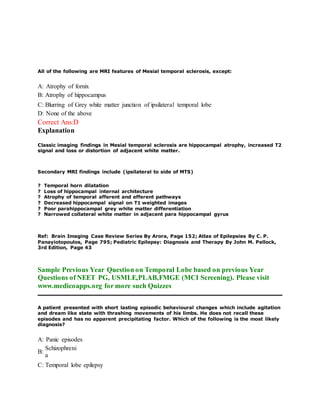
- 1. All of the following are MRI features of Mesial temporal sclerosis, except: A: Atrophy of fornix B: Atrophy of hippocampus C: Blurring of Grey white matter junction of ipsilateral temporal lobe D: None of the above Correct Ans:D Explanation Classic imaging findings in Mesial temporal sclerosis are hippocampal atrophy, increased T2 signal and loss or distortion of adjacent white matter. Secondary MRI findings include (ipsilateral to side of MTS) ? Temporal horn dilatation ? Loss of hippocampal internal architecture ? Atrophy of temporal afferent and efferent pathways ? Decreased hippocampal signal on T1 weighted images ? Poor parahippocampal grey white matter differentiation ? Narrowed collateral white matter in adjacent para hippocampal gyrus Ref: Brain Imaging Case Review Series By Arora, Page 152; Atlas of Epilepsies By C. P. Panayiotopoulos, Page 795; Pediatric Epilepsy: Diagnosis and Therapy By John M. Pellock, 3rd Edition, Page 43 Sample Previous Year Question on Temporal Lobe based on previous Year Questions of NEET PG, USMLE,PLAB,FMGE (MCI Screening). Please visit www.medicoapps.org for more such Quizzes A patient presented with short lasting episodic behavioural changes which include agitation and dream like state with thrashing movements of his limbs. He does not recall these episodes and has no apparent precipitating factor. Which of the following is the most likely diagnosis? A: Panic episodes B: Schizophreni a C: Temporal lobe epilepsy
- 2. D: Dissociative disorder Correct Ans:C Explanation The patient in the question has most likely suffered an attack of temporal lobe epilepsy. The loss of consciousness need not always be present but it is associated with loss of memory for the episode. In temporal lobe seizure, the patient usually experiences auras or warning signs, epigastric discomfort, olfactory hallucination, sensation of deja vu. A dream like state is often a feature of TLE. There may be loss of consciousness abnormal movement of mouth, and rarely abnormal movement of body. Frontal lobe epilepsy is also associated with episodes of agitation known as ‘intermittent explosive disorder’. Ref: Clinical Neuroanatomy By Stephen G. Waxman, 26th Edition, Chapter 19 Sample Previous Year Question on Temporal Lobe based on previous Year Questions of NEET PG, USMLE,PLAB,FMGE (MCI Screening). Please visit www.medicoapps.org for more such Quizzes Gustatory hallucinations are most commonly associated with: A: Temporal lobe epilepsy B: Grand mal epilepsy C: Anxiety disorders D: Tobacco dependence Correct Ans:A Explanation Gustatory hallucinations are most commonly associated with temporal lobe lesions, especially uncinate gyrus seizures. Patients report experiencing bitter, sweet, salty, tobacco-like, metallic or indescribable strange tastes. They are found in 4% of seizure patients with temporal lobe foci. Ref: Clinical neuropsychology 4th Ed By Kenneth M.Heilman, Page 488. Sample Previous Year Question on Temporal Lobe based on previous Year Questions of NEET PG, USMLE,PLAB,FMGE (MCI Screening). Please visit www.medicoapps.org for more such Quizzes Gustatory hallucinations are most commonly associated with:
- 3. A: Temporal lobe epilepsy B: Grand mal epilepsy C: Anxiety disorders D: Tobacco dependence Correct Ans:A Explanation Mesial temporal lobe epilepsy is the epilepsy most commonly associated with gustatory hallucinations. This syndrome produces seizures with visual, auditory, gustatory and visceral hallucinations. Ictal gustatory hallucinations are the experience of a taste due to a focal seizure in the absence of an environmental stimulus of the sensation. Common gustatory hallucinations includes metallic, rusty, bitter, and salty tastes. Ref: Atlas of Epilepsies, Volume 3 edited by S.R. Benbadis page 451. Sample Previous Year Question on Temporal Lobe based on previous Year Questions of NEET PG, USMLE,PLAB,FMGE (MCI Screening). Please visit www.medicoapps.org for more such Quizzes Gustatory hallucinations are most commonly associated with: A: Temporal lobe epilepsy B: Grand mal epilepsy C: Anxiety disorders D: Tobacco dependence Correct Ans:A Explanation Gustatory hallucinations are most commonly associated with temporal lobe lesions, especially uncinate gyrus seizures. Patients report experiencing bitter, sweet, salty, tobacco-like, metallic or indescribable strange tastes. They are found in 4% of seizure patients with temporal lobe foci. Ref: Clinical neuropsychology 4th Ed By Kenneth M.Heilman, Page 488. Sample Previous Year Question on Temporal Lobe based on previous Year Questions of NEET PG, USMLE,PLAB,FMGE (MCI Screening). Please visit www.medicoapps.org for more such Quizzes
- 4. A 60 year old man presents to a physician because of difficulty in reading and coming down stairs, which he attributes to an inability to "look down." Physical examination reveals that the patient looks around by moving his head rather than his eyes and also shows a distinctive axial rigidity of neck, trunk, and proximal limb muscles. He shows poverty of movement and dysarthric speech. Mentally, the patient responds very slowly but has better memory and intellect than are initially apparent. Which of the following pathologic findings of the brain would most likely be present? A: Depigmentation of the substantia nigra and locus ceruleus B: Diffuse cortical atrophy with relative sparing of primary motor and sensory areas C: Selective frontal and temporal lobe atrophy D: Widespread neuronal loss and gliosis in subcortical sites Correct Ans:D Explanation The disease is progressive supranuclear palsy, a degenerative disorder characterized by ophthalmoplegia, pseudobulbar palsy, axial dystonia, and bradykinesia. The presentation described in the question is typical. The pathologic changes consist of widespread neuronal loss and gliosis in subcortical sites with sparing of the cerebral and cerebellar cortices. Pigmented neurons in the substantia nigra (pars compacta) and locus ceruleus selectively degenerate in Parkinson's disease. In Alzheimer's disease, there is diffuse cortical atrophy, especially over the association cortex of frontal, temporal, and parietal lobes, with relative sparing of primary sensory and motor areas.Selective frontal and temporal lobe atrophy is characteristic of Pick's disease. Ref: Wyatt C., Butterworth IV J.F., Moos P.J., Mackey D.C., Brown T.G. (2008). Chapter 11. Neuropathology. In C. Wyatt, J.F. Butterworth IV, P.J. Moos, D.C. Mackey, T.G. Brown (Eds),Pathology: The Big Picture. Sample Previous Year Question on Temporal Lobe based on previous Year Questions of NEET PG, USMLE,PLAB,FMGE (MCI Screening). Please visit www.medicoapps.org for more such Quizzes All of the following are pathologic features of brain in AIDS, EXCEPT: A: Perivascular giant cell invasion B: Microglial nodules C: Vasculitis D: Temporal lobe infarction Correct Ans:C Explanation Pathologic features of brain in AIDS includes: Gross:
- 5. Encephalitis: frontal /temporal cortical atrophy Histology: Meningitis with mild lymphocytic infiltrates and scanty perivascular inflammation Features of giant cell (HIV) encephalitis are: Presence of microglial nodules Pericapillary aggregates of multinucleated giant cells having generous or scanty neoplasm Perivascular lymphocytic cuffing In leukoencephalopathy: there is diffuse diffuse demyelination and infiltration of macrophages and multinucleated giant cells. Ref: Neuropathology,A Volume in the High Yield Pathology Series (Expert Consult ... edited by Anthony T. Yachnis page 235. Sample Previous Year Question on Temporal Lobe based on previous Year Questions of NEET PG, USMLE,PLAB,FMGE (MCI Screening). Please visit www.medicoapps.org for more such Quizzes A 9 year old boy with Fallot's tetralogy, had high grade fever followed by focal seizure 2 days prior to hospital admission. His blood counts were increased and predominantly polymorphs. CT scan of the head showed a focal lesion suggestive of an abscess. Where would be the commonest location of brain abscess in this patient? A: Cerebellum B: Thalamu s C: Temporal lobe D: Parietal lobe Correct Ans:D Explanation Cyanotic congenital heart diseases like TOF are a significant risk factor for brain abscess. The mortality associated with the condition is very high. Focal ischemia in the brain, particularly the parietal lobe is the cause. Otogenic : Temporal lobe and cerebellum ; Paranasal sinuses : Frontal Lobe. Intracranial abscesses can originate from infection of contiguous structures (eg, otitis media, dental infection, mastoiditis, sinusitis) secondary to hematogenous spread from a remote site (especially in patients with cyanotic congenital heart disease), after skull trauma or surgery, and, rarely, following meningitis. In at least 15% of cases, no source can be identified.
- 6. Sample Previous Year Question on Temporal Lobe based on previous Year Questions of NEET PG, USMLE,PLAB,FMGE (MCI Screening). Please visit www.medicoapps.org for more such Quizzes An adolescent is brought to the emergency department following an episode of myoclonic jerks at morning after waking up. His consciousness was not impaired. His EEG shows generalized 3-4 Hz spike and slow wave complexes. Most probable diagnosis is? A: Generalized tonic clonic seizure B: Absent seizure C: Temporal lobe epilepsy D: Juvenile myoclonic epilepsy Correct Ans:D Explanation Juvenile myoclonic epilepsy is a subtype of idiopathic generalized epilepsy with onset usually between 8 and 20 years of age. Myoclonic jerks, especially in the morning, are of variable intensity ranging from simple twitching (“flying saucer syndrome”) to falls; consciousness is not impaired in it. It is precipitated by alcohol and sleep deprivation. Patients will have normal intelligence. The typical interictal EEG abnormality consists of a generalized 4- to 6-Hz spike or polyspike and slow-wave discharges lasting 1-20 seconds. Usually, 1-3 spikes precede each slow wave. Also know: In Generalized tonic clonic seizure EEG shows a normal background with generalized epileptiform discharges such as spike or polyspike wave complexes at 2.5 to 4 Hz. During absence seizures there is an abrupt onset of bilaterally synchronous and symmetrical 3 Hz spike-wave discharge, irrespective of whether typical absences are simple or complex. Ictal recordings from patients with typical temporal lobe epilepsy usually exhibit 5-7 Hz, rhythmic, sharp theta activity, maximal in the sphenoidal and the basal temporal electrodes on the side of seizure origin. Ref: A-Z of Neurological Practice: A Guide to Clinical Neurology By Andrew J. Larner, Alasdair J Coles, Neil J. Scolding, Roger A Barker, 2011, Page 368 ; Clinical Electroencephalography by Misra,2005, Page 188 Sample Previous Year Question on Temporal Lobe based on previous Year Questions of NEET PG, USMLE,PLAB,FMGE (MCI Screening). Please visit www.medicoapps.org for more such Quizzes Which of the following sites is not involved in a posterior cerebral artery infarct?
- 7. A: Midbrai n B: Thalamus C: Temporal lobe D: Anterior Cortex Correct Ans:D Explanation The cortical branches of posterior cerebral artery supplies the inferolateral surface of the temporal lobe and the lateral and medial surfaces of the occipital lobe (visual cortex). The central branches supply the deep masses of gray matter within the cerebral hemisphere and midbrain. The anterior cortex is supplied by the anterior and middle cerebral arteries. Ref: Snell’s Clinical Anatomy 7th Edition, Page 814 Sample Previous Year Question on Temporal Lobe based on previous Year Questions of NEET PG, USMLE,PLAB,FMGE (MCI Screening). Please visit www.medicoapps.org for more such Quizzes The area of the brain resistant to Neurofibrillary tangles of Alzheimer's disease is: A: Visual association areas B: Entorhinal cortex C: Temporal lobe D: Lateral geniculate body Correct Ans:D Explanation Lateral geniculate body of the thalamus is resistant to the neurofibrillary tangles of Alzheimer’s disease. So lateral geniculate body is the single best answer of choice. Ref: Harrisons Principles of Internal Medicine, 16th Edition, Pages 2398-2401.
- 8. Sample Previous Year Question on Temporal Lobe based on previous Year Questions of NEET PG, USMLE,PLAB,FMGE (MCI Screening). Please visit www.medicoapps.org for more such Quizzes The most characteristic EEG finding in complex partial (psychomotor) seizures is: A: Spikes over the temporal lobes B: Diffuse Slowing C: Generalized Spike and wave pattern D: Multifocal spikes Correct Ans:A Explanation Partial complex seizures (psychomotor seizures or temporal lobe seizures) are characterized clinically by automatisms – repetitive complex but purposeless and inappropriate motor activities. There may or may not be changes in the consciousness and responsiveness. There may be bizarre behavior, strange feelings, confusion, and fear. The most common electroencephalographic finding is spike waves over the temporal lobes. Ref: Greenberg D.A., Aminoff M.J., Simon R.P. (2012). Chapter 12. Seizures & Syncope. In D.A. Greenberg, M.J. Aminoff, R.P. Simon (Eds), Clinical Neurology, 8e. Sample Previous Year Question on Temporal Lobe based on previous Year Questions of NEET PG, USMLE,PLAB,FMGE (MCI Screening). Please visit www.medicoapps.org for more such Quizzes The most characteristic EEG finding in complex partial (psychomotor) seizures is: A: Spikes over the temporal lobes B: Diffuse Slowing C: Generalized Spike and wave pattern D: Multifocal spikes Correct Ans:A Explanation Partial complex seizures (psychomotor seizures or temporal lobe seizures) are characterized clinically by automatisms – repetitive complex but purposeless and inappropriate motor activities. There may or may not be changes in the consciousness and responsiveness. There may be bizarre behavior, strange feelings, confusion, and fear. The most common electroencephalographic finding is spike waves over the temporal lobes. Ref: Greenberg D.A., Aminoff M.J., Simon R.P. (2012). Chapter 12. Seizures & Syncope. In D.A. Greenberg, M.J. Aminoff, R.P. Simon (Eds), Clinical Neurology, 8e.
- 9. Sample Previous Year Question on Temporal Lobe based on previous Year Questions of NEET PG, USMLE,PLAB,FMGE (MCI Screening). Please visit www.medicoapps.org for more such Quizzes A leukemia patient who has undergone multiple courses of chemotherapy develops herpes simplex encephalitis. Which of the following would you expect a CT scan of the patient's brain to show? A: Generalized volume loss B: Volume loss selectively in the basal ganglia C: Volume loss selectively in the brainstem D: Volume loss selectively in the temporal and frontal lobes Correct Ans:D Explanation Herpes simplex can cause a necrotizing, hemorrhagic acute encephalitis that may rapidly produce death. The encephalitis characteristically involves the lower portions of the cerebral cortex, notably the temporal lobes and the base of the frontal lobes, possibly because the infection spreads from the oropharynx. Ref: Ropper A.H., Samuels M.A. (2009). Chapter 33. Viral Infections of the Nervous System, Chronic Meningitis, and Prion Diseases. In A.H. Ropper, M.A. Samuels (Eds), Adams and Victor's Principles of Neurology, 9e. Sample Previous Year Question on Temporal Lobe based on previous Year Questions of NEET PG, USMLE,PLAB,FMGE (MCI Screening). Please visit www.medicoapps.org for more such Quizzes Blunt injury to the eye can lead to hyphaema, detachment (due to haemarrhage), dislocation of lens, damage to macula, damage to drainage angle of the eye, orbital fracture or cataract as a late effect. Out of all the options given vitreous detachment is the best suited. Ref: BC of eyes: Volume 1 - Page 31. A: Frontal lobe tumors B: Craniopharyngioma C: Pitutary tumor D: Temporal lobe tumor Correct Ans:D Explanation
- 10. Among the options given, temporal lobe tumor compress the inferior retinal fibers resulting in the visual field defect called superior quadrantopia (pie in the sky). If a pituitary adenoma compress the optic chiasma from below, initially it result in upper bitemporal quadrantopsia followed by bitemporal hemianopsia. Craniopharyngioma compress optic chiasma from above, and initially result in lower bitemporal quadrantopsia followed by bitemporal hemianopsia Sample Previous Year Question on Temporal Lobe based on previous Year Questions of NEET PG, USMLE,PLAB,FMGE (MCI Screening). Please visit www.medicoapps.org for more such Quizzes Careful testing of the visual fields in a patient complaining of difficulty reading demonstrates a central scotoma involving one visual field. This defect is most likely due to a lesion involving which of the following structures? A: Macula B: Optic chiasm C: Optic radiations in the parietal lobe D: Optic radiations in the temporal lobe Correct Ans:A Explanation The probable location of lesions producing visual defects is a favorite topic for examiners (and is also well worth knowing if you have occasion to work up such a patient). Here is a list that may help you sort through these problems: Central scotoma ~ macula Ipsilateral blindness ~ optic nerve Bitemporal hemianopia ~ optic chiasm Homonymous hemianopia ~ optic tract Upper homonymous quadrantanopia ~ temporal optic radiations Lower homonymous quadrantanopia ~ parietal optic radiations Also, cortical lesions produce defects similar to those of the optic radiations, but may spare the macula. Ref: Sterns G.K., Faye E.E. (2011). Chapter 24. Low Vision. In P. Riordan-Eva, E.T. Cunningham, Jr. (Eds), Vaughan & Asbury's General Ophthalmology, 18e. Sample Previous Year Question on Temporal Lobe based on previous Year Questions of NEET PG, USMLE,PLAB,FMGE (MCI Screening). Please visit www.medicoapps.org for more such Quizzes
- 11. Careful testing of the visual fields in a patient complaining of difficulty reading demonstrates a central scotoma involving one visual field. This defect is MOST likely due to a lesion involving which of the following structures? A: Macula B: Optic chiasm C: Optic radiations in the parietal lobe D: Optic radiations in the temporal lobe Correct Ans:A Explanation Here is a list that may help you sort through these problems: Central scotoma ~ macula Ipsilateral blindness ~ optic nerve Bitemporal hemianopia ~ optic chiasm Homonymous hemianopia ~ optic tract Upper homonymous quadrantanopia ~ temporal optic radiations Lower homonymous quadrantanopia ~ parietal optic radiations Also, cortical lesions produce defects similar to those of the optic radiations, but may spare the macula. Ref: Sterns G.K., Faye E.E. (2011). Chapter 24. Low Vision. In P. Riordan-Eva, E.T. Cunningham, Jr. (Eds), Vaughan & Asbury's General Ophthalmology, 18e. Sample Previous Year Question on Temporal Lobe based on previous Year Questions of NEET PG, USMLE,PLAB,FMGE (MCI Screening). Please visit www.medicoapps.org for more such Quizzes