SlideShare ist ein Scribd-Unternehmen logo
1 von 2
Downloaden Sie, um offline zu lesen
I case _ report


Extraction, immediate implant
placement and guided bone
regeneration using a flapless
approach
author_Barry K. Bartee, DDS, MD, USA


                                                                            tical incisions to achieve primary closure. Because of
                                                                            the unique features of Cytoplast® dense PTFE mem-
                                                                            branes and the ability of the membranes to remain
                                                                            exposed in the oral cavity without risk of infection,
                                                                            the soft tissue architecture, keratinized tissue width
                                                                            and position of the mucogingival junction adjacent
                                                                            to the socket can be preserved. Using the minimally
                                                                            invasive tunneling technique described in this arti-
                                                                            cle facilitates minimally invasive socket reconstruc-
                                                                            tion avoiding vertical incisions and incision of the
                                                                            interdental papillae. The technique described can be
                                                                            used for immediate implant placement, as in this
                                                                            case, or for socket preservation using particulate
                                                                            graft material if a staged approach is desired.


Fig. 16


                               _Introduction
                                  Esthetic requirements for        Fig. 1                     Fig. 4
                               implant supported restorations
                               include the presence of natural
                               soft tissue contours, and to the
                               extend possible, the full pres-
                               ence of the interdental papillae.
                               Regardless of the technique
                               used for tooth extraction, some     Fig. 2                     Fig. 5
                               degree of soft and hard tissue
                               loss is inevitable. The use of
                               guided tissue regeneration
                               membranes over sockets has
                               been shown to be one method to
                               preserve bone after extraction,
                               but most techniques involve the
                                                                   Fig. 3                     Fig. 6
                               use of large flaps and even ver-


24 I implants        3_ 2009
case _ report      I


 Fig. 7                                Fig. 8                                Fig. 9


 Fig. 10                               Fig. 11                               Fig. 12


 Fig. 13                               Fig. 14                               Fig. 15


_Case Presentation                                         the interdental papillae, taking care to keep the edge
                                                           of the material a minimum of 1.0 mm away from ad-
    This is a 60-year-old female who presented with        jacent tooth roots (Fig. 12). A single 3-0 PTFE suture
a crown-root fracture of a non-vital maxillary right       is placed to further stabilize the membrane. The
central incisor. The crown was temporarily stabi-          membrane is intentionally left exposed, as primary
lized with composite resin bonded to the adjacent          closure is not required in this technique (Fig. 13).
teeth (Fig. 1). Extraction of the tooth and immedi-            Figure 14 shows the surgical site at three weeks.
ate implant placement was planned. To minimize             The exposed membrane is easily removed by grasp-
soft and hard tissue recession, a flapless, minimally      ing with a tissue forcep. Topical anesthesia may be
invasive extraction technique was employed (Fig.           used, but local anesthesia is not necessary.
2). The tooth root was extracted using only an in-             The site at six weeks after implant placement
trasulcular incision. A #15 blade was used to sever        (three weeks after membrane removal), reveals ker-
the periodontal ligament and create space for root         atinized mucosa forming across the former extrac-
luxation and elevation (Fig. 3). Next, a subperiosteal     tion site (Fig. 15).
pocket was created on the buccal and palatal aspect            Figure 16 shows the clinical view following
of the socket using a micro periosteal elevator (Fig.      placement of the implant abutment and acrylic pro-
4). Following luxation and initial elevation of the        visional restoration.
root with the micro elevator, the tooth was removed
with forceps (Fig. 5). The interdental papillae were       _Summary
carefully undermined and elevated. This can be
done with a small periosteal elevator or curette (Fig.        The flapless technique described provides a min-
6). All remaining soft tissue was removed from the         imally invasive approach to extraction with socket
interior and margins of the socket with a sharp            grafting or immediate implant placement. Because
curette (Fig. 7). The implant osteotomy was done in        the interdental papilla remains intact, there is less
the standard fashion, with the implant being placed        disruption of blood supply. As a result, there is a      _contact implants
against the palatal wall of the socket (Fig. 8). The gap   greater potential for maintenance of soft tissue vol-
between the facial aspect of the implant and the           ume. In addition, the use of a dense PTFE membrane
buccal wall was filled with a combination of auto-                                                                  Barry K. Bartee, DDS, PA
                                                           improves the predictability of immediate implant
genous bone chips harvested from the implant os-                                                                    3234 64th Street
                                                           placement, excluding the requirement for primary
teotomy combined with allograft bone (Fig. 9). A                                                                    Lubbock,Texas 79413
                                                           closure and resultant disruption of soft tissue archi-
textured, high-density PTFE barrier membrane is                                                                     USA
                                                           tecture._
placed. The membrane is trimmed, then placed into                                                                   Phone: +1-806-792-0030
                                                                                                                    Fax: +1-806-792-8730
the superiosteal pocket on the palatal aspect (Fig.           Cytoplast® is a registered trademark of Os-
                                                                                                                    E-mail: dr2bkb@aol.com
10). The membrane is then tucked under the facial          teogenics Biomedical, Inc.
flap (Fig. 11). Next, the membrane is tucked under            © 2008 Osteogenics Biomedical, Inc. BBFY0607


                                                                                                                    implants
                                                                                                                         3    _ 2009   I 25

Weitere ähnliche Inhalte

Was ist angesagt?

Guided bone regeneration
Guided bone regenerationGuided bone regeneration
Guided bone regenerationBhaumik Thakkar
 
IMPORTANT ANATOMICAL CONSIDERATIONS IN PERIODONTAL SURGERY AND IMPLANTS
IMPORTANT ANATOMICAL CONSIDERATIONS IN PERIODONTAL SURGERY AND IMPLANTSIMPORTANT ANATOMICAL CONSIDERATIONS IN PERIODONTAL SURGERY AND IMPLANTS
IMPORTANT ANATOMICAL CONSIDERATIONS IN PERIODONTAL SURGERY AND IMPLANTSShrutiPatil123
 
Guided tissue regeneration
Guided tissue regenerationGuided tissue regeneration
Guided tissue regenerationAbdullah Karamat
 
Guided tissue regeneration
Guided tissue regenerationGuided tissue regeneration
Guided tissue regenerationPeriowiki.com
 
Non bonegraft associated regeneration. Dr. kinjal ghelani
Non bonegraft associated regeneration.   Dr. kinjal ghelaniNon bonegraft associated regeneration.   Dr. kinjal ghelani
Non bonegraft associated regeneration. Dr. kinjal ghelanikinjalgabani
 
Approaches to ridge augmentation
Approaches to ridge augmentationApproaches to ridge augmentation
Approaches to ridge augmentationR Viswa Chandra
 
Regenerative materials
Regenerative materialsRegenerative materials
Regenerative materialsAbhishek Gupta
 
Root conditioning agents
Root conditioning agentsRoot conditioning agents
Root conditioning agentsmythreyeethakur
 
Management of soft tissue aesthetics in implant- D
Management of soft tissue aesthetics in implant- DManagement of soft tissue aesthetics in implant- D
Management of soft tissue aesthetics in implant- DAjay Singh
 
Advanced soft tissue & hard tissue grafting Clinical Training
Advanced soft tissue & hard tissue grafting Clinical TrainingAdvanced soft tissue & hard tissue grafting Clinical Training
Advanced soft tissue & hard tissue grafting Clinical TrainingDr. Rajat Sachdeva
 
“PASS” Principles for Predictable Bone Regeneration
 “PASS” Principles for Predictable Bone Regeneration “PASS” Principles for Predictable Bone Regeneration
“PASS” Principles for Predictable Bone RegenerationDr. Arjun Hari Rijal
 
Osseointegration/ orthodontic continuing education
Osseointegration/ orthodontic continuing educationOsseointegration/ orthodontic continuing education
Osseointegration/ orthodontic continuing educationIndian dental academy
 
Peri Implant Anatomy, Function and Biology
Peri Implant Anatomy, Function and BiologyPeri Implant Anatomy, Function and Biology
Peri Implant Anatomy, Function and BiologyNavneet Randhawa
 
Periodontal regeneration
Periodontal regenerationPeriodontal regeneration
Periodontal regenerationVijeta12
 

Was ist angesagt? (20)

Guided bone regeneration
Guided bone regenerationGuided bone regeneration
Guided bone regeneration
 
IMPORTANT ANATOMICAL CONSIDERATIONS IN PERIODONTAL SURGERY AND IMPLANTS
IMPORTANT ANATOMICAL CONSIDERATIONS IN PERIODONTAL SURGERY AND IMPLANTSIMPORTANT ANATOMICAL CONSIDERATIONS IN PERIODONTAL SURGERY AND IMPLANTS
IMPORTANT ANATOMICAL CONSIDERATIONS IN PERIODONTAL SURGERY AND IMPLANTS
 
Guided tissue regeneration
Guided tissue regenerationGuided tissue regeneration
Guided tissue regeneration
 
Guided tissue regeneration
Guided tissue regenerationGuided tissue regeneration
Guided tissue regeneration
 
Non bonegraft associated regeneration. Dr. kinjal ghelani
Non bonegraft associated regeneration.   Dr. kinjal ghelaniNon bonegraft associated regeneration.   Dr. kinjal ghelani
Non bonegraft associated regeneration. Dr. kinjal ghelani
 
Connective tissue grafts
Connective tissue grafts Connective tissue grafts
Connective tissue grafts
 
Dental Soft tissue regeneration
Dental Soft tissue regeneration Dental Soft tissue regeneration
Dental Soft tissue regeneration
 
Im409 22 24 Haenssler
Im409 22 24 HaensslerIm409 22 24 Haenssler
Im409 22 24 Haenssler
 
Approaches to ridge augmentation
Approaches to ridge augmentationApproaches to ridge augmentation
Approaches to ridge augmentation
 
Regenerative Periodontal Surgery
Regenerative Periodontal Surgery Regenerative Periodontal Surgery
Regenerative Periodontal Surgery
 
Regenerative materials
Regenerative materialsRegenerative materials
Regenerative materials
 
Root conditioning agents
Root conditioning agentsRoot conditioning agents
Root conditioning agents
 
14
1414
14
 
Management of soft tissue aesthetics in implant- D
Management of soft tissue aesthetics in implant- DManagement of soft tissue aesthetics in implant- D
Management of soft tissue aesthetics in implant- D
 
Advanced soft tissue & hard tissue grafting Clinical Training
Advanced soft tissue & hard tissue grafting Clinical TrainingAdvanced soft tissue & hard tissue grafting Clinical Training
Advanced soft tissue & hard tissue grafting Clinical Training
 
“PASS” Principles for Predictable Bone Regeneration
 “PASS” Principles for Predictable Bone Regeneration “PASS” Principles for Predictable Bone Regeneration
“PASS” Principles for Predictable Bone Regeneration
 
Osseointegration/ orthodontic continuing education
Osseointegration/ orthodontic continuing educationOsseointegration/ orthodontic continuing education
Osseointegration/ orthodontic continuing education
 
Bone graft
Bone graftBone graft
Bone graft
 
Peri Implant Anatomy, Function and Biology
Peri Implant Anatomy, Function and BiologyPeri Implant Anatomy, Function and Biology
Peri Implant Anatomy, Function and Biology
 
Periodontal regeneration
Periodontal regenerationPeriodontal regeneration
Periodontal regeneration
 

Andere mochten auch

Bone physiology/ dental implant courses
Bone physiology/ dental implant coursesBone physiology/ dental implant courses
Bone physiology/ dental implant coursesIndian dental academy
 
Applied anatomy,physiology for dental implants
Applied anatomy,physiology for dental implantsApplied anatomy,physiology for dental implants
Applied anatomy,physiology for dental implantsAnil Goud
 
Bone grafts and Bone Substitutes/ dental implant courses
Bone grafts and Bone Substitutes/ dental implant coursesBone grafts and Bone Substitutes/ dental implant courses
Bone grafts and Bone Substitutes/ dental implant coursesIndian dental academy
 
Localized bone augmentation and implant site development
Localized bone augmentation and implant site developmentLocalized bone augmentation and implant site development
Localized bone augmentation and implant site developmentPalm Immsombatti
 
Bone cure guided bone regeneration membrane
Bone cure guided bone regeneration membraneBone cure guided bone regeneration membrane
Bone cure guided bone regeneration membraneLilach Yona
 
Guided tissue regeneration
Guided tissue regenerationGuided tissue regeneration
Guided tissue regenerationParth Thakkar
 

Andere mochten auch (10)

Bone physiology/ dental implant courses
Bone physiology/ dental implant coursesBone physiology/ dental implant courses
Bone physiology/ dental implant courses
 
Applied anatomy,physiology for dental implants
Applied anatomy,physiology for dental implantsApplied anatomy,physiology for dental implants
Applied anatomy,physiology for dental implants
 
Bone grafts and Bone Substitutes/ dental implant courses
Bone grafts and Bone Substitutes/ dental implant coursesBone grafts and Bone Substitutes/ dental implant courses
Bone grafts and Bone Substitutes/ dental implant courses
 
Physiology of bone
Physiology of bonePhysiology of bone
Physiology of bone
 
Localized bone augmentation and implant site development
Localized bone augmentation and implant site developmentLocalized bone augmentation and implant site development
Localized bone augmentation and implant site development
 
Histology of bone
Histology of boneHistology of bone
Histology of bone
 
Bones
BonesBones
Bones
 
Bone cure guided bone regeneration membrane
Bone cure guided bone regeneration membraneBone cure guided bone regeneration membrane
Bone cure guided bone regeneration membrane
 
Bone physiology
 Bone physiology Bone physiology
Bone physiology
 
Guided tissue regeneration
Guided tissue regenerationGuided tissue regeneration
Guided tissue regeneration
 

Ähnlich wie Im309 24 25 Sybron

connectivetissuegraftswithnonincisedpapillaesurgicalapproachforperiodontalrec...
connectivetissuegraftswithnonincisedpapillaesurgicalapproachforperiodontalrec...connectivetissuegraftswithnonincisedpapillaesurgicalapproachforperiodontalrec...
connectivetissuegraftswithnonincisedpapillaesurgicalapproachforperiodontalrec...MohammadEissaAhmadi
 
SOCKET PRESERVATION TECHNIQUE- A Case Presentation.pptx
SOCKET PRESERVATION TECHNIQUE- A Case Presentation.pptxSOCKET PRESERVATION TECHNIQUE- A Case Presentation.pptx
SOCKET PRESERVATION TECHNIQUE- A Case Presentation.pptxAshokKp4
 
Dentistry News example
Dentistry News exampleDentistry News example
Dentistry News exampleMartin Nielsen
 
Guided tissue regeneration
Guided tissue regenerationGuided tissue regeneration
Guided tissue regenerationRajashree Dhar
 
Soft tissue considerations for implant placement
Soft tissue considerations for implant placementSoft tissue considerations for implant placement
Soft tissue considerations for implant placementGanesh Nair
 
Clinical management of edentulous maxillectomy patient / dental courses
Clinical management of edentulous maxillectomy patient / dental coursesClinical management of edentulous maxillectomy patient / dental courses
Clinical management of edentulous maxillectomy patient / dental coursesIndian dental academy
 
Clinical management of edentulous maxillectomy/ dentistry site
Clinical management of edentulous maxillectomy/ dentistry siteClinical management of edentulous maxillectomy/ dentistry site
Clinical management of edentulous maxillectomy/ dentistry siteIndian dental academy
 
Papilla preservation flap
Papilla preservation flapPapilla preservation flap
Papilla preservation flapVidya Vishnu
 
EAO Stockholm easygraft
EAO Stockholm easygraftEAO Stockholm easygraft
EAO Stockholm easygraftMinas Leventis
 
Impression techniques in implants
Impression techniques in implantsImpression techniques in implants
Impression techniques in implantsMohammad Algraisi
 
DENTAL IMPLANTOLOGY - OSSEOINTEGRATION.pptx
DENTAL IMPLANTOLOGY - OSSEOINTEGRATION.pptxDENTAL IMPLANTOLOGY - OSSEOINTEGRATION.pptx
DENTAL IMPLANTOLOGY - OSSEOINTEGRATION.pptxMostafaElGendy37
 
Single Visit Replacement of Central Maxillary Using Fiber-Reinforced Composi...
 Single Visit Replacement of Central Maxillary Using Fiber-Reinforced Composi... Single Visit Replacement of Central Maxillary Using Fiber-Reinforced Composi...
Single Visit Replacement of Central Maxillary Using Fiber-Reinforced Composi...Abu-Hussein Muhamad
 
Implant placement in posterior maxilla by Dr. Ajay Singh
Implant placement in posterior maxilla by Dr. Ajay SinghImplant placement in posterior maxilla by Dr. Ajay Singh
Implant placement in posterior maxilla by Dr. Ajay SinghAjay Singh
 

Ähnlich wie Im309 24 25 Sybron (20)

Bone grafting.pptx
Bone grafting.pptxBone grafting.pptx
Bone grafting.pptx
 
connectivetissuegraftswithnonincisedpapillaesurgicalapproachforperiodontalrec...
connectivetissuegraftswithnonincisedpapillaesurgicalapproachforperiodontalrec...connectivetissuegraftswithnonincisedpapillaesurgicalapproachforperiodontalrec...
connectivetissuegraftswithnonincisedpapillaesurgicalapproachforperiodontalrec...
 
Im409 06 08 Boutros
Im409 06 08 BoutrosIm409 06 08 Boutros
Im409 06 08 Boutros
 
SOCKET PRESERVATION TECHNIQUE- A Case Presentation.pptx
SOCKET PRESERVATION TECHNIQUE- A Case Presentation.pptxSOCKET PRESERVATION TECHNIQUE- A Case Presentation.pptx
SOCKET PRESERVATION TECHNIQUE- A Case Presentation.pptx
 
Dentistry News example
Dentistry News exampleDentistry News example
Dentistry News example
 
Reconstructive periodontal therapy
Reconstructive periodontal therapyReconstructive periodontal therapy
Reconstructive periodontal therapy
 
Guided tissue regeneration
Guided tissue regenerationGuided tissue regeneration
Guided tissue regeneration
 
4
44
4
 
21 palermo, minetti 2
21   palermo, minetti 221   palermo, minetti 2
21 palermo, minetti 2
 
Soft tissue considerations for implant placement
Soft tissue considerations for implant placementSoft tissue considerations for implant placement
Soft tissue considerations for implant placement
 
Clinical management of edentulous maxillectomy patient / dental courses
Clinical management of edentulous maxillectomy patient / dental coursesClinical management of edentulous maxillectomy patient / dental courses
Clinical management of edentulous maxillectomy patient / dental courses
 
Clinical management of edentulous maxillectomy/ dentistry site
Clinical management of edentulous maxillectomy/ dentistry siteClinical management of edentulous maxillectomy/ dentistry site
Clinical management of edentulous maxillectomy/ dentistry site
 
Papilla preservation flap
Papilla preservation flapPapilla preservation flap
Papilla preservation flap
 
Implant Protocol For Maxillary Dentures
Implant Protocol For Maxillary DenturesImplant Protocol For Maxillary Dentures
Implant Protocol For Maxillary Dentures
 
EAO Stockholm easygraft
EAO Stockholm easygraftEAO Stockholm easygraft
EAO Stockholm easygraft
 
Impression techniques in implants
Impression techniques in implantsImpression techniques in implants
Impression techniques in implants
 
DENTAL IMPLANTOLOGY - OSSEOINTEGRATION.pptx
DENTAL IMPLANTOLOGY - OSSEOINTEGRATION.pptxDENTAL IMPLANTOLOGY - OSSEOINTEGRATION.pptx
DENTAL IMPLANTOLOGY - OSSEOINTEGRATION.pptx
 
the Dentist Oct 2015
the Dentist Oct 2015the Dentist Oct 2015
the Dentist Oct 2015
 
Single Visit Replacement of Central Maxillary Using Fiber-Reinforced Composi...
 Single Visit Replacement of Central Maxillary Using Fiber-Reinforced Composi... Single Visit Replacement of Central Maxillary Using Fiber-Reinforced Composi...
Single Visit Replacement of Central Maxillary Using Fiber-Reinforced Composi...
 
Implant placement in posterior maxilla by Dr. Ajay Singh
Implant placement in posterior maxilla by Dr. Ajay SinghImplant placement in posterior maxilla by Dr. Ajay Singh
Implant placement in posterior maxilla by Dr. Ajay Singh
 

Mehr von Virgil Koszegi

Mehr von Virgil Koszegi (6)

Im0210 stoianov Part 2
Im0210 stoianov Part 2Im0210 stoianov Part 2
Im0210 stoianov Part 2
 
Im0110 stoianov Part1
Im0110 stoianov Part1Im0110 stoianov Part1
Im0110 stoianov Part1
 
Clinical Case Nr 2
Clinical Case Nr 2Clinical Case Nr 2
Clinical Case Nr 2
 
40 Years of DGZI
40 Years of DGZI40 Years of DGZI
40 Years of DGZI
 
Hvarkongres
HvarkongresHvarkongres
Hvarkongres
 
Dgzi Events
 Dgzi Events Dgzi Events
Dgzi Events
 

Im309 24 25 Sybron

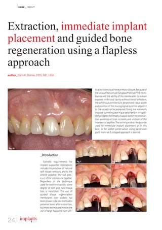
  • 1. I case _ report Extraction, immediate implant placement and guided bone regeneration using a flapless approach author_Barry K. Bartee, DDS, MD, USA tical incisions to achieve primary closure. Because of the unique features of Cytoplast® dense PTFE mem- branes and the ability of the membranes to remain exposed in the oral cavity without risk of infection, the soft tissue architecture, keratinized tissue width and position of the mucogingival junction adjacent to the socket can be preserved. Using the minimally invasive tunneling technique described in this arti- cle facilitates minimally invasive socket reconstruc- tion avoiding vertical incisions and incision of the interdental papillae. The technique described can be used for immediate implant placement, as in this case, or for socket preservation using particulate graft material if a staged approach is desired. Fig. 16 _Introduction Esthetic requirements for Fig. 1 Fig. 4 implant supported restorations include the presence of natural soft tissue contours, and to the extend possible, the full pres- ence of the interdental papillae. Regardless of the technique used for tooth extraction, some Fig. 2 Fig. 5 degree of soft and hard tissue loss is inevitable. The use of guided tissue regeneration membranes over sockets has been shown to be one method to preserve bone after extraction, but most techniques involve the Fig. 3 Fig. 6 use of large flaps and even ver- 24 I implants 3_ 2009
  • 2. case _ report I Fig. 7 Fig. 8 Fig. 9 Fig. 10 Fig. 11 Fig. 12 Fig. 13 Fig. 14 Fig. 15 _Case Presentation the interdental papillae, taking care to keep the edge of the material a minimum of 1.0 mm away from ad- This is a 60-year-old female who presented with jacent tooth roots (Fig. 12). A single 3-0 PTFE suture a crown-root fracture of a non-vital maxillary right is placed to further stabilize the membrane. The central incisor. The crown was temporarily stabi- membrane is intentionally left exposed, as primary lized with composite resin bonded to the adjacent closure is not required in this technique (Fig. 13). teeth (Fig. 1). Extraction of the tooth and immedi- Figure 14 shows the surgical site at three weeks. ate implant placement was planned. To minimize The exposed membrane is easily removed by grasp- soft and hard tissue recession, a flapless, minimally ing with a tissue forcep. Topical anesthesia may be invasive extraction technique was employed (Fig. used, but local anesthesia is not necessary. 2). The tooth root was extracted using only an in- The site at six weeks after implant placement trasulcular incision. A #15 blade was used to sever (three weeks after membrane removal), reveals ker- the periodontal ligament and create space for root atinized mucosa forming across the former extrac- luxation and elevation (Fig. 3). Next, a subperiosteal tion site (Fig. 15). pocket was created on the buccal and palatal aspect Figure 16 shows the clinical view following of the socket using a micro periosteal elevator (Fig. placement of the implant abutment and acrylic pro- 4). Following luxation and initial elevation of the visional restoration. root with the micro elevator, the tooth was removed with forceps (Fig. 5). The interdental papillae were _Summary carefully undermined and elevated. This can be done with a small periosteal elevator or curette (Fig. The flapless technique described provides a min- 6). All remaining soft tissue was removed from the imally invasive approach to extraction with socket interior and margins of the socket with a sharp grafting or immediate implant placement. Because curette (Fig. 7). The implant osteotomy was done in the interdental papilla remains intact, there is less the standard fashion, with the implant being placed disruption of blood supply. As a result, there is a _contact implants against the palatal wall of the socket (Fig. 8). The gap greater potential for maintenance of soft tissue vol- between the facial aspect of the implant and the ume. In addition, the use of a dense PTFE membrane buccal wall was filled with a combination of auto- Barry K. Bartee, DDS, PA improves the predictability of immediate implant genous bone chips harvested from the implant os- 3234 64th Street placement, excluding the requirement for primary teotomy combined with allograft bone (Fig. 9). A Lubbock,Texas 79413 closure and resultant disruption of soft tissue archi- textured, high-density PTFE barrier membrane is USA tecture._ placed. The membrane is trimmed, then placed into Phone: +1-806-792-0030 Fax: +1-806-792-8730 the superiosteal pocket on the palatal aspect (Fig. Cytoplast® is a registered trademark of Os- E-mail: dr2bkb@aol.com 10). The membrane is then tucked under the facial teogenics Biomedical, Inc. flap (Fig. 11). Next, the membrane is tucked under © 2008 Osteogenics Biomedical, Inc. BBFY0607 implants 3 _ 2009 I 25