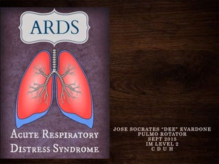
ARDS Acute Respiratory Syndrome
•Als PPTX, PDF herunterladen•
20 gefällt mir•1,657 views

ARDS, Acute Respiratory Syndrome, Wet Lung, Stiff Lung, patient, Cebu, CDUH, Evardone

Empfohlen
Weitere ähnliche Inhalte
Was ist angesagt?
Was ist angesagt? (20)
Andere mochten auch
Andere mochten auch (20)
Ähnlich wie ARDS Acute Respiratory Syndrome
Ähnlich wie ARDS Acute Respiratory Syndrome (20)
Kürzlich hochgeladen
Kürzlich hochgeladen (20)
ARDS Acute Respiratory Syndrome
- 1. JOSE SOCRATES “DEE” EVARDONE PULMO ROTATOR SEPT 2015 IM LEVEL 2 C D U H
- 2. ACTUAL CASE
- 3. Patient profile V.I. Filipino male, 48 years old, Married (-) DM (-) HPN NON-smoker Occasional alcohol drinker Prev hosp- 6/2015- acid related peptic disease,
- 5. HISTORY OF PRESENT ILLNESS • 10 years PTA, onset of intermittent abdominal pain, epigastric area mixed with burning and colicky • Admitted 4x due to this condition • no UTZ nor UGIE was done, managed as Acid related disease, takes Buscopan as his reliever • 1 day PTA, recurrence of the abdominal pain, peri- umbilical, epigastric and substernal in location, VAS of 9/10 • Px consulted a physician and was immediately referred here • (-) weight loss • (-) melena • (-) vomiting • (-) diarrhea • (-) fever
- 6. PE in the ER Conscious, coherent, NIRD Warm, good turgor and mobility, CRT <2 seconds, No rashes, Anicteric sclerae, pink palpebral conjunctivae Equal chest expansion, clear breath sounds Distinct heart sounds, normal rate, regular rhythm Flat, no scars/lesion, Hypoactive bowel sounds, soft, (+) tenderness on all quadrants, direct and indirect, no organomegaly, Strong peripheral pulses, no edema, Neurologic: Unremarkable BP: 110/80mmHg, HR: 73 bpm, RR: 20c Temp: 37.2°C GUT: (-) KPS
- 7. on admission IMPRESSION -Acute Abdomen probably 2 to Bleeding PUD DIAGNOSTICS 1. CBC, UA, CXR, Xray of the Abdomen, amylase, lipase, sgpt, TBDB THERAPEUTICS 1. Hydration with PNSS 2. PPI 3. NPO Differential -Cholelithiases -Cholecystitis -Acute Pancreatitis
- 8. CBC Adm Hgb 18.0 Hct 55 WBC Seg Bas Eos Lymph Mono 19,000 80 0 0 12 8 Platelet 120 MCV MCH MCHC 87.8 28.7 32.1 CHEM Amylase 2,450 Lipase 17,420 Creatinine 2.28 CrClear 33 ml/min LDH 4360 AST 237 ALT 55 Alk Phos 35 BUN 28 Potassium 5.1 Sodium 133 BUN/Crea 12.28 CHEM TB 1.2 DB 0.6 IB 0.6 Albumin 3.2 Globulin 2.7
- 10. 3rd HOSPITAL DAY
- 11. 4th HOSPITAL DAY
- 12. SOURCES
- 13. INTRODUCTION
- 14. • distinct type of hypoxemic respiratory failure • acute abnormality of both lungs • First recognized in the 1960s • Military clinicians in Vietnam called it Shock Lung • Civilian clinicians called it ADULT respiratory distress syndrome • Clinical hallmarks of ARDS are: – hypoxemia and – bilateral radiographic opacities • Pathological hallmark is diffuse alveolar damage • ARDS is an acute, diffuse, inflammatory lung injury that leads to: – increased pulmonary vascular permeability, – increased lung weight, and – a loss of aerated tissue
- 15. • 60 cases /100,1000 population • 10% of all ICU admissions involve acute respiratory failure, 20% of these meet ARDS criteria (2% in all ICU px)
- 24. DEFINITION
- 25. Acute Respiratory Distress Syndrome ARDS (clinical syndrome) severe dyspnea of rapid onset hypoxemia diffuse pulmonary infiltrates RESPIRATORY FAILURE Acute respiratory distress syndrome (ARDS) is a clinical syndrome of severe dyspnea of rapid onset, hypoxemia, and diffuse pulmonary infiltrates leading to respiratory failure.
- 26. ETIOLOGY
- 30. DIAGNOSTICS • Excluding cardiogenic pulmonary edema a. Brain natriuretic peptide (BNP) b. Echocardiography c. Right heart catheterization • Excluding other causes of hypoxemic respiratory failure a. Noninvasive respiratory sampling i. tracheobronchial aspiration or ii. mini-bronchoalveolar lavage (mini-BAL) b. Flexible bronchoscopy c. Lung biopsy a. The results of the biopsy resulted in the addition of specific therapy in 60 percent of patients b. withdrawal of unnecessary therapy in 37 percent
- 32. Acute Respiratory Distress Syndrome
- 34. Acute Respiratory Distress Syndrome Exudative Phase
- 35. A R D S
- 39. Acute Respiratory Distress Syndrome Exudative Phase plasma proteins aggregate hyaline membrane whorls Alveolar edema collapse intrapulmonary shunting and hypoxemia DYSPNEA Pulmonary Hypertension Hypercapnia within 12–36 h after the initial insult can be delayed by 5–7 days
- 41. Exudative Phase
- 45. Proliferative Phase • usually lasts from day 7 to day 21 • Most patients recover rapidly and are liberated from mechanical ventilation during this phase • Some patients develop progressive lung injury and early changes of pulmonary fibrosis • Histologically: 1. the initiation of lung repair, 2. the organization of alveolar exudates, and 3. a shift from a neutrophil- to a lymphocyte-predominant pulmonary infiltrate 4. type II pneumocytes proliferate along alveolar basement membranes • synthesize new pulmonary surfactant and • differentiate into type I pneumocytes
- 47. Fibrotic Phase • many patients with ARDS recover lung function 3–4 weeks • require long-term support on mechanical ventilators and/or supplemental oxygen • extensive alveolar-duct and interstitial fibrosis • emphysema-like changes, with large bullae • The physiologic consequences include – an increased risk of pneumothorax, – reductions in lung compliance, and – increased pulmonary dead space • substantial burden of excess morbidity • associated with increased mortality risk
- 48. Fibrotic Phase
- 50. TREATMENT
- 51. • Recent reductions in ARDS mortality rates are largely the result of general advances in the care of critically ill patients caringforthesepatients requirescloseattentionto the recognition and treatment of underlying medical and surgical disorders the minimization of procedures and their complications prophylaxis against venous thromboembolism, gastrointestinal bleeding, aspiration, excessive sedation, and central venous catheter infections prompt recognition of nosocomial infections provision of adequate nutrition
- 52. 1.MANAGEMENT OF MECHANICAL VENTILATION 2.FLUID MANAGEMENT 3.NEUROMUSCULAR BLOCKADE 4.GLUCOCORTICOIDS 5.OTHER THERAPIES
- 53. 1.MANAGEMENT OF MECHANICAL VENTILATION Ventilator-Induced Lung Injury • mechanical ventilation can aggravate lung injury • require two processes: 1. repeated alveolar overdistention 2. recurrent alveolar collapse • (6 mL/kg of predicted body weight) VS (12 mL/kg predicted body weight) • Mortality rate 31% vs 40% This improvement in survival represents the most substantial ARDS-mortality benefit that has been demonstrated for any therapeutic intervention to date
- 54. 1. MANAGEMENT OF MECHANICAL VENTILATION Prevention of Alveolar Collapse • the presence of alveolar and interstitial fluid and the loss of surfactant can lead to a marked reduction of lung compliance • Without an increase in end-expiratory pressure, significant alveolar collapse can occur at end-expiration • 12–15 mmHg in ARDS, is a theoretical “optimal PEEP” for alveolar recruitment • Keep the lung open • Three large randomized trials have investigated the utility of PEEP-based strategies to keep the lung open, improvement in lung function was evident but overall mortality rates were not altered significantly
- 55. A R D S
- 56. 1. MANAGEMENT OF MECHANICAL VENTILATION Prevention of Alveolar Collapse • Until more data become available on the clinical utility of high PEEP, it is advisable to set PEEP to minimize Fio2 and optimize Pao2 • inverse-ratio ventilation (I:E >1:1) –can improve oxygenation and –can help reduce Fio2 to ≤0.60 –no benefit in ARDS mortality risk PRONE POSITIONING: • improved arterial oxygenation • hazardous
- 57. A R D S
- 58. Permissive Hypercapnia and Tracheal Gas Insufflation • The use of lower VTs (to avoid VILI) often results in respiratory acidemia, an effect that has been termed “permissive hypercapnia” • The theoretical detrimental effects of hypercapnia include: – myocardial depression, – increased pulmonary vascular resistance, and – decreased renal blood flow • most clinically important adverse effect of hypercapnia is elevated intracranial pressure from increased cerebral blood flow • data suggesting that hypercapnia has protective effects, including attenuation of free radical–mediated lung injury and pulmonary inflammation • TGI is a technique whereby a gas flow is introduced via a small catheter placed in the endotracheal tube, with its tip near the carina. • TGI has been proposed as an adjunct to permissive hypercapnia, as the insufflated gas improves CO2 clearance from the anatomic dead space and ventilator tubing. A R D S
- 61. 2. FLUID MANAGEMENT • Increased pulmonary vascular permeability leading to interstitial and alveolar edema fluid rich in protein Low left atrial filling pressure: 1. minimizes pulmonary edema and prevents further decrements in arterial oxygenation and lung compliance; 2. improves pulmonary mechanics; shortens ICU stay and the duration of mechanical ventilation; and 3. is associated with a lower mortality rate in both medical and surgical ICU patients • aggressive attempts to reduce left atrial filling pressures with fluid restriction and diuretics
- 62. 3. NEUROMUSCULAR BLOCKADE •In severe ARDS, sedation alone can be inadequate for the patient ventilator synchrony •Cisatracurium besylate for 48h •blockade increased the rate of survival and ventilator-free days without increasing ICU- acquired paresis •however, these results must be replicated prior to their widespread application in clinical practice
- 63. 4. GLUCOCORTICOIDS •Few studies have shown any benefit. •Current evidence does not support the use of high-dose glucocorticoids in the care of ARDS patients.
- 64. •ANTIOXIDANTS •GRANULOCYTE-MONOCYTE COLONY STIMULATING FACTOR •INHALED VASODILATORS –Nitric oxide –Prostacyclin •ANTI-INFLAMMATORY THERAPIES –Glucocorticoids –Macrolide antibiotics •BETA AGONISTS •MESENCHYMAL STEM CELLS Novel therapies
- 65. 1. N-acetylcysteine 2. Procysteine (L-2-oxothiazolidine-4-carboxylate) 3. Glutamine 4. Antioxidant preparations (selenium, beta carotene, zinc, vitamin E and C) 5. Lisophylline 6. Intravenous prostaglandin E1 7. Neutrophil elastase inhibitors 8. Ibuprofen 9. Activated protein C 10. Ketoconazole 11. Statins 12. Surfactant INEFFECTIVE OR HARMFUL THERAPIES
- 69. • Recent mortality estimates for ARDS range from 26% to 44% • mortality in ARDS is largely attributable to nonpulmonary causes, with sepsis and nonpulmonary organ failure accounting for >80% of deaths • The major risk factors for ARDS mortality are nonpulmonary – Age – Preexisting organ dysfunction from chronic medical illness 1. chronic liver disease, 2. cirrhosis, 3. chronic alcohol abuse, 4. chronic immunosuppression, 5. sepsis, 6. chronic renal disease, 7. failure of any nonpulmonary organ, and 8. increased APACHE III scores MORTALITY
- 70. • direct lung injury – nearly twice as likely to die vs indirect • Predicted mortality risks: 1. An early (within 24 h of presentation) elevation in pulmonary dead space (>0.60) 2. Severe arterial hypoxemia (Pao2/Fio2, <100 mmHg) • little additional value in predicting ARDS mortality rom other measures of the severity of lung injury: a. the level of PEEP (≥10 cm H2O), b. respiratory system compliance (≤40 mL/cm H2O), c. the extent of alveolar infiltrates on chest radiography, and d. the corrected expired volume per minute (≥10 L/min) MORTALITY
- 71. • majority of patients recover nearly normal lung function • usually recover maximal lung function within 6 months • One year after endotracheal extubation, >1/3 will have a normal Spirometry values and diffusion capacity • Unlike mortality risk, recovery of lung function is strongly associated with the extent of lung injury in early ARDS • Associated with less recovery of pulmonary function: 1. Low static respiratory compliance, 2. high levels of required PEEP, 3. longer durations of mechanical ventilation, and 4. high lung injury scores Physical function after 5 years – despite normal lung function 1. Exercise limitation 2. Decreased physical quality of life Functional Recovery in ARDS Survivors
- 72. KEYPOINTS defined by acute hypoxemia, bilateral infiltrates on chest radiograph, and the absence of left atrial hypertension Direct and Indirect Lung injury The pathogenesis of ARDS is not clear, but increased permeability of the alveolar-capillary membrane due to neutrophilic infiltration of the lungs is thought to be an important factor ARDS is a syndrome, not a specific diagnosis; clinicians must, therefore, look for the cause in order to institute specific therapy The mortality rate for ARDS has fallen since the 1980s and is now about 40%. Mechanical ventilation is lifesaving but, when inappropriately applied, can induce or aggravate lung injury Most deaths in patients with ARDS are from multiorgan failure rather than hypoxia per se. No pharmacologic therapies have been shown to improve survival in ARDS; Many survivors of ARDS suffer from a reduced quality of life and cognitive impairment
- 73. 4th HOSPITAL DAY
- 74. 6th HOSPITAL DAY
- 76. THANK YOU
Hinweis der Redaktion
- Impression was Dengue fever since the patient has nospecific fever and nonproductive cough Pending extensive workup wasn’t taken for it may distort the results
- A distinct type of hypoxemic respiratory failure characterized by acute abnormality of both lungs was first recognized during the 1960s. Military clinicians working in surgical hospitals in Vietnam called it shock lung, while civilian clinicians referred to it as adult respiratory distress syndrome [1]. Subsequent recognition that individuals of any age could be afflicted led to the current term, acute respiratory distress syndrome (ARDS). ARDS is an acute, diffuse, inflammatory lung injury that leads to increased pulmonary vascular permeability, increased lung weight, and a loss of aerated tissue [2]. Clinical hallmarks of ARDS are hypoxemia and bilateral radiographic opacities, while the pathological hallmark is diffuse alveolar damage (ie, alveolar edema with or without focal hemorrhage, acute inflammation of the alveolar walls, and hyaline membranes). ARDS is associated with a variety of risk factors and etiologies. These conditions are grouped together under the term ARDS because the clinical, physiological features, pathological features, and management are similar regardless of the inciting event.
- The annual incidence of ARDS is estimated to be as high as 60 cases/100,000 population. Approximately 10% of all intensive care unit (ICU) admissions involve patients with acute respiratory failure; ~20% of these patients meet the criteria for ARDS
- Annual pooled weighted mortality from 1981 to 2006 for all included studies, by year of conduct. The solid points show data for observational studies; the open points show data for randomized controlled trials. Upper and lower confidence intervals are shown for observational studies and randomized controlled trials, respectively. Interpolation lines are included to facilitate visual interpretation and do not indicate trends between point estimates.
- Respiratory Unit. Figure 39-7 shows the respiratory unit (also called “respiratory lobule”), which is composed of a respiratory bronchiole, alveolar ducts, atria, and alveoli. There are about 300 million alveoli in the two lungs, and each alveolus has an average diameter of about 0.2 millimeter.
- The alveolar walls are extremely thin, and between the alveoli is an almost solid network of interconnecting capillaries, shown in Figure 39-8. Indeed, because of the extensiveness of the capillary plexus, the flow of blood in the alveolar wall has been described as a “sheet” of flowing blood. Thus, it is obvious that the alveolar gases are in very close proximity to the blood of the pulmonary capillaries. Further, gas exchange between the alveolar air and the pulmonary blood occurs through the membranes of all the terminal portions of the lungs, not merely in the alveoli themselves. All these membranes are collectively known as the respiratory membrane, also called the pulmonary membrane.
- Figure 39-8 A, Surface view of capillaries in an alveolar wall. B, Cross-sectional view of alveolar walls and their vascular supply.
- Type I pneumocyte Type I pneumocytes (or squamous alveolar cells) cover approximately 90–95% of the alveolar surface, and are involved in the gas exchange between the alveoli and blood. Type II pneumocyte Type II pneumocytes cover a minority of the alveolar surface. Their function is to secrete surfactant, which decreases surface tension within the alveoli. They are also capable of cellular division, giving rise to more Type I pneumocytes when the lung tissue is damaged.
- Note the following different layers of the respiratory membrane: 1. A layer of fluid lining the alveolus and containing surfactant that reduces the surface tension of the alveolar fluid 2. The alveolar epithelium composed of thin epithelial cells 3. An epithelial basement membrane 4. A thin interstitial space between the alveolar epithelium and the capillary membrane 5. A capillary basement membrane that in many places fuses with the alveolar epithelial basement membrane 6. The capillary endothelial membrane
- An osmotic gradient favoring fluid reabsorption from the interstitium is maintained by retention of serum proteins within the intravascular space. Fluid which does leak into the interstitium is transported to lymphatics from which it is returned to the circulation. Finally, tight junctions between alveolar epithelial cells prevent leakage of fluid into the alveolar space. Arrows represent lymphatic movement; small circles represent protein.
- Acute respiratory distress syndrome (ARDS) is a clinical syndrome of severe dyspnea of rapid onset, hypoxemia, and diffuse pulmonary infiltrates leading to respiratory failure. ARDS is caused by diffuse lung injury from many underlying medical and surgical disorders. The lung injury may be direct, as occurs in toxic inhalation, or indirect, as occurs in sepsis (Table 322-1).
- While many medical and surgical illnesses have been associated with the development of ARDS, most cases (>80%) are caused by a relatively small number of clinical disorders: severe sepsis syndrome and/or bacterial pneumonia (~40–50%), trauma, multiple transfusions, aspiration of gastric contents, and drug overdose. Among patients with trauma, the most frequently reported surgical conditions in ARDS are Pulmonary contusion, multiple bone fractures, and chest wall trauma/flail chest, whereas head trauma, near-drowning, toxic inhalation, and burns are rare causes. The risks of developing ARDS are increased in patients with more than one predisposing medical or surgical condition.
- By expert consensus, ARDS is defined by three categories based on the degrees of hypoxemia (Table 322-2). These stages of mild, moderate, and severe ARDS are associated with mortality risk and with the duration of mechanical ventilation in survivors.
- Excluding cardiogenic pulmonary edema — An absence of cardiac exam abnormalities (eg, an S3 or S4 gallop, new or changed murmur), elevated right-sided filling pressures (eg, elevated jugular venous pressure), and certain radiographic abnormalities (eg, pulmonary venous congestion, Kerley B lines, cardiomegaly, and pleural effusions), helps distinguish ARDS from cardiogenic pulmonary edema. Several additional diagnostic tests may also be helpful, including measurement of plasma brain natriuretic peptide levels, echocardiography, and right heart catheterization: ●Brain natriuretic peptide (BNP) – A plasma BNP level below 100 pg/mL favors ARDS, but higher levels neither confirm heart failure nor exclude ARDS [27,28]. This derives from an observational study of patients with ARDS (n = 33) or cardiogenic pulmonary edema (n = 21) [27]. ●Echocardiography – Many clinicians use transthoracic echocardiography as the first-line diagnostic test if cardiogenic pulmonary edema cannot be excluded by clinical evaluation and measurement of the BNP level. While severe aortic or mitral valve dysfunction, severe diastolic dysfunction, or a severely reduced left ventricular ejection fraction favors cardiogenic pulmonary edema, the latter is insufficient to confirm primary cardiogenic pulmonary edema because some precipitants of ARDS (eg, septic shock) can cause an acute, severe cardiomyopathy that develops concomitantly with ARDS [29,30]. In addition, cardiogenic pulmonary edema cannot be excluded on the basis of an echocardiogram, since diastolic dysfunction and volume overload may exist even if the left heart function appears normal. ●Right heart catheterization – There is ample evidence that there is generally no value to routine right heart catheterization for either the diagnosis or management of ARDS [31,32]. However, pulmonary artery catheterization may be considered if primary cardiogenic pulmonary edema cannot be excluded on the basis of the clinical evaluation, plasma BNP measurement, and echocardiogram. Excluding other causes of hypoxemic respiratory failure — Potentially treatable causes of ARDS and alternative forms of acute hypoxemic respiratory failure with bilateral infiltrates should be considered once cardiogenic pulmonary edema has been excluded. If such conditions cannot be identified on the basis of the clinical context and accompanying symptoms and signs, additional diagnostic testing should be performed: ●Noninvasive respiratory sampling – The lower respiratory tract can be sampled via tracheobronchial aspiration or mini-bronchoalveolar lavage (mini-BAL). Tracheobronchial aspiration is performed by advancing a catheter through the endotracheal tube until resistance is met and then applying suction, while mini-BAL is performed by advancing a catheter through the endotracheal tube until resistance is met, infusing sterile saline through the catheter, and then aspirating. Regardless of the technique, the specimen that is obtained may be evaluated via microscopic analysis (eg, Gram stain, cytology) and microbiologic culture; these studies may identify pneumonia or rapidly progressive cancer as the correct diagnosis. ●Flexible bronchoscopy – Flexible bronchoscopy can obtain lower respiratory samples for microscopic analysis and microbiologic culture if the noninvasive techniques are unsuccessful. It can also identify abnormalities that may not be detected with noninvasive sampling. Therefore, flexible bronchoscopy is a reasonable next step whenever noninvasive sampling is nondiagnostic. ●Lung biopsy – Surgical lung biopsy may be considered when alternative causes of acute hypoxemic respiratory failure cannot be excluded on the basis of the clinical context, symptoms, signs, and bronchoscopy [33,34]. The safety of lung biopsy in selected patients with hypoxemic respiratory failure was demonstrated by a retrospective study of 57 patients with ARDS who underwent open lung biopsy [33]. The patients had a mean ratio of arterial oxygen tension to fraction of inspired oxygen (PaO2/FiO2) of 145 mmHg and the rate of major complications was 7 percent, with no deaths attributed to the biopsy. Although the complication rate was 39 percent, most were tolerable (eg, persistent air leaks). The results of the biopsy resulted in the addition of specific therapy in 60 percent of patients and the withdrawal of unnecessary therapy in 37 percent. Consider the following examples of findings that suggest a specific etiology for acute hypoxemic respiratory failure. Frothy bloody secretions throughout the airways, increasing red blood cells in serial bronchoalveolar lavage (BAL) specimens, and hemosiderin-laden macrophages in the BAL fluid suggest diffuse alveolar hemorrhage. A large number of eosinophils in the BAL fluid suggest idiopathic acute eosinophilic pneumonia. And, recovery of lipid-laden macrophages or recognizable food particles suggest aspiration pneumonitis.
- The natural history of ARDS is marked by three phases—exudative, proliferative, and fibrotic—that each have characteristic clinical and pathologic features (Fig. 322-1). Diagram illustrating the time course for the development and resolution of ARDS. The exudative phase is notable for early alveolar edema and neutrophil-rich leukocytic infiltration of the lungs, with subsequent formation of hyaline membranes from diffuse alveolar damage. Within 7 days, a proliferative phase ensues with prominent interstitial inflammation and early fibrotic changes. Approximately 3 weeks after the initial pulmonary injury, most patients recover. However, some patients enter the fibrotic phase, with substantial fibrosis and bullae formation.
- Schematic representation of the time course of the acute respiratory distress syndrome (ARDS). During the early (or exudative) phase, the lesion is characterized by high permeability pulmonary edema followed by the formation of hyaline membranes. After seven to ten days, a proliferative phase may develop, with marked interstitial inflammation, fibrosis, and disordered healing. Redrawn from Katzenstein AA, Askin FB. Surgical Pathology of Non-neoplastic Lung Disease. Saunders, Philadelphia, 1982. Graphic 77051 Version 2.0
- In this phase (Fig. 322-2), alveolar capillary endothelial cells and type I pneumocytes (alveolar epithelial cells) are injured, with consequent loss of the normally tight alveolar barrier to fluid and macromolecules. Edema fluid that is rich in protein accumulates in the interstitial and alveolar spaces. Significant concentrations of cytokines (e.g., interleukin 1, interleukin 8, and tumor necrosis factor α) and lipid mediators (e.g., leukotriene B4) are present in the lung in this acute phase. In response to proinflammatory mediators, leukocytes (especially neutrophils) traffic into the pulmonary interstitium and alveoli.
- Activated neutrophils exit the bloodstream and transmigrate across thevalveolar-capillary membrane, releasing cytokines, proteases, reactive oxygen species, and othervcompounds. Although crucial to host defensevagainst pathogens, the compounds secreted or released by the neutrophil have the capacity to damage the tissue of the host.
- Breakdown of the capillary endothelial barrier allows leakage of serum proteins into the interstitial space, undoing the osmotic gradient which normally promotes fluid reabsorption. Arrows represent lymphatic movement; small circles represent protein.
- The quantity of fluid pouring from the capillaries overwhelms the capacity of the interstitium and the lymphatics, resulting in interstitial edema. Arrows represent lymphatic movement; small circles represent protein.
- Breakdown of the alveolar epithelial barrier allows leakage of edema fluid into the alveolar space. Arrows represent lymphatic movement; small circles represent protein.
- In addition, condensed plasma proteins aggregate in the air spaces with cellular debris and dysfunctional pulmonary surfactant to form hyaline membrane whorls. Pulmonary vascular injury also occurs early in ARDS, with vascular obliteration by microthrombi and fibrocellular proliferation (Fig. 322-3). Alveolar edema predominantly involves dependent portions of the lung, with diminished aeration and atelectasis. Collapse of large sections of dependent lung markedly decreases lung compliance. Consequently, intrapulmonary shunting and hypoxemia develop and the work of breathing increases, leading to dyspnea. The pathophysiologic alterations in alveolar spaces are exacerbated by microvascular occlusion that results in reductions in pulmonary arterial blood flow to ventilated portions of the lung (and thus in increased dead space) and in pulmonary hypertension. Thus, in addition to severe hypoxemia, hypercapnia secondary to an increase in pulmonary dead space is prominent in early ARDS. The exudative phase encompasses the first 7 days of illness after exposure to a precipitating ARDS risk factor, with the patient experiencing the onset of respiratory symptoms. Although usually presenting within 12–36 h after the initial insult, symptoms can be delayed by 5–7 days. Dyspnea develops, with a sensation of rapid shallow breathing and an inability to get enough air. Tachypnea and increased work of breathing result frequently in respiratory fatigue and ultimately in respiratory failure. Laboratory values are generally nonspecific and are primarily indicative of underlying clinical disorders. The chest radiograph usually reveals alveolar and interstitial opacities involving at least three-quarters of the lung fields. While characteristic for ARDS, these radiographic findings are not specific and can be indistinguishable from cardiogenic pulmonary edema (Chap. 326). Unlike the latter, however, the chest x-ray in ARDS rarely shows cardiomegaly, pleural effusions, or pulmonary vascular redistribution. Chest CT in ARDS reveals extensive heterogeneity of lung involvement Because the early features of ARDS are nonspecific, alternative diagnoses must be considered. In the differential diagnosis of ARDS, the most common disorders are cardiogenic pulmonary edema, diffusepneumonia, and alveolar hemorrhage. Less common diagnoses to consider include acute interstitial lung diseases (e.g., acute interstitial pneumonitis; Chap. 315), acute immunologic injury (e.g., hypersensitivity pneumonitis; Chap. 310), toxin injury (e.g., radiation pneumonitis; Chap. 263), and neurogenic pulmonary edema (Chap. 47e).
- A representative anteroposterior chest x-ray in the exudative phase of ARDS shows diffuse interstitial and alveolar infiltrates that can be difficult to distinguish from left ventricular failure.
- This phase of ARDS usually lasts from day 7 to day 21. Most patients recover rapidly and are liberated from mechanical ventilation during this phase. Despite this improvement, many patients still experience dyspnea, tachypnea, and hypoxemia. Some patients develop progressive lung injury and early changes of pulmonary fibrosis during the proliferative phase. Histologically, the first signs of resolution are often evident in this phase, with the initiation of lung repair, the organization of alveolar exudates, and a shift from a neutrophil- to a lymphocyte-predominant pulmonary infiltrate. As part of the reparative process, type II pneumocytes proliferate along alveolar basement membranes. These specialized epithelial cells synthesize new pulmonary surfactant and differentiate into type I pneumocytes
- While many patients with ARDS recover lung function 3–4 weeks after the initial pulmonary injury, some enter a fibrotic phase that may require long-term support on mechanical ventilators and/or supplemental oxygen. Histologically, the alveolar edema and inflammatory exudates of earlier phases are now converted to extensive alveolar-duct and interstitial fibrosis. Marked disruption of acinar architecture leads to emphysema-like changes, with large bullae. Intimal fibroproliferation in the pulmonary microcirculation causes progressive vascular occlusion and pulmonary hypertension. The physiologic consequences include an increased risk of pneumothorax, reductions in lung compliance, and increased pulmonary dead space. Patients in this late phase experience a substantial burden of excess morbidity. Lung biopsy evidence for pulmonary fibrosis in any phase of ARDS is associated with increased mortality risk.
- The normal alveolus (left) and the injured alveolus in the acute phase of acute lung injury and the acute respiratory distress syndrome (right). In the acute phase of the syndrome (right), there is sloughing of both the bronchial and alveolar epithelial cells, with the formation of protein-rich hyaline membranes on the denuded basement membrane. Neutrophils are shown adhering to the injured capillary endothelium and transmigrating through the interstitium into the air space, which is filled with protein-rich edema fluid. In the air space, an alveolar macrophage is secreting cytokines—i.e., interleukins 1, 6, 8, and 10 (IL-1, -6, -8, and -10) and tumor necrosis factor α (TNF-α)—that act locally to stimulate chemotaxis and activate neutrophils. Macrophages also secrete other cytokines, including IL-1, -6, and -10. IL-1 can also stimulate the production of extracellular matrix by fibroblasts. Neutrophils can release oxidants, proteases, leukotrienes, and other proinflammatory molecules, such as platelet-activating factor (PAF). A number of antiinflammatory mediators are also present in the alveolar milieu, including the IL-1-receptor antagonist, soluble TNF-α receptor, autoantibodies to IL-8, and cytokines such as IL-10 and IL-11 (not shown). The influx of protein rich edema fluid into the alveolus has led to the inactivation of surfactant. MIF, macrophage inhibitory factor.
- GENERAL PRINCIPLES Recent reductions in ARDS mortality rates are largely the result of general advances in the care of critically ill patients Thus, caring for these patients requires close attention to: (1) the recognition and treatment of underlying medical and surgical disorders (e.g., sepsis, aspiration, trauma); (2) the minimization of procedures and their complications; (3) prophylaxis against venous thromboembolism, gastrointestinal bleeding, aspiration, excessive sedation, and central venous catheter infections; (4) prompt recognition of nosocomial infections; and (5) provision of adequate nutrition
- Ventilator-Induced Lung Injury Despite its life-saving potential, mechanical ventilation can aggravate lung injury. Experimental models have demonstrated that ventilator-induced lung injury appears to require two processes: repeated alveolar overdistention and recurrent alveolar collapse. As is clearly evident from chest CT ARDS is a heterogeneous disorder, principally involving dependent portions of the lung with relative sparing of other regions. Because compliance differs in affected versus more “normal” areas of the lung, attempts to fully inflate the consolidated lung may lead to overdistention of and injury to the more normal areas. Ventilator-induced injury can be demonstrated in experimental models of acute lung injury, with high-tidal-volume (VT) ventilation resulting in additional, synergistic alveolar damage. A large-scale, randomized controlled trial sponsored by the National Institutes of Health and conducted by the ARDS Network compared low VT ventilation (6 mL/kg of predicted body weight) to conventional VT ventilation (12 mL/kg predicted body weight). The mortality rate was significantly lower in the low VT patients (31%) than in the conventional VT patients (40%). This improvement in survival represents the most substantial ARDS-mortality benefit that has been demonstrated for any therapeutic intervention to date.
- Prevention of Alveolar Collapse In ARDS, the presence of alveolar and interstitial fluid and the loss of surfactant can lead to a marked reduction of lung compliance. Without an increase in end-expiratory pressure, significant alveolar collapse can occur at end-expiration, with consequent impairment of oxygenation. In most clinical settings, positive end-expiratory pressure (PEEP) is empirically set to minimize Fio2 (inspired O2 percentage) and maximize Pao2 (arterial partial pressure of O2). On most modern mechanical ventilators, it is possible to construct a static pressure–volume curve for the respiratory system. The lower inflection point on the curve represents alveolar opening (or “recruitment”). The pressure at this point, usually 12–15 mmHg in ARDS, is a theoretical “optimal PEEP” for alveolar recruitment. Titration of the PEEP to the lower inflection point on the static pressure–volume curve has been hypothesized to keep the lung open, improving oxygenation and protecting against lung injury. Three large randomized trials have investigated the utility of PEEP-based strategies to keep the lung open. In all three trials, improvement in lung function was evident but overall mortality rates were not altered significantly. Until more data become available on the clinical utility of high PEEP, it is advisable to set PEEP to minimize Fio2 and optimize Pao2 Measurement of esophageal pressures to estimate transpulmonary pressure may help identify an optimal PEEP in some cases. Oxygenation can also be improved by increasing mean airway pressure with inverse-ratio ventilation. In this technique, the inspiratory time (I) is lengthened so that it is longer than the expiratory time (E)— that is, I:E > 1:1. With diminished time to exhale, dynamic hyperinflation leads to increased end-expiratory pressure, similar to ventilator-prescribed PEEP. This mode of ventilation has the advantage of improving oxygenation with lower peak pressures than are required for conventional ventilation. Although inverse-ratio ventilation can improve oxygenation and can help reduce Fio2 to ≤0.60, thus avoiding possible oxygen toxicity, no benefit in ARDS mortality risk has been demonstrated. Recruitment maneuvers that transiently increase PEEP to “recruit” atelectatic lung can also increase oxygenation, but a mortality benefit has not been established. In several randomized trials, mechanical ventilation in the prone position improved arterial oxygenation, but its effect on survival and other important clinical outcomes remains uncertain. Moreover, unless the critical-care team is experienced in “proning,” repositioning critically ill patients can be hazardous, leading to accidental endotracheal extubation, loss of central venous catheters, and orthopedic injury.
- FIGURE 90-5 n Potential mechanisms by which mechanical ventilation might cause or contribute to multisystem organ failure (MSOF). mΦ, macrophage.
- Until more data become available on the clinical utility of high PEEP, it is advisable to set PEEP to minimize Fio2 and optimize Pao2 Measurement of esophageal pressures to estimate transpulmonary pressure may help identify an optimal PEEP in some cases. Oxygenation can also be improved by increasing mean airway pressure with inverse-ratio ventilation. In this technique, the inspiratory time (I) is lengthened so that it is longer than the expiratory time (E)— that is, I:E > 1:1. With diminished time to exhale, dynamic hyperinflation leads to increased end-expiratory pressure, similar to ventilator-prescribed PEEP. This mode of ventilation has the advantage of improving oxygenation with lower peak pressures than are required for conventional ventilation. Although inverse-ratio ventilation can improve oxygenation and can help reduce Fio2 to ≤0.60, thus avoiding possible oxygen toxicity, no benefit in ARDS mortality risk has been demonstrated. Recruitment maneuvers that transiently increase PEEP to “recruit” atelectatic lung can also increase oxygenation, but a mortality benefit has not been established. In several randomized trials, mechanical ventilation in the prone position improved arterial oxygenation, but its effect on survival and other important clinical outcomes remains uncertain. Moreover, unless the critical-care team is experienced in “proning,” repositioning critically ill patients can be hazardous, leading to accidental endotracheal extubation, loss of central venous catheters, and orthopedic injury.
- *See text for formula to calculate predicted body weight. FIO2, fractional concentration of oxygen in inspired gas; I : E ratio, ratio of inspired to expired gas; PaO2, arterial oxygen pressure; PEEP, positive endexpiratory pressure; SpO2, pulse oximetry. Data from Anonymous: Ventilation with lower tidal volumes as compared with traditional tidal volumes for acute lung injury and the acute respiratory distress syndrome. The Acute Respiratory Distress Syndrome Network (comment). N Engl J Med 342:1301–1308, 2000.
- Permissive Hypercapnia and Tracheal Gas Insufflation The use of lower VTs (to avoid VILI) often results in respiratory acidemia, an effect that has been termed “permissive hypercapnia.” The theoretical detrimental effects of hypercapnia include myocardial depression, increased pulmonary vascular resistance, and decreased renal blood flow. Perhaps the most clinically important adverse effect of hypercapnia is elevated intracranial pressure from increased cerebral blood flow. However, there are data suggesting that hypercapnia has protective effects, including attenuation of free radical–mediated lung injury and pulmonary inflammation; other studies have reported an injurious effect on alveolar epithelial cells.200 Given the uncertainty about permissive hypercapnia, it is worth remembering that the only large randomized, controlled trial to show a reduction in mortality in ARDS treated respiratory acidosis aggressively. In the absence of other data, following the protocol used in that study seems prudent. One approach that has been used to decrease high levels of arterial carbon dioxide pressure (PaCO2) is tracheal gas insufflation (TGI). TGI is a technique whereby a gas flow is introduced via a small catheter placed in the endotracheal tube, with its tip near the carina. TGI has been proposed as an adjunct to permissive hypercapnia, as the insufflated gas improves CO2 clearance from the anatomic dead space and ventilator tubing. However, the gas flow has the potential to increase alveolar volume and pressure and may increase PEEP.201 Although many case reports describe the use of TGI,202 there have been no randomized studies of TGI in patients with ARDS. Because of technical and monitoring issues related to TGI, routine use of TGI in ARDS is not recommended.
- Increased pulmonary vascular permeability leading to interstitial and alveolar edema fluid rich in protein is a central feature of ARDS. In addition, impaired vascular integrity augments the normal increase in extravascular lung water that occurs with increasing left atrial pressure. Maintaining a low left atrial filling pressure minimizes pulmonary edema and prevents further decrements in arterial oxygenation and lung compliance; improves pulmonary mechanics; shortens ICU stay and the duration of mechanical ventilation; and is associated with a lower mortality rate in both medical and surgical ICU patients. Thus, aggressive attempts to reduce left atrial filling pressures with fluid restriction and diuretics should be an important aspect of ARDS management, limited only by hypotension and hypoperfusion of critical organs such as the kidneys.
- In severe ARDS, sedation alone can be inadequate for the patient ventilator synchrony required for lung-protective ventilation. This clinical problem was recently addressed in a multicenter, randomized, placebo-controlled trial of early neuromuscular blockade (with cisatracurium besylate) for 48 h. In severe ARDS, early neuromuscular blockade increased the rate of survival and ventilator-free days without increasing ICU-acquired paresis. These promising findings support the early administration of neuromuscular blockade if needed to facilitate mechanical ventilation in severe ARDS; however, these results must be replicated prior to their widespread application in clinical practice. Cisatracurium (formerly recognized as 51W89,[1] and marketed as Nimbex) is a neuromuscular-blocking drug or skeletal muscle relaxant in the category of non-depolarizing neuromuscular-blocking drugs, used adjunctively in anesthesia to facilitate endotracheal intubation and to provide skeletal muscle relaxation duringsurgery or mechanical ventilation. It is a bisbenzyltetrahydroisoquinolinium agent with an intermediate duration of action. Cisatracurium is one of the ten isomers of the parent molecule, atracurium.[2] Moreover, cisatracurium represents approximately 15% of the atracurium mixture
- Many attempts have been made to treat both early and late ARDS with glucocorticoids, with the goal of reducing potentially deleterious pulmonary inflammation. Few studies have shown any benefit. Current evidence does not support the use of high-dose glucocorticoids in the care of ARDS patients.
- A, recommended therapy based on strong clinical evidence from randomized clinical trials; B, recommended therapy based on supportive but limited clinical data; C,recommended only as alternative therapy on the basis of indeterminate evidence; D, not recommended on the basis of clinical evidence against efficacy of therapy. Abbreviations: ARDS, acute respiratory distress syndrome; ECMO, extracorporeal membrane oxygenation; NO, nitric oxide; NSAIDs, nonsteroidal anti-inflammatory drugs; PEEP, positive end-expiratory pressure; PGE1, prostaglandin E1.
- A, recommended therapy based on strong clinical evidence from randomized clinical trials; B, recommended therapy based on supportive but limited clinical data; C, recommended only as alternative therapy on the basis of indeterminate evidence; D, not recommended on the basis of clinical evidence against efficacy of therapy. Abbreviations: ARDS, acute respiratory distress syndrome; ECMO, extracorporeal membrane oxygenation; NO, nitric oxide; NSAIDs, nonsteroidal anti-inflammatory drugs; PEEP, positive end-expiratory pressure; PGE1, prostaglandin E1.
- Recent mortality estimates for ARDS range from 26% to 44%. There is substantial variability, but a trend toward improved ARDS outcomes appears evident. Of interest, mortality in ARDS is largely attributable to nonpulmonary causes, with sepsis and nonpulmonary organ failure accounting for >80% of deaths. Thus, improvement in survival is likely secondary to advances in the care of septic/infected patients and those with multiple organ failure The major risk factors for ARDS mortality are nonpulmonary. Advanced age is an important risk factor. Patients >75 years of age have a substantially higher mortality risk (~60%) than those <45 (~20%). Moreover, patients >60 years of age with ARDS and sepsis have a threefold higher mortality risk than those <60. Other risk factors include preexisting organ dysfunction from chronic medical illness—in particular, chronic liver disease, cirrhosis, chronic alcohol abuse, chronic immunosuppression, sepsis, chronic renal disease, failure of any nonpulmonary organ, and increased APACHE III scores
- Patients with ARDS arising from direct lung injury (including pneumonia, pulmonary contusion, and aspiration Table 322-1) are nearly twice as likely to die as those with indirect causes of lung injury, while surgical and trauma patients with ARDS— especially those without direct lung injury—have a higher survival rate than other ARDS patients. An early (within 24 h of presentation) elevation in pulmonary dead space (>0.60) and severe arterial hypoxemia (Pao2/Fio2, <100 mmHg) predict increased mortality risk from ARDS; however, there is surprisingly little additional value in predicting ARDS mortality from other measures of the severity of lung injury, including the level of PEEP (≥10 cm H2O), respiratory system compliance (≤40 mL/cm H2O), the extent of alveolar infiltrates on chest radiography, and the corrected expired volume per minute (≥10 L/min).
- While it is common for patients with ARDS to experience prolonged respiratory failure and remain dependent on mechanical ventilation for survival, it is a testament to the resolving powers of the lung that the majority of patients recover nearly normal lung function. Patients usually recover maximal lung function within 6 months. One year after endotracheal extubation, more than one-third of ARDS survivors have normal spirometry values and diffusion capacity. Most of the remaining patients have only mild abnormalities in pulmonary function. Unlike mortality risk, recovery of lung function is strongly associated with the extent of lung injury in early ARDS. Low static respiratory compliance, high levels of required PEEP, longer durations of mechanical ventilation, and high lung injury scores are all associated with less recovery of pulmonary function. Of note, when physical function is assessed 5 years after ARDS, exercise limitation and decreased physical quality of life are often documented despite normal or nearly normal pulmonary function. When caring for ARDS survivors, it is important to be aware of the potential for a substantial burden of psychological problems in patients and family caregivers, including significant rates of depression and posttraumatic stress disorder.
- KEY POINTS • ARDS is a syndrome defined by acute hypoxemia, bilateral infiltrates on chest radiograph, and the absence of left atrial hypertension. • ARDS has been associated with diseases that directly involve the lungs (e.g., pneumonia) and also with diseases that do not obviously involve the lungs (e.g., pancreatitis). • The pathogenesis of ARDS is not clear, but increased permeability of the alveolar-capillary membrane due to neutrophilic infiltration of the lungs is thought to be an important factor. • ARDS is a syndrome, not a specific diagnosis; clinicians must, therefore, look for the cause in order to institute specific therapy. • The mortality rate for ARDS has fallen since the 1980s and is now about 40%. • Mechanical ventilation is lifesaving but, when inappropriately applied, can induce or aggravate lung injury. • Most deaths in patients with ARDS are from multiorgan failure rather than hypoxia per se. • No pharmacologic therapies have been shown to improve survival in ARDS; thus, management consists of treating the underlying cause, lung-protective ventilation, conservative management of fluids, and excellent supportive care. • Many survivors of ARDS suffer from a reduced quality of life and cognitive impairment.