
Empfohlen
Weitere ähnliche Inhalte
Was ist angesagt?
Was ist angesagt? (20)
Andere mochten auch
Andere mochten auch (20)
Ähnlich wie Muscle contraction Higher Level Biology IB
Ähnlich wie Muscle contraction Higher Level Biology IB (20)
Kürzlich hochgeladen
Kürzlich hochgeladen (20)
Muscle contraction Higher Level Biology IB
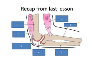
- 1. Recap from last lesson 2 3 5 6 7 1 4
- 2. Muscle contraction 11.2.5 Describe the structure of striated muscle fibres, including the myofibrils with light and dark bands, mitochondria, the sarcoplasmic reticulum, nuclei and the sarcolemma 11.2.6 Draw and label a diagram to show the structure of the sarcomere, including Z lines, actin filaments, myosin filaments with heads, and the resultant light and dark bands 11.2.7 Explain how skeletal muscles contract, including the release of calcium ions from the sarcoplasmic reticulum, the formation of cross-bridges, the sliding of actin and myosin
- 4. Types of muscle • Skeletal or striated muscle • Smooth Muscle • Cardiac Muscle
- 6. Striated Muscle • Also called skeletal muscle because it is responsible for skeletal movement. • Muscles made of thousands of cells called muscle fibres • Connective tissues, blood vessels and nerves • Muscle cells contain multiple nuclei that lie just inside the membrane called sarcolemma. • Sarcolemma has multiple tunnel-like extensions called T tubules (transverse tubules)
- 7. Striated Muscle • Cytoplasm is called the sarcoplasm • Sarcoplasm contains a large numbers of glycosomes that store glycogen. • Myoglobin is also found in the sarcoplasm • Myofibrils run the length of the cell • Myofibrils are the contractile elements of the muscle
- 8. Myofibrils are made up of sarcomeres, and sarcomeres are the units that allow movement. • I bands only actin • H band only myosin You need to be able to draw and label these diagrams
- 9. Myofibril structure Actin • Thin filaments -(8nm in diameter) • Contains myosin-binding sites • Individual molecules for helical structures • Includes two regulatory proteins, tropomyosin and troponin Myosin • Thick filaments (16nm in diameter) • Contains myosin heads that have actin binding sites • Individual molecules form a common shaft-like region with outward protruding heads • Heads are referred to as cross- bridges and contain ATP- binding sites and ATPase enzymes
- 10. Step 1 Sliding Filament Theory • Action potential reaches neuromuscular junction. • Neurotransmitter called acetylcholine released into the gap • Ach binds to receptors on Sarcolemma • Sodium ion channels open and Sarcolemma membrane depolarizes. • Muscle action potential generated.
- 11. Step 2 Sliding Filament Theory • Action potential moves along T- tubule. • Calcium ions are released into the Sarcoplasm from the Sarcoplasmic reticulum
- 12. Step 3 Sliding Filament Theory • Calcium ions released into sarcoplasm • Calcium ions bind to the troponin on the actin myofilaments
- 13. Step 4 Sliding Filament Theory • Calcium ions bind to the troponin on the actin myofilaments • Myosin binding sites are exposed • Heads contain ATPase which splits ATP and releases energy.
- 14. Step 5 Sliding Filament Theory • Myosin binds to myosin binding sites helped by tropomyosin • Myosin-actin cross bridge rotate to produce the power • ATP binds resulting in the detachment of the myosin from the actin • No further action potentials cause calcium levels to drop. Troponin/Tropomyosin complex falls back into original shape – blocking myosin binding sites and muscle relaxes.
- 15. starter • Troponin • Tropmyosin • Actin • Myosin • Calcium • Sarcomere • Sarcolemma • Sarcoplasm
- 16. Very Simply – this happens
- 17. The contraction cycle 1. Myosin heads activated by the splitting of ATP. 2. Myosin heads attach to myosin binding sites on actin. 3. Cross bridge formed and ADP released, energy lost and myosin bends towards the centre of sarcomere. 4. Myosin binds to ATP and causes the head to detach from the actin attachment site
- 18. Longer answer question Use the mark scheme, your notes and simple diagrams to illustrate what happens from start to finish when a muscle contract.
- 19. Mark scheme for exam Q muscles / fibres / myofibrils contain (repeating) units called sarcomeres; muscle / sarcomeres contain actin filaments and myosin filaments; actin fibres are thin and myosin fibres are thick; arriving action potential causes release of Ca2+; from sarcoplasmic / endoplasmic reticulum; Ca2+ binds to troponin; causing troponin and tropomyosin to move (on actin); exposing binding sites on actin / for myosin; ATP binds to myosin heads releasing them / breaking cross bridges; ATP hydrolysed / split into ADP + Pi; ATP / energy causes myosin heads to change shape / swivel / become cocked; myosin heads bind / form cross-bridges to (exposed) actin binding sites; myosin heads swivel / move actin (releasing ADP + Pi); myosin filaments move actin filaments towards centre of sarcomere; sliding of filaments / actin and myosin shortens the sarcomere; 9 max (Plus up to [2] for quality)